Molecular Biology of the Cell — Volume 3 - Alberts B., Bray D., Lewis J., Raff M., Roberts K., Watson J. 1994
From Cells to Multicellular Organisms
Cellular Mechanisms of Development
Principles of Spatial Structure Formation
The various Organs of an animal do not arise from a chaotic accumulation of Cells; the Spatial Organization of different Cell types is predetermined from the outset. The interactions coordinating this process of pattern formation can be remarkably diverse. In some cases, such as in mosaic eggs, the spatial organization may be established within a single cell, and the resulting Intracellular Distribution of cytoplasmic determinants is faithfully reproduced in the arrangement of the embryo's daughter cells. In other cases, such as mesoderm induction in Xenopus or vulval development in nematodes, a defined Structure emerges As a result of interactions between individual cells. Consequently, all types of spatial signals—both intracellular and intercellular—play a crucial role in establishing spatial organization. It can be said that these signals provide cells with positional information, guiding their further specialization.
In this section, we examine the various ways in which positional information is generated and utilized. We will also see how, during The Development of many organisms, individual cells acquire precise molecular "addresses" or positional values. Thus, an early embryonic cell may "know" relatively little—for instance, simply how close it is located to the HEAD or the tail. As for its descendants in the adult animal's body, many cell generations later they may "know" that they are bone cells situated at the tip of the third joint of a forelimb. Such specialization is built up gradually through mechanisms based on cellular memory: early spatial signals provide only a rather rough outline of the overall coordinates, whereas subsequent local signals supply cells with finer address refinements tied to local coordinates.
16.4.1. Asymmetry determined by environmental spatial organization can be amplified through positive feedback [34, 35]
The formation of spatial organization begins with the creation of asymmetry: the future head must differ from the tail, the dorsal side from the ventral. We have already seen that in most animal species, eggs are asymmetric (see Section 16.2.2). This asymmetry often arises within the egg cell during its development in the Ovary. In insects, for example, the oocyte (the future egg cell) is located at one pole of the follicle, where it contacts nurse cells on one side, receiving stored nutrients from them via cytoplasmic bridges (see Section 15.3.4). In all likelihood, the asymmetric delivery of stored nutrients to the oocyte creates chemical differences across different Regions of the egg Cytoplasm, corresponding to the future head and tail of the embryo (see Fig. 16-39).
Class="center">
Fig. 16-39. Control of Drosophila egg polarity by the environment surrounding the oocyte in the ovary. Normally, nurse cells supply stored nutrients to one of the oocyte poles, which corresponds to the future head. In dicephalic mutants, the oocyte is located in the center of the follicle and is symmetrical with respect to the nurse cells. As a consequence, a symmetrical egg is produced, giving rise to an embryo with heads at both ends.
Initially, the asymmetry may be quite subtle: cells subsequently employ their own internal mechanisms to amplify this weak asymmetry and become polarized. For instance, in the egg of the marine alga Fucus, the rhizoid, or rootlet, forms at only one end. Normally, the rhizoid grows downward and away from light. Thus, gravity and light together determine the side on which it develops. This polarity is driven by an ionic current of Ca2+ through The Cell (see Section 20.5.6): it is believed that random light or gravity cues induce a slight asymmetry in the distribution of Proteins involved in transporting Ca2+ ions across The Plasma Membrane. This asymmetry is subsequently amplified by positive feedback mechanisms (see Fig. 20-66, Section 20.5.7).
In a similar manner, positive feedback can operate at other stages in the development of Multicellular Organisms. For example, when a polarized cell divides, the daughter cells inherit the weakly expressed asymmetry and, by amplifying it, become more strongly polarized. This polarization is thus reinforced across successive cell generations. Therefore, in principle, Intercellular signaling is not strictly required for The Emergence of complex structures in multicellular organisms (Fig. 16-40). It is possible that certain pattern-formation processes relying on autonomous cell behavior can be explained by the operation of such a mechanism.
However, as a rule, spatial pattern formation in multicellular organisms also depends on intercellular signals. Here, too, positive feedback plays a vital role in amplifying initially weak asymmetries, generating localized groups of cells with vastly different properties. These groups of cells may, in turn, emit signals that influence the development of neighboring cells. A simple example is found in the primitive Organism Hydra. The body of the hydra is tube-shaped, with a "head" bearing tentacles at one end and a "FOOT" at the other (see Fig. 1-33). Hydra is well known for its remarkable ability to regenerate its entire body after the removal of any part. Numerous head-amputation experiments performed on hydra suggest that the regeneration process is driven by a local autocatalytic process that amplifies the cells' weak tendency to acquire "head" properties. All Cells of the body participate in creating this missing structure by producing an inhibitory signal that diffuses along the longitudinal body axis, thereby preventing the formation of a head in other regions of the organism (Fig. 16-41).

Fig. 16-40. A complex multicellular structure can, in principle, arise through a series of asymmetric cell divisions without any intercellular interactions (aside from those maintaining Cell Adhesion). The internal polarity inherited by each daughter cell through successive cell divisions may prove entirely sufficient to direct the next cycle of intracellular pattern formation. The daughter cells generated in subsequent divisions will likewise be distinct, and their internal polarization will not diminish.
16.4.2. A localized signaling region can frequently establish a morphogen gradient [36]
The long-range influence exerted by the head over the rest of the hydra body is a widespread phenomenon. In many developing systems, small patches of tissue are able to acquire special properties that make them a source of a signal capable of spreading through adjacent tissue and controlling its behavior. Specifically, the signal may consist of diffusing molecules secreted by the signaling region. Suppose that as the signaling molecule diffuses through neighboring Tissues, it undergoes degradation. In this case, the highest concentration of the signaling substance will be near the source. With increasing distance from the source, The amount of this substance will decrease, thereby establishing a concentration gradient across the tissue (see Fig. 16-42). At varying distances from the signal source, cells will be exposed to different levels of this substance depending on its concentration, and will consequently acquire different properties. A hypothetical substance of this kind—whose concentration is read by cells to determine their position relative to a source—is called a morphogen. The signaling region is able, through the morphogen, to control pattern formation across large fields of surrounding tissue. Morphogen gradients provide a simple and effective means of supplying cells with positional information, which is why morphogens have taken center stage in our attention. It should be added here that embryonic cells can acquire positional information through other means as well. Fig. 16-21 illustrates one such alternative, where short-range induction interactions form the underlying mechanism. The Formation of the activator in this region triggers The production of an inhibitor that diffuses over long distances and suppresses activator synthesis elsewhere. For example, when a hydra is decapitated, the remaining tissue normally regenerates a head; however, if a head from another hydra is grafted onto the amputation site or at some distance from it, no new additional head is formed.

Fig. 16-41. Mechanism of spatial pattern generation via an autocatalytic process.
As a result of an autocatalytic process, the "activator" substance accumulates to a very high level in a small region whose Location is determined by the system's subtle initial asymmetry; this high concentration relative to certain reference points, or "landmarks," is termed a morphogen. Morphogen gradients provide a simple and effective means of supplying cells with positional information, which is why morphogens have become a central focus of our attention. It should be added here that embryonic cells are capable of acquiring positional information through alternative mechanisms. Fig. 16-21 illustrates one such alternative, where short-range inductive interactions underlie the mechanism.

Fig. 16-42. If a substance is produced at a specific point and undergoes degradation as it diffuses from that point, a concentration gradient of this substance is established, peaking at the point of origin. Such a substance can act as a morphogen, whose local concentration controls cell behavior according to their distance from the source.
Although numerous Examples of signaling regions are known that (from our perspective) could exert long-range effects via a morphogen gradient, in practice we currently have only a few instances of the direct chemical identification of a morphogen. As a rule, it is only known that when THE POSITION OF a putative signaling region is altered—following transplantation or by other means—The structure of the adjacent tissue changes accordingly. Observations of this kind do not reveal the extent to which changes in individual cell structures are determined by a direct response to the morphogen versus THE CONTRIBUTION OF interacting responding cells. Presumably, in most cases, the initial morphogen gradient triggers a broad-acting pulse that initiates spatial pattern formation, while local intercellular interactions participate in refining the details. Accumulating evidence indicates that the body plan of insects arises in precisely this manner, as will be discussed below. Here, we examine what type of spatial organization can be generated by the action of a morphogen gradient within a field of cells, each responding independently.
16.4.3. A cell's response threshold dictates a strictly defined character of its determination despite a smooth morphogen gradient [37]
In the presence of a smooth morphogen concentration gradient, one might expect cell properties in different regions to change gradually as well. Such subtle differences do indeed occur in certain tissues. Of greatest interest, however, is the emergence of sharp qualitative distinctions—such as those between Cartilage and Muscle cells, which lack transitional forms. Within a population of initially homogeneous cells, a threshold response to a smoothly varying signal can give rise to steep differences between cells: in each responding cell, The Effect of a small incremental change in the signal can be amplified via positive feedback. Consequently, cells exposed to a signal whose intensity varies only slightly will choose different developmental pathways depending on whether they experience a suprathreshold or subthreshold intensity. Furthermore, multiple thresholds of intensity may exist for individual signals, allowing a single variable to control multiple binary choices. Once a cell has definitively embarked on a pathway leading to one of its stable states under METABOLISM/18.html">The Influence of a given factor, it will continue to develop in that chosen direction even in the absence of the factor that originally controlled the choice. Along this pathway, temporary, position-dependent influences can elicit a "memorization" effect regarding the cues the cell has experienced. A cell's choice of a specific state depends on the positional information retained in its cellular memory. This record, embodied in a specific cellular property, can be termed its "positional value."
16.4.4. Embryonic fields are very small, meaning that the basic Features of the adult body plan must be determined quite early with the participation of cellular memory [38]
Regardless of The Nature of the mechanisms providing cells with positional information, their range of action is rather limited: they are typically effective only within small domains (morphogenetic fields) measuring about 1 mm in length, which corresponds to roughly 100 cell diameters or fewer. It follows that There is a limit to the level of detail that can be laid down within such a restricted space. For this reason, the ultimate positional value of a cell must be established through successive elements of positional information recorded at various Selection/3.html">Stages of development. Consequently, a determination mechanism based on cellular memory is absolutely essential for the development of large, complexly organized animals. The distinction between the head and the tail must be laid down while the corresponding primordia are still no longer than 1 mm. By the time the animal reaches a length of a centimeter or a meter, the events that generated these differences will be "ancient history"; for these distinctions to be maintained, cells must possess a robust memory.
Thus, the overall body plan is established very early, while increasingly fine details are added later as the individual organ primordia reach sizes suitable for recording additional positional information (Fig. 16-43).
We will examine how this occurs in greater detail using limb development as an example.
16.4.5. During limb development, positional information is progressively refined [39]
The development of limbs and many other organs (such as Teeth, vertebrae, or Skin) involves a remarkably limited set of differentiation pathways. A limb, for instance, consists of only a few cell types: muscle, cartilage, bone, and Connective Tissue cells. Yet, these few differentiated cell types are arranged in space in an exceptionally complex manner. Furthermore, the differences between forelimbs and hindlimbs do not arise from the presence of additional tissue types; rather, each limb is characterized by a unique spatial arrangement of tissues. Transplantation experiments with developing chick limbs have demonstrated that the internal differences between limb cells, which dictate the resulting structures, are determined long before overt differentiation begins.
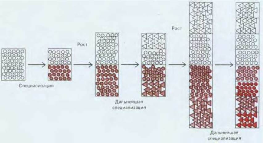
Fig. 16-43. When the embryo is small, it begins to subdivide into distinct regions with unique properties that correspond to the major body PARTS OF THE adult organism. The cells in each such region—whose boundaries are often quite difficult to define—acquire broad positional values (represented here as triangles or circles). As the embryo grows, these regions expand as well, and subregions begin to emerge within them, ultimately producing an increasingly detailed pattern of positional values.
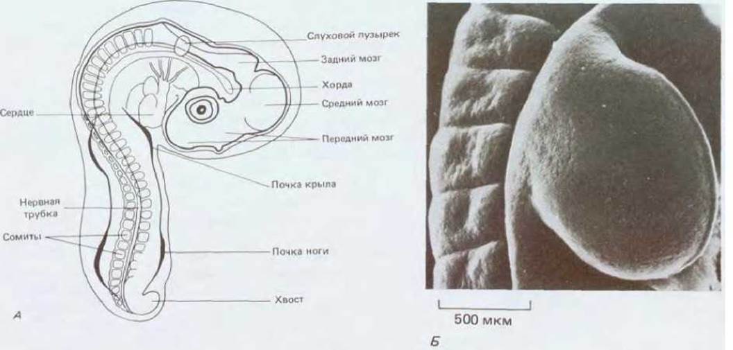
Fig. 16-44. A. A chick embryo after three days of incubation, showing the positions of the early limb buds. B. A wing bud and adjacent somites after another day of development; the bud has grown into a paddle-shaped outgrowth (approximately 1 mm long, 1 mm wide, and 0.5 mm thick). Scanning electron micrograph of the dorsal surface of the wing. (A - from W. H. Freeman, B. Bracegirdle, An Atlas of Embryology, London, Heinemann, 1967; B - courtesy of Paul Martin.)
Legs and wings in the chick embryo arise almost simultaneously as small, elongated buds protruding from the lateral body wall (Fig. 16-44). The cells of both pairs of limb buds appear identical and are undifferentiated at this stage; nor is there any morphological indication of the future skeletal pattern (Fig. 14-20, Section 14.2). At this stage, a small piece of undifferentiated tissue can be excised from the base of a leg bud (tissue that would normally form the lower leg) and grafted onto the tip of a wing bud. As the graft develops, it forms a digit rather than a lower leg or a corresponding tip-of-the-wing structure (Fig. 16-45). This experiment demonstrates, first, that the cells of the early leg bud are already determined as leg cells, and second, that despite this determination, they have not yet acquired detailed positional values along the distal axis of the bud and can respond to wing positional signals by forming structures characteristic of the tip of the limb rather than its base. We can conclude that the final differentiation of vertebrate cells is determined by a series of sequential positional information signals recorded by cellular memory at different times. A cell's terminal state emerges as the outcome of a specific sequence of developmental decisions.

Fig. 16-45. Future thigh tissue, when grafted to the margin of a chick wing bud, gives rise to digits. (J. W. Saunders et al., Dev. Biol., 1:281-301, 1959.)
16.4.6. Having acquired positional values, similar cells become non-equivalent [40]
Although forelimb and hindlimb buds are composed of identical types of differentiated cells, it is abundantly clear that these cells are non-equivalent. They differ in their internal state, which is dictated by distinct positional values. Limb bud cells retain the positional memory of whether they belong to a leg or a wing even after differentiation; consequently, differentiated cells of the same type (such as cartilage cells) are non-equivalent. This implies that adult cells are actually far more diverse than traditional classifications of vertebrate cell types would suggest (see Chapter 17, Supplementary Material).
Researchers have accumulated evidence pointing to the existence of such subtle differences among differentiated cells of the same type located in different regions of the vertebrate body. Examples of cell non-equivalence in the skin and Nervous system will be discussed later in this chapter (Section 16.6.4) and in Chapter 19 (Section 19.7.9). One of the most striking examples comes from studies of limb regeneration in urodele amphibians, which have demonstrated that cell non-equivalence applies not only to forelimbs versus hindlimbs, but also to cells located at different levels along the proximodistal axis of a single limb.
If a limb is amputated at any level from its base in a newt or axolotl, the missing part is fully restored. A mound of outwardly undifferentiated cells covered by epidermis forms at the cut surface of the stump; this is known as the regeneration blastema. Through growth and differentiation, the blastema gives rise precisely to those structures that normally lie distal to the plane of amputation. If the hand is removed, a hand regenerates; if both the forearm and hand are removed, the missing forearm and hand are restored. Remarkably, it does not matter where the blastema develops: whether it forms normally at the proximal end of the stump or is experimentally induced at the distal end of an amputated limb segment (Fig. 16-46). In either case, the blastema cells produce only those structures that normally lie distal to the level of the cut, although in the latter case, a mirror-image duplication of the existing segment is formed. Presumably, cells located at different levels along the limb axis have memorized distinct positional values and rely on these values to execute the program that restores missing parts.
16.4.7. Cells in different regions are provided with the same positional information but interpret it differently [36, 41]
The experiment schematically illustrated in Fig. 16-45 offers insight into how complex, detailed positional information is generated within chick limb cells. It demonstrates that the signals providing cells with positional information along the limb axis are fundamentally identical in both wings and legs. When leg cells are transplanted to the tip of a wing bud, they accurately read the signals indicating a distal position and thus participate in forming digits. However, interpreting this information in their own fashion, they form toes rather than wing digits. Mechanisms based on cellular memory can thus be deployed to supply positional information to cells in various distinct fields. As a result, a specific structure develops in each field through the integration of information encoded by the cells' unique developmental history and extrinsic intercellular signals.

A regeneration blastema forms on both surfaces; in both cases, the blastema cells give rise to the limb structures that normally lie distal to the level of the cut
Fig. 16-46. As shown by this experimental design, the Nature of the structures formed during blastema regeneration in a salamander is determined by the level at which the limb was amputated, rather than by which structures remain in the stump. Both the forearm and hand regenerate from both distal and proximal stump tissues.
These considerations apply not only to the signals encoding spatial organization along the long (proximodistal) axis of the limb, but also to the anteroposterior (thumb-to-little-finger) axis. These two axes (Fig. 16-47) correspond to different components of positional values, much like a Cartesian coordinate system, which in turn are established through distinct mechanisms of positional information readout. The regulation of anteroposterior patterning is of particular interest because it represents one of the few instances where a morphogen has been successfully identified chemically.
16.4.8 Retinoic acid is a putative morphogen in the vertebrate limb bud [42]
At the posterior margin of the chick limb bud lies a small group of cells with unique properties, which become apparent when these cells are grafted into the anterior margin of another limb bud (Fig. 16-48). Under the Influence of the graft, the recipient bud begins to expand in width within the first day, ultimately forming a grossly altered limb. This results in a duplication of all skeletal elements and their associated soft tissues, producing a second set of structures organized in mirror Symmetry with respect to the original anteroposterior axis. This second set of limb structures is derived almost entirely from the recipient bud's own cells, rather than from the graft tissue. It appears that the host cells are instructed by the graft to form a duplicated, reversed set of structures. The region of the limb bud that serves as the source of the graft has been named the zone of polarizing activity (ZPA), or simply the polarizing region.
A polarizing region is present in both leg and wing buds, and remarkably, the polarizing regions from either pair of limbs—and even from different vertebrate species—are functionally interchangeable. Regardless of the graft's origin (leg or wing, chick or mouse), it invariably induces the formation of an extra chick wing outgrowth composed of host embryonic tissues, which then develops additional chick digits. Consequently, the signal emitted by polarizing regions is conserved across different limbs and species. Limb cells interpret this signal according to their own genome and developmental history.
In a normal wing, three digits correspond to digits 2, 3, and 4 of a five-fingered hand and are designated accordingly. Digit 4 forms closest to the polarizing region, digit 3 somewhat farther away, and digit 2 farthest away. The same rule applies to tissues placed at varying distances from a polarizing region graft, as if the pattern of resulting digits reflects exposure to a graded morphogen secreted by the polarizing tissue (Fig. 16-49). The graded nature of the signal emitted by polarizing cells can be demonstrated by grafting varying numbers of cells: a graft of about 30 cells induces an extra digit 2, a graft of 80 cells induces an extra digit 3, and a graft of 130 cells induces an extra digit 4.
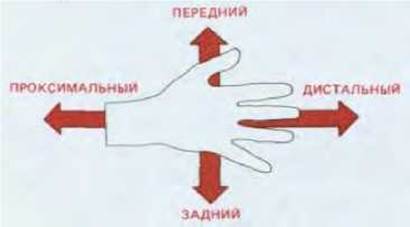
Fig. 16-47. Proximodistal and anteroposterior axes of the vertebrate limb.
The effect of transplanted cells from the polarizing region can be replicated by implanting an inert carrier soaked in retinoic acid. Retinoic acid diffuses from the carrier, establishing a concentration gradient along the limb bud, where the peak concentration is at least three times higher than the minimum concentration; additional digits begin to form at a concentration of approximately 20 nM. Utilizing highly sensitive chromatographic techniques, researchers have demonstrated that a natural retinoic acid gradient of precisely this magnitude exists within a normal limb bud, with the highest concentration localized in the cells of the polarizing region. It is highly probable that retinoic acid itself serves as the natural morphogen (Fig. 16-50).
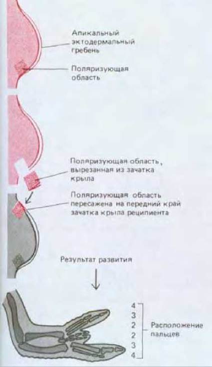
Fig. 16-48. A graft from the zone of polarizing activity induces a mirror-image duplication of wing structures in the recipient.

Fig. 16-49. The distance between the transplanted piece of the zone of polarizing activity and the host's corresponding zone determines the type of structures formed by the wing bud cells. The numbers 2, 3, and 4 indicate which digit develops from each region of the bud.

Fig. 16-50. Chemical Structure of retinoic acid.
The retinoic acid receptor has recently been identified. It turned out to be a protein homologous to steroid and thyroid Hormone Receptors; it binds to specific DNA sequences and regulates the Transcription of specific genes. The cDNA encoding this receptor has been isolated and sequenced. Currently, both DNA probes and corresponding Antibodies are used to detect these receptors in tissues such as limb buds.
16.4.9. Growth is controlled by a pattern of positional values that can be altered through intercalation [43]
Up to this point, our Discussion of pattern formation and positional values has overlooked one crucial aspect of the process: the regulation of growth, which is essential for embryonic parts and organs to attain their appropriate sizes. In some cases, these mechanisms rely on autonomous cellular programs triggered during the Cytology/cytology/16.html">Early stages of specific organ rudiments. However, in many other instances, growth and the structure of positional values are determined as a direct result of ongoing intercellular interactions and are closely linked to them. By studying regulatory processes that occur in various organisms when tissue fragments with different positional values are swapped, researchers have discovered that cells from these fragments grow and adapt to local conditions. These experiments lead to a simple, general rule, best illustrated by studies on limb regeneration in cockroaches.
Cockroaches belong to insects that do not undergo a radical metamorphosis during their development from larva to adult; instead, development proceeds gradually through a series of juvenile stages. The limbs of young cockroaches are well differentiated, but unlike human limb cells, these differentiated cells retain The ability to respond to signals governing the spatial ORGANIZATION OF THE limb. If this organization is disrupted, it is restored. These features make the experimental subject ideal for investigating the systems that control spatial pattern formation by performing surgeries on young individuals after embryonic development is complete.
We will describe experiments that examined The behavior of the epidermal layer and cuticle, which cover the body of the cockroach and form the outer surface of the limb. The growth of the exoskeleton is linked to successive molts, in which the young insect sheds its old cuticle and replaces it with a new, larger one. The cuticular material is secreted by the underlying epidermal cells, which form a single cell layer. The positional values of these cells determine the STRUCTURE OF THE cuticle they produce; if these values are altered experimentally, the outcome can be detected by examining the cuticle after a molt. Regeneration is observed only in young individuals, as adult insects neither grow nor molt.
A cockroach leg consists of several segments arranged (from base to tip) in the following order: coxa, trochanter, Femur, Tibia, tarsus. The tarsus, in turn, consists of several small segments and terminates in a pair of claws (Fig. 16-51). If two legs severed at different levels of the tibia are removed, the distal part of one leg can be transplanted onto the proximal stump of the other. This yields a "composite" leg missing the middle section of the tibia. Nevertheless, following a molt, a seemingly normal limb develops: the lost middle section of the tibia regenerates (Fig. 16-52, A). The surgery can be modified to produce an even more striking result. The tibia of one leg is cut near its proximal end, and the tibia of another near its distal end. The long severed tibia of one leg is joined to the long stump of the other, resulting in an elongated limb with a duplicated middle section (Fig. 16-52, B). After molting, this leg is found to have elongated even further: a third middle section has formed between the two preexisting middle sections. As shown in Fig. 16-52, B, the bristles in this newly formed region point in the opposite direction relative to the bristles on the rest of the tibia.

Fig. 16-51. A cockroach leg. With each successive molt, the leg grows, but its basic structure remains unchanged.

Fig. 16-52. Following the joining of mismatched parts of a cockroach tibia, intercalation of new tissue (shown in light tone) takes place, filling the gap in The sequence of positional values (numbered 1 to 10). In the first case (A), intercalation restores the missing region, whereas In the second case (B), a third, middle section of the tibia forms between the two already present. The orientation of the bristles indicates the polarity of the intercalating tissue. In both cases, the continuity of the positional value sequence is restored.
Many operations of this type have been performed. All of them point to the existence of a system of positional values whose presence renders cells located in different positions non-equivalent. The character of a positional value is closely tied to the control of cell proliferation. It is conventional to designate a positional value with a number that varies from a maximum at one end of a limb segment to a minimum at the other. In the surgeries described above, epidermal cells with dramatically different positional values are brought into contact. Consequently, cell proliferation is triggered at the junction site, and the new cells acquire values that smoothly, without abrupt jumps, bridge the gap between the positional values of the cells brought together by the operation (Fig. 16-52). This result can be generalized into the rule of intercalation: discontinuities in a smooth gradient of positional values induce local cell proliferation, and newly generated cells acquire intermediate positional values, thereby restoring structural continuity. Proliferation ceases only once the gap is filled with cells possessing all the missing positional values, which restores the normal spatial distribution of cells. This entire process is known as intercalary regeneration.
The rule of intercalation and its corollary—that growth continues until a specific distribution of positional values is achieved—constitutes a vital organizing principle for the systems in which it operates. Starting from a roughly defined and miniature structure (for example, driven by a morphogen gradient), this rule dictates the generation of a complete set of positional values and regulates the growth of each structural part to a definitive size. All of this is necessary to ensure that the resulting structure is qualitatively, and thus topologically, correct. When applied to two- and three-dimensional structures, the rule of intercalation proves valid for a wide range of phenomena, encompassing both normal regeneration of amputated body parts and abnormal effects (such as the appearance of extra limbs following certain grafts). In all likelihood, this rule holds true for many processes of Organogenesis and regeneration not only in insects, but also in crustaceans and amphibians. For instance, in Drosophila, early errors in structural specification are corrected in accordance with this rule (Fig. 16-60). Even in mammals, whose lost structures do not regenerate in adulthood, growth and spatial organization during the Embryonic period may still be governed by the rule of intercalation. The molecular mechanisms underlying this crucial growth control pathway remain unknown for now.
The emergence of various cell types in the embryo is strictly ordered in space. This process typically begins with the establishment of egg asymmetry and continues through intercellular interactions during Embryogenesis. At each stage of this process, an initially subtle asymmetry is amplified by positive feedback, resulting in a well-defined spatial organization. One can say that the signals coordinating pattern formation supply cells with positional information. In the simplest case, a graded concentration of a diffusing morphogen can control cellular properties based on distance from the morphogen source; during chick limb development, retinoic acid likely acts as the morphogen controlling the formation of elements along the thumb-to-little-finger axis. Discrete differences in cellular properties may correspond to threshold responses to the morphogen.
A cell's spatial characteristics are determined by the positional information it receives over time. While the embryo is small, this pattern is quite rough, but as the embryo grows, refining details are added. Cells of early forelimb and hindlimb buds in vertebrate embryos acquire distinct positional values, making The properties of cells from different limbs non-equivalent long before differentiation. Refinements in the structure of each organ emerge after a more detailed internal network of positional information is established within each of these rudiments. This detailed positional information system is likely very similar in homologous organs such as forelimbs and hindlimbs. Equipped with a cellular memory, cells from different fields interpret this refining positional information differently, according to their developmental history.
The structure of positional values in many animals is closely linked to the control of cell proliferation via a simple rule of intercalation. This rule was formulated through studies of limb regeneration in insects and amphibians. It states that a disruption in the continuity of positional values triggers local cell proliferation, and newly generated cells acquire intermediate positional values that restore the continuity of the disrupted structure. This exact same mechanism may also operate during normal embryonic development, correcting inaccuracies in the initial positional information profiles.
Last update: 12/08/2026
Editorial and Educational Adaptation: This material has been compiled based on the primary/original source text. The project team performed an editorial review, corrected technical inaccuracies, structured sections, and adapted the content for an educational format.
What was processed:
- elimination of formatting defects (OCR errors, structural breaks, corrupted characters);
- editorial organization of content;
- standardization of terminology in accordance with academic sources;
- verification of factual statements against the original source text.
All mentions of the author, publication year, and origin of the primary text have been preserved in accordance with the source.