BOTANY, VOLUME 2 - PLANT PHYSIOLOGY - 2007
7. DEVELOPMENTAL PHYSIOLOGY
7.3. Cellular Basis of Development
7.3.1. METABOLISM and Intracellular Protein Sorting
Following successful Transcription on The pathway of genetic expression, the information contained in mRNA is utilized to synthesize Proteins. This process takes place on Ribosomes and is referred to as Translation. Here, The Genetic Code (see 7.3.1.1) is translated into a collinear sequence of Amino Acids. Protein folding and, where necessary, protein modification are two processes that may occur either co-translationally during Protein Synthesis or post-translationally after synthesis has been completed, ultimately yielding a functional protein (see 7.3.1.2). The cellular protein pool is the result of a regulated balance between PROTEIN SYNTHESIS AND protein degradation (see 7.3.1.3).
In eukaryotes, protein synthesis occurs in the Cytoplasm, Plastids (if present), and Cell/35.html">Mitochondria. Proteins synthesized within plastids or mitochondria remain within their respective Organelles, whereas proteins synthesized in the cytoplasm are directed to various compartments or secreted from The Cell. Proper intracellular protein sorting is crucial for establishing and maintaining compartmentation in eukaryotes (see 7.3.1.4).
Among the numerous amino acids found in nature, generally only 20 so-called Proteinogenic Amino Acids are used for protein synthesis during translation (see Fig. 1.11). As noted above, their sequence within a protein is determined by The base sequence of the DNA or mRNA, respectively.
The informational content of DNA or mRNA, expressed by four "characters" ("code letters"), must be translated during protein synthesis into a polypeptide "text" comprising a maximum of 20 equally informative "characters." Naturally, using 4 nucleic acid "characters" to encode 20 amino acids is only possible through combinations. If a base doublet composed of 4 different bases were to serve as a "code word" of the genetic code (i.e., corresponding to a specific amino acid), a maximum of 42 = 16 different amino acids could be encoded. If three consecutive NUCLEOTIDES (a triplet) constitute a code word, then 43 = 64 different Amino acids can be encoded. It has been demonstrated that the genetic code indeed consists of successive, non-overlapping base triplets—codons—that correspond to individual amino acids (Table 7.3). The genetic code is universal (i.e., it is valid for Viruses, Bacteria, plants, animals, and humans), though not entirely invariable, which is why it is referred to as the standard code. Exceptions (Table 7.4) are discussed below. Codons are written in the 5' -> 3' direction, corresponding to the direction of mRNA movement during translation.
Class="center">Table 7.3. Standard Genetic Code
UUU |
Phe |
UCU |
Ser |
UAU |
Tyr |
UGU |
Cys |
UUC |
Phe |
UCC |
Ser |
UAC |
Tyr |
UGC |
Cys |
UUA |
Leu |
UCA |
Ser |
UAA |
Stop |
UGA |
Stop |
UUG |
Leu |
UCG |
Ser |
UAG |
Stop |
UGG |
Trp |
CUU |
Leu |
CCU |
Pro |
CAU |
His |
CGU |
Arg |
CUC |
Leu |
CCC |
Pro |
CAC |
His |
CGC |
Arg |
CUA |
Leu |
CCA |
Pro |
CAA |
Gln |
CGA |
Arg |
CUG |
Leu |
CCG |
Pro |
CAG |
Gln |
CGG |
Arg |
AUU |
Ile |
ACU |
Thr |
AAU |
Asn |
AGU |
Ser |
AUC |
Ile |
ACC |
Thr |
AAC |
Asn |
AGC |
Ser |
AUA |
Ile |
ACA |
Thr |
AAA |
Lys |
AGA |
Arg |
AUG |
Met |
ACG |
Thr |
AAG |
Lys |
AGG |
Arg |
GUU |
Val |
GCU |
Ala |
GAU |
Asp |
GGU |
Gly |
GUC |
Val |
GCC |
Ala |
GAC |
Asp |
GGC |
Gly |
GUA |
Val |
GCA |
Ala |
GAA |
Glu |
GGA |
Gly |
GUG |
Val |
GCG |
Ala |
GAG |
Glu |
GGG |
Gly |
Triplets are given in the 5' -> 3' direction (According to the three-letter amino acid code, Fig. 1.11). Bold type: stop codons or the Methionine codon, which in an appropriate context (see Fig.
7.11) designates the initiation site of translation.
While only a single corresponding codon exists for Tryptophan and methionine, all Other Amino Acids are represented by 2 to 6 codons, meaning that the genetic code is degenerate. This degeneracy primarily involves the base at the third position of the codon. This feature confers an evolutionary advantage, as not every point mutation (substitution of one base for another) leads to A change in the Amino Acid Sequence of the corresponding protein. It is noteworthy that amino acids encoded by multiple codons are more frequently found in proteins, and that UC-containing triplets encode hydrophobic amino acids, whereas AG-containing triplets encode hydrophilic ones; consequently, the former are located on the left/upper side and the latter on the right/lower side of the codon table (see Table 7.3). Finally, base triplet families (sharing the first base) specify amino acids that exhibit similarities in their Biosynthesis and, consequently, in their Structure. The correspondence between codons and amino acids is therefore not random, suggesting the coevolution of codons and amino acids. As a molecular mechanism, the structural complementarity between certain Ribonucleic Acids and specific amino acid molecules in an "RNA world" is currently discussed.
Along with codons specifying Amino acid sequences, the code also contains "punctuation marks": the initiation codon 5'-AUG-3', which simultaneously encodes methionine, as well as three stop codons: 5'-UAA-3', 5'-UAG-3', and 5'-UGA-3', designated as "ochre", "amber", and "opal"1, respectively; these mark the start point and the end of the translatable region of the mRNA.
1 These names derive from the eye color phenotype of the fruit fly Drosophila melanogaster, in which Mutations in the proteins responsible for eye color introduce corresponding stop codons that prematurely terminate translation. — Note by the Editor.
Table 7.4. Selected deviations from the standard genetic code
Codon 5' -> 3' |
Standard code meaning |
Alternative coding |
|
Chondriome: UGA |
Stop |
Tryptophan |
|
AUA |
Isoleucine |
Methionine |
Saccharomyces cerevisiae |
CGG |
Tryptophan |
Zea mays |
|
Plastid genome: AUA |
Isoleucine |
Start |
Heterocapsa triquetra (dinoflagellates) |
UUG |
Leucine |
Start |
Heterocapsa triquetra (dinoflagellates) |
|
UGA |
Stop |
Tryptophan |
Mycoplasma spec. |
Nuclear genomes: CUG |
Leucine |
Candida cylindracea |
|
UAA, UAG |
Stop |
Glutamine |
Certain Protozoa, Acetabularia |
UGA |
Stop |
Selenocysteine |
Sequence context-dependent in certain pro- and eukaryotes (e.g., Chlamydomonas) |
Fig. 7.13. Encoding of amino acid information on DNA, mRNA, and tRNA. mRNA codons possess a base sequence identical to the corresponding triplets of the coding DNA strand (except that U replaces T in mRNA). Complementary triplets of the template strand used for mRNA synthesis are termed codogenes; their base sequence is essentially identical to tRNA anticodons (again with U replacing T in RNA). However, anticodons sometimes contain rare bases resulting from secondary modification of primary bases, and non-standard Base Pairs eventually occur (see text)

Over time, deviations from the standard triplet code have been discovered (see Table 7.4). Notably, in most PROKARYOTES AND EUKARYOTES, the stop codon 5'-UGA-3' encodes selenocysteine, the twenty-first proteinogenic amino acid, which in plants has thus far been detected only in the polypeptide chain of Glutathione peroxidase in Chlamydomonas reinhardtii. This "recoding" depends on the conformation of a stem-loop structure in the mRNA formed by internal base pairing, which is located immediately 3' to the UGA triplet in prokaryotes, or within the 3'-untranslated region of the mRNA in eukaryotes.
tRNAs containing 74 to 94 nucleotides serve as intermediate molecules that ultimately enable the translation of mRNA triplet sequences into amino acid sequences (see Figs. 1.10, A; 7.13); of these, bacteria possess 30–45 different types, and eukaryotes up to 50. Each tRNA carries a complementary triplet, the anticodon, within its anticodon loop (see Fig. 1.10, A), which engages in complementary base pairing with the codon on the ribosome, thereby delivering the codon-specified amino acid to the protein synthesis machinery. All tRNAs feature a 5'-CCA-3' base sequence at their protruding 3'-end and, in their "charged" state, always carry a single characteristic amino acid linked via an ester bond between its carboxyl group and the 2'- or 3'-hydroxyl group of the terminal ribose. tRNA "charging" is catalyzed by Aminoacyl-tRNA synthetases (Fig. 7.14), with a specific enzyme existing for each amino acid. Numerous contacts between the respective synthetase, The amino acid, and the acceptor tRNA—which encompasses the anticodon and various other Structural elements of the tRNA (including rare bases!; see Fig. 1.10, A, B)—ensure that only matching tRNAs and amino acids react to form aminoacyl-tRNA. Additionally, as a proofreading function, aminoacyl-tRNA synthetases possess esterase activity that hydrolytically removes incorrectly attached aminoacyl residues. This esterase activity is significantly weaker against the cognate amino acid for a given tRNA. Experiments have demonstrated that the translation error rate in Escherichia coli is approximately 1 per 104 incorporated amino acids. Since there are more distinct tRNAs than proteinogenic amino acids, several isoaccepting tRNAs exist for many amino acids.
Fig. 7.14. Aminoacyl-tRNA: A — Synthesis of aminoacyl-tRNA. The amino acid is first activated by ATP to form aminoacyl-adenylate; class II aminoacyl-tRNA synthetases transfer the activated amino acid with the release of AMP to the 3'-OH group of the ribose at the 3'-end of the tRNA; class I Enzymes transfer it to the 2'-OH group; B — 3'-end of an aminoacyl-tRNA whose formation is catalyzed by a class II aminoacyl-tRNA synthetase; puromycin is a structural analogue of the 3'-end of a tRNA charged with Tyrosine or phenylalanine; the amide bond of puromycin is not cleaved by peptidyl transferase, so that binding of puromycin to the ribosomal acceptor site leads to premature termination of protein synthesis

The synthesis of rare tRNA bases, which can account for up to 10% of tRNA bases and occur predominantly in paired loop regions (see Fig. 1.10, A), proceeds via post-transcriptional modification, primarily in the cytoplasm, involving methylation, reduction, or the attachment of a dimethylallyl moiety to an adenine residue via a cytoplasmic dimethylallyl transferase. In the latter case, N6(Δ2-isopentenyl)adenine (IPA)—a compound that in its free form acts as a cytokinin and regulates plant development (see 7.6.2)—typically arises at the 3'-position adjacent to the anticodon as an integral component of the tRNA.
Selenocysteine tRNA is formed through secondary modification, where serine is first attached to the tRNA and then converted into selenocysteine by the enzyme selenocysteine synthase, with selenophosphate acting as the selenium donor. In bacteria, secondary modification also generates N-formylmethionine tRNA from methionine tRNA, which serves as the initiator amino acid at the 5'-AUG-3' start codon during Translation initiation instead of methionine.
The loop-induced conformational bend of the anticodon causes the first Base of the anticodon and the third base of the mRNA codon to undergo somewhat imprecise base pairing (see Fig. 7.13). Consequently, alternative base pairs differing from the standard ones (G with C, or A with U; see Fig. 1.6) can also form. This phenomenon is known as wobble1. For example, G-U pairs (forming 2 Hydrogen Bonds) are possible. The guanine derivative inosine (I) at this anticodon position can even pair with 3 different bases (A, U, C) via 2 hydrogen bonds. In rare cases, unusual bases appear even in the middle position of the anticodon triplet (such as pseudouridine,  , which pairs with A; see Fig. 1.10). Wobbling reduces the total number of tRNAs required to decode all triplets. Mitochondrial tRNAs frequently form pairs with all 4 base types at the third codon position. This "super-wobble" significantly decreases the required number of tRNAs in mitochondria.
1 In addition to literal movement, the term "wobble" conveys nuances of unreliability, instability, and unforeseen contingency. — Ed. note.
7.3.1.2. Protein Biosynthesis
The process of protein biosynthesis can be divided into:
✵ the initiation phase,
✵ the elongation phase,
✵ the termination phase.
Below, we will limit our Discussion to protein biosynthesis on eukaryotic 80S ribosomes (see 2.2.4), while highlighting key differences from prokaryotic translation on 70S ribosomes.
The initiation phase of translation begins at the 5' cap of the mRNA (see Fig. 7.12)1 with the assembly of a pre-initiation complex. This complex comprises the small (40S) ribosomal subunit, an initiator tRNA charged with methionine (distinct from the methionine tRNA that recognizes internal 5'-AUG-3' codons within the Open Reading Frame), and other proteins known as initiation factors. In plants, translation initiation also involves the poly(A) tail at the 3' end of the mRNA and the poly(A)-binding protein; the longer the poly(A) stretch, the higher the frequency of translation initiation. The resulting initiation complex scans the mRNA in the 5' —> 3' direction to locate the start codon. This start codon differs from "internal" methionine codons due to its flanking sequence context (the Kozak consensus sequence, see Fig. 7.11). Once the initiation complex reaches this position, the large (60S) ribosomal subunit binds, and protein synthesis commences.
1 It should be emphasized that eukaryotic translation initiation requires a free, cap-modified 5' mRNA terminus. The ribosome then synthesizes a single protein encoded closest to the 5' end. Even if an mRNA carried information for two distinct proteins, the eukaryotic ribosome would be unable to translate both open reading frames. This property shapes eukaryotic genome structure: each promoter drives a single Gene, and translated mRNAs are invariably monocistronic. — Ed. note.
In contrast, prokaryotic translation initiation begins with The formation of an initiation complex at a ribosome-binding site located 3 to 10 bases upstream of the start codon in the 5' direction (the Shine-Dalgarno sequence: 5'-AGGAGGU-3' or variants thereof)1. Prokaryotes also utilize a specialized initiator tRNA, though it carries N-formylmethionine rather than methionine.
1 Crucially, prokaryotic translation initiation does not require a modified 5' mRNA terminus; prokaryotic protein synthesis starts internally, allowing a single RNA molecule to encode multiple proteins. Polycistronic mRNAs, in turn, make it possible to group several genes into a single Operon. — Ed. note.
The elongation phase proceeds very similarly in pro- and eukaryotes (Fig. 7.15) and requires a different set of proteins known as elongation factors. Upon binding of the 60S subunit, two tRNA-binding sites are established on the ribosome: the P (peptidyl) site, initially occupied by the initiator tRNA, and the A (aminoacyl) site, which accepts the second aminoacyl-tRNA complementary to the triplet following the start codon. Codon-anticodon recognition occurs on the 40S subunit, while peptide bond formation takes place on the 60S subunit via the catalytic activity of peptidyl transferase (a function of the 28S rRNA in eukaryotes or the 23S rRNA in prokaryotes). As the tRNA is released, the carboxyl group of the first amino acid reacts with the amino group of the second amino acid still attached to its tRNA. Consequently, the nascent protein features a free amino group at the beginning of its amino acid sequence (or an N-formylamino group in prokaryotes). For this reason, the starting end of the polypeptide is called the N-terminus, while the terminal region possessing a free carboxyl group is termed the carboxyl end or C-terminus. Following the peptidyl transferase reaction, the first tRNA dissociates from the ribosome, and the next tRNA—carrying the attached dipeptide—shifts to the P site, carrying the base-paired mRNA along with it (translocation). The vacated A site now exposes the subsequent triplet, allowing the corresponding aminoacyl-tRNA to bind, thereby initiating the next peptidyl transferase reaction, and so on. Polypeptide chain elongation proceeds at a rate of ~25 amino acids per second in eukaryotes and ~50 amino acids per second in bacteria. Due to the bulk of the ribosome, the N-terminus of the emerging protein chain only emerges from the ribosome once approximately 35–40 Amino acids have been linked together.
Fig. 7.15. Schematic diagram of ribosomal translation. The onset of polypeptide synthesis on an 80S ribosome is depicted. The start codon (light grey) is recognized by its flanking sequence context (Kozak sequence, see Fig. 7.11) and forms base pairs with the anticodon of the initiator MET-tRNA. The mRNA is read triplet by triplet in the 5' —> 3' direction. Two peptide bonds have already formed, and their corresponding tRNAs have left the ribosome. A third tRNA carrying the attached peptide chain occupies the P (peptidyl) site, while a fourth aminoacyl-tRNA—in this example, charged with tyrosine—has bound to the A (aminoacyl) site and completed codon-anticodon pairing prior to the peptidyl transferase reaction. The peptide chain is shown without regard to its steric conformation (see Fig. 1.12).

When the ribosome reaches a stop codon, the A site is bound by one of three termination factors (depending on the peptide being synthesized). The polypeptide is released upon Cleavage from the tRNA at the P site, causing the translation complex to dissociate. In eukaryotes, the initiation, elongation, and termination phases of translation are energy-dependent, utilizing GTP as an energy source. In prokaryotes, GTP is required for initiation and elongation, but not for termination.
Like transcription, translation is subject to regulation. Nuclear Gene Expression is controlled primarily at the transcriptional level and less frequently at the translational level. Translational control plays a vital role under conditions such as oxygen deprivation or cellular stress. Conversely, in plastid genes, translational control serves as a major mechanism of gene expression regulation (see Fig. 7.6). This system relies on nuclear-encoded RNA-binding proteins and specific sequence elements located upstream of the coding region on the mRNA, to which these regulatory proteins bind.
Translation can be blocked by various inhibitors. The antibiotic puromycin (see Fig. 7.14), owing to its structural similarity to phenylalanine or tyrosine tRNA, competes for the ribosomal tRNA-binding sites and causes premature termination of nascent protein chains, which are subsequently released as peptidyl-puromycin. Chloramphenicol inhibits the peptidyl transferase activity of the 50S subunit in 70S ribosomes, but not that of the 60S subunit in 80S ribosomes; consequently, it inhibits translation exclusively in bacteria, plastids, and mitochondria, while leaving cytoplasmic translation unaffected. Cycloheximide, on the other hand, inhibits the peptidyl transferase of the 60S subunit rather than the 50S subunit, thereby suppressing cytoplasmic protein synthesis.
The polypeptide chain released by the ribosome is not yet biologically active and achieves its functional conformation only through subsequent Processing steps. These invariably include protein folding, frequently chemical modifications and proteolytic processing, and, in rare instances, protein splicing.
Although the ultimate (native) conformation of a protein is dictated by its amino acid sequence, only small proteins fold spontaneously (albeit slowly) in vitro. Inside the cell, the majority of proteins require helper proteins—folding assistants known as chaperones (see 1.3.2.2)—to reach their native conformation. Small proteins with molecular weights up to ~60 kDa that fold into a single autonomous domain are assisted by chaperonins, which are high-molecular-weight complexes found in pro- and eukaryotes, as well as in plastids and mitochondria. Chaperonins enclose individual polypeptide chains within a central cavity and release them only after folding is complete (see Fig. 7.18). This folding process requires ATP. In plastids and mitochondria, protein folding is mediated by the chaperonin Hsp60, a high-molecular-weight cylindrical complex composed of 14 subunits of the HSP60 protein arranged in two stacked rings of seven subunits each. HSP60 is a heat Shock protein1, with the number 60 denoting the molecular weight (in kDa) of an individual complex subunit. Its synthesis is strongly upregulated following heat shock (a rapid Temperature increase above 32 °C) to refold thermally denatured proteins.
1 The designation HSP stands for heat shock protein. Although Russian literature sometimes uses the abbreviation БТШ, this translation adheres to international standards. — Ed. note.
Larger proteins and multidomain proteins containing independently folding units rely on chaperones as folding assistants. The most widespread chaperone is also a heat shock protein, HSP70. It binds in monomeric form to hydrophobic segments of unfolded or partially unfolded proteins. It is believed that ATP Hydrolysis is required solely to release the polypeptide chain from the chaperone, whereas the actual folding occurs spontaneously. Chaperones bind to polypeptide chains concurrently with their synthesis, i.e., cotranslationally. This prevents the aggregation of proteins synthesized on Polysomes (see 2.2.4)—adjacent ribosomes translating the same mRNA molecule at intervals of only about 80 nucleotides. Chaperones and chaperonins frequently cooperate within the cell. For instance, during protein import into plastids and mitochondria (see 7.3.1.4), polypeptide chains are protected against premature folding through binding to cytoplasmic HSP70 chaperones. Upon translocation across organellar membranes, they are handed off to plastid or mitochondrial HSP70 isoforms and subsequently transferred to HSP60-type chaperones located in the stroma or matrix for final folding (see Fig. 7.18).
Protein processing via chemical modifications and/or proteolysis may also occur co-translationally or post-translationally. Examples are provided in Section 7.3.1.4, and chemical modifications involved in the REGULATION OF ENZYMATIC Activity were discussed previously (see 6.1.7.2, Figs. 6.12, 6.69, 6.71).
It has only recently been discovered that, in rare cases, proteins reach their final functional form via protein splicing (see 7.2.2.2). In this process, an internal sequence (the intein, from internal protein sequence) and external segments (exteins, from external protein segments)1 are excised from a precursor protein, and the remaining portions are ligated to form the mature protein. Often, this process is autocatalytic, meaning it is carried out by the protein precursor itself. A fascinating example is found in the 69 kDa subunit of the vacuolar type V H+-translocating ATPase of Yeast (see Fig. 6.5): during autocatalytic splicing, a 50 kDa intein is excised from the 119 kDa precursor. This intein possesses intrinsic enzymatic activity, acting as a sequence-specific endonuclease that recognizes specific sites during the insertion of intein-encoding DNA into The Genome. Thus, the intein and its corresponding DNA represent a mobile genetic element (see 7.2.1.1).
1 In fact, these names are adopted from English-language literature. — Ed. note.
The amount of protein in a cell is determined not only by its rate of synthesis, but also by its rate of degradation. A wealth of evidence indicates that protein degradation is likewise a tightly controlled cellular process. Eukaryotic (and archaebacterial) Cells contain a protease with a molecular mass of 600 — 900 kDa, known as the proteasome (see 1.3.2.3; Fig. 1.16), which occurs in the cytoplasm and nucleus of eukaryotes and non-specifically degrades proteins into short Peptides (~6 — 9 amino acids). However, only those proteins are hydrolyzed that have first been tagged by the covalent attachment of multiple (4 or more) ubiquitin molecules, thereby marking them for destruction. Ubiquitin is a 76-amino-acid protein widely distributed across all eukaryotes1. Specific enzymes covalently transfer ubiquitin in an ATP-dependent reaction to a Lysine residue of the protein substrate (the reaction pathway is illustrated schematically in Fig. 7.44). Ubiquitination may proceed constitutively or in an induced manner, i.e., depending on the state of the protein (such as phosphorylated/dephosphorylated; moreover, misfolded proteins are rapidly ubiquitinated). In this case, the N-terminal amino acid together with internal lysine residues (these structural elements, taken together, are also referred to as the N-degron) determine the biological half-life of the protein. For instance, proteins bearing arginine or lysine as their N-terminal amino acid undergo rapid degradation via the proteasome.
1 The protein's name derives from the Latin ubiquis — widespread everywhere, ubiquitous. In the German text, ubiquitäres. — Ed. note.
Alongside the ubiquitin-proteasome system, ATP-dependent Protein Hydrolysis mediated by Clp proteases is of major importance. They are so named because casein-degrading proteases from Escherichia coli (caseinolytic proteases) were the first of this protease class to be discovered. Clp proteases are found in plants within the cytoplasm, nucleus, plastids, and mitochondria, as well as in bacteria and animals.
Relatively little is known as yet about protein degradation in Mitochondria and Plastids. Vacuolar proteases, which frequently occur in large amounts, might also participate in the hydrolysis of the cell's own proteins in plants. These processes are likewise poorly understood. Nevertheless, vacuolar proteases are thought to perform defensive Functions: upon pathogen attack, they are released from ruptured cells and damage the invading microorganism (see 9.3.4).
7.3.1.4. Protein Sorting in the Cell: Biogenesis of Cellular Organelles
Proteins synthesized within mitochondria or plastids remain within their respective organelle, whereas nucleus-encoded and thus cytoplasmically synthesized proteins are either secreted or must reach specific cellular compartments to perform their functions. Consequently, the accurate distribution of nucleus-encoded proteins is a crucial process for the biogenesis of cellular organelles and the maintenance of compartmentalization (Fig. 7.16).
Information regarding the destination within the cell where a protein must be delivered is embedded within the protein itself—ultimately, within The nucleotide sequence of its gene. The signals directing the localization of a protein product within the cell are segments of the polypeptide chain located at the N- or C-terminus, or within the amino acid sequence, which interact with specific receptors. In this context, the conformation and accessibility of the topogenic signal1 are far more critical than the amino acid sequence itself. This explains why, in many cases, topogenic signals cannot be identified solely from the amino acid sequence, or why proteins destined for the exact same cellular compartment may possess completely different amino acid sequences within their topogenic signal regions.
1 Russian scientific tradition lacks established terminology for this concept, so we have retained the original German term topogenes Signal. English-language literature also uses the terms "localization signal" or "targeting signal" (directing a protein to a specific compartment). — Ed. note.
Fig. 7.16. Schematic Overview of the major pathways for the Intracellular Distribution of nucleus-encoded proteins. The processes are described in detail in the text. C and N denote the C- and N-termini of the protein, respectively. NLS stands for nuclear localization signal; PTS for peroxisomal targeting signal; SRP for signal recognition particle
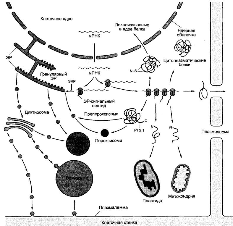
Translation of all nuclear mRNAs initiates exclusively in the cytoplasm. Proteins whose translation is completed on The Endoplasmic reticulum (ER) possess an N-terminal signal peptide consisting of 16 — 30 amino acids, with a central core of 4 to 12 hydrophobic amino acids. As soon as this signal peptide emerges from the ribosome (when the growing peptide chain is approximately 70 amino acids long), it is bound by the ribonucleoprotein complex known as SRP (signal recognition particle), which halts translation. The resulting complex comprising the SRP, ribosome, nascent polypeptide chain, and mRNA then binds to the SRP receptor on the ER membrane surface. Here, upon release of the SRP and GTP hydrolysis, the beginning of the polypeptide chain is handed over to the translocation complex (the translocon). Polypeptide elongation continues while the chain is simultaneously (co-translationally) threaded through the hydrophilic pore of the translocon into the ER lumen, accompanied by Cleavage of the signal sequence. Protein domains fold co-translationally as well; disulfide bridges form where necessary, and oligoglycan chains attach to specific asparagine residues to yield N-Glycosides. ER Membrane Proteins are synthesized in a similar fashion. However, transmembrane domains—amino acid regions spanning about 20 hydrophobic residues folded into an α-Helix (or occasionally a β-sheet)—are laterally released from the translocon into The Lipid Bilayer, thereby anchoring the protein in the ER membrane. The details of these well-studied protein synthesis pathways at the ER are omitted here for reasons of space.
Proteins synthesized at the ER are transported via vesicular flow (see Fig. 7.16) mediated by dictyosomes to the Plasmalemma or vacuoles, or alternatively they remain in the ER. In the absence of other topogenic signals, proteins delivered to the ER lumen are secreted via the Golgi apparatus. Membrane proteins lacking other topogenic signals end up in The Plasma Membrane via the same route. Additional topogenic signals have been identified for all other destination compartments. For instance, proteins retained in the ER are characterized by a C-terminal retention signal—the amino acid sequence -Lys-Asp-Glu-Leu(COOH) or a related sequence. Proteins destined for vacuoles or the tonoplast also carry specific signal sequences. These are typically N-terminal sequences 12—16 amino acids in length, almost invariably containing the -Asn-Pro-Ile-Arg- motif. Conversely, proteins deposited in storage vacuoles (typical of seeds) carry C-terminal or, less frequently, internal motifs functioning as topogenic signals. In all known instances, vacuolar signal sequences are proteolytically cleaved within the target compartment. For example, toxic ricin in Ricinus communis is split into two Polypeptides following the removal of an internal signal sequence; these polypeptides are ultimately re-linked covalently via disulfide bridges to form the mature A and B chains of ricin. Protein-storing vacuoles are formed only partially via Golgi-derived vesicles; in addition, vesicles containing Reserve Proteins bud off directly from the ER.
If an ER signal peptide is not generated at the onset of translation, protein synthesis continues in the cytoplasm with the formation of polysomes, and the resulting polypeptide chain is released into the cytoplasm. Subsequent intracellular sorting of these proteins—which are transported to plastids, mitochondria, Peroxisomes (or glyoxysomes), and the Cell Nucleus, or alternatively leave the cell via plasmodesmata—is likewise mediated by a diverse array of topogenic signals, the deciphering of which has been progressing rapidly in recent years. In the absence of such signals, the protein remains in the cytoplasm where it was synthesized. Below, we briefly outline only the typical pathways, bearing in mind that numerous variations exist for specific cases.
Peroxisomal biogenesis has been studied most thoroughly in baker's yeast. The principles governing the formation of peroxisomes and glyoxysomes in higher plants are expected to be similar. Peroxisomes arise through the post-translational import of fully folded proteins into preperoxisomes, which bud off as vesicles from the ER and expand in volume as their protein content increases. Proteins destined for import into these organelles possess one of Two Types of amino acid sequences: PTS1 (C-terminal) or PTS2 (N-terminal) (peroxisomal targeting sequence). PTS2 consists of nine amino acids, whereas PTS1 comprises only three. A typical example is the tripeptide -Ser-Lys-Leu-(COOH), which is not cleaved off following transport. The details of the import mechanism—which is postulated to involve either a large pore or an endocytosis-like process—remain unknown. In any case, colloidal gold particles up to 9 nm in diameter coated with peroxisomal proteins are imported into the organelle along with the protein.
During the germination of fat-storing seeds, A large number of glyoxysomes are initially produced (lipid mobilization, see 6.12); these subsequently disappear upon the onset of Photosynthesis and are replaced by peroxisomes (Photorespiration, see 6.5.6). It has been demonstrated that this transition involves the direct remodeling of glyoxysomes into peroxisomes. Specifically, using-specific Antibodies, organelles were identified that contained both typical glyoxysomal and peroxisomal enzymes (enzymes characteristic of a particular compartment). Because glyoxysomes and peroxisomes share numerous enzymes (such as catalase and enzymes of fatty acid β-oxidation), converting glyoxysomes into peroxisomes is highly efficient, though the precise mechanism of this transformation remains unclear.
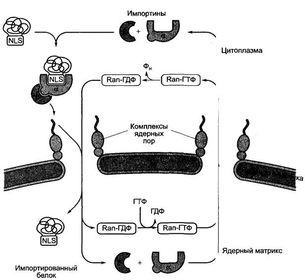
Proteins destined for the cell nucleus (e.g., Histones, transcription factors, Cell Cycle proteins) are likewise transported in their already folded state. In the cytoplasm, an internal signal sequence consisting of 10 — 38 predominantly basic amino acids (NLS, nuclear localization signal), which may be monopartite or bipartite, is bound by the NLS receptor, importin α (Fig. 7.17). Importin α then associates with importin β, which in turn interacts with nuclear pore proteins (for structure, see 2.2.3.4, Fig. 2.26), thereby guiding the imported protein to the nuclear pores. Here, driven by ATP hydrolysis, the protein-importin α+β complex passes into the nuclear matrix, where it dissociates. Importin α and importin β are recycled back to the cytoplasm for subsequent rounds of transport. Translocation of the importin-protein complex through the nuclear pore is mediated by the GDP-bound form of the GTP-binding protein Ran (Ran-GDP); the recycling of importins α and β back to the cytoplasm requires the GTP-bound form of Ran (Ran-GTP) (Ran stands for *Ras nuclear*, as it was the first Ras-family protein localized to The Nucleus; *rat adenosarcoma* refers to the rat tumor tissue in which the first GTP-binding protein of this family was discovered).
Fig. 7.17. Simplified diagram of importin-dependent Protein transport from the cytoplasm through nuclear pores into the cell nucleus. The import complex binds to the nuclear pore complex (see Fig. 2.26) and translocates through the nuclear pore via an ATP- and Ran-GTP-dependent mechanism. Importins are exported back to the cytoplasm in their GTP-bound form by the Ran protein.
Modification (such as phosphorylation) of an NLS-bearing protein can alter its recognition by the NLS receptor. This represents one of The regulatory mechanisms controlling the nuclear import of certain transcription factors, whose phosphorylation state is modulated by environmental and other cellular signals. In its inactive state, the red-light photoreceptor Phytochrome (see 7.7.2.4) is localized in the cytoplasm; upon illumination—which alters the phosphorylation state of the protein—it translocates into the cell nucleus (see Fig. 7.86).
Protein import into mitochondria and plastids is also well understood. The following discussion will be restricted to protein import into Chloroplasts, while highlighting the differences inherent to mitochondrial transport. We will focus solely on the primary transport pathway (Fig. 7.18).
Fig. 7.18. Schematic diagram of protein import into chloroplasts. At contact sites, the inner and outer plastid membranes interact via protein Components of the translocation machinery (TOC complex, TIC complex), shown here in a simplified form. Proteins are imported in an unfolded state if they possess an N-terminal signal sequence known as a transit peptide. The phosphorylated transit peptide is first bound by the TOC86 protein (molecular mass 86 kDa) upon dephosphorylation and is subsequently transferred to the translocation pore formed by TOC75. For further details, see the text

Proteins destined for import into chloroplasts carry a *transit peptide* at their N-terminus, whereas those imported into mitochondria bear a region termed the *leader sequence*, which is proteolytically cleaved after import. Mitochondrial leader sequences, 15 — 35 amino acids in length, are invariably positively charged and form an amphipathic α-helix, with hydrophobic residues clustered on one side and hydrophilic residues on the opposite side. In contrast, plastid transit peptides are longer (30 — 100 amino acids; for instance, the transit peptide of EPSP synthase consists of 77 amino acids). They are rich in polar amino acids yet contain few or no positively charged residues, and they do not form amphipathic α-helices. Unlike mitochondrial leader sequences, however, they are phosphorylated at serine and/or Threonine residues. These distinctive features underlie the accurate sorting of proteins to either chloroplasts or mitochondria. The translocation process occurs post-translationally in both cases and proceeds in a very similar manner: the imported proteins are unfolded in both instances, meaning they exhibit relatively few distinct structural features.
During protein import into the matrix or stroma, respectively, two membranes must be crossed. Transport takes place at contact sites, where signal sequence receptors and translocation complexes are located. A distinction is made between the translocon of the outer chloroplast membrane (TOC) and the translocon of the inner chloroplast membrane (TIC). Similarly, mitochondria possess TOM and TIM systems whose proteins, however, are not homologous to TOC and TIC.
According to current understanding, the unfolded conformation of proteins destined for import is maintained by cytoplasmic chaperones (see 7.3.1.2), primarily HSP70. Following the binding of the signal sequence to a receptor protein (in chloroplasts, an 86 kDa protein of the TOC complex), the polypeptide chain is pulled through the translocation apparatus in an ATP- and GTP-dependent reaction. In chloroplasts, TOC75 also participates in protein import, forming a pore through which ions can also pass. Prior to translocation, the transit peptide is dephosphorylated. On the stromal side, the signal sequence is proteolytically cleaved, and the mature, biologically active protein is folded with the aid of a plastid HSP70 isoform and the Hsp60 chaperonin (see 7.3.1.2). The entire process of importing stromal proteins from the cytoplasm requires energy: ATP must be present in the cytoplasm, the intermembrane space, and the stroma (see Fig. 7.18). Mitochondrial protein import additionally requires an electrical potential gradient across The inner mitochondrial membrane.1
1 Recently, chloroplast proteins have been discovered that are imported only when the thylakoid membrane possesses a high potential gradient (∆μH+). — Editorial note.
Certain chloroplast proteins, such as plastocyanin (see 6.4.4) which are localized in the thylakoid lumen, must be transported from the cytoplasm across three membranes. The plastocyanin precursor features a dual N-terminal signal sequence: a 38-amino-acid transit peptide that is cleaved in the stroma, followed by a second signal peptide, 28 amino acids long, which is released after the removal of the transit peptide and facilitates Transport Across the thylakoid membrane. In some proteins, transport across the thylakoid membrane depends on the pH gradient between the stroma and the thylakoid lumen.
7.3.2. CELL CYCLE AND Cell Cycle Control
Plant development is based on Cell Division, cell growth, and Cell Differentiation. The additional cell migration characteristic of animals plays no role in plants, and apoptosis, which is important in animal development, has a subordinate role here. Apoptosis refers to the regulated degradation and subsequent death of cells during organ development, such as during digit formation. Onset of apoptosis can be recognized by the fragmentation of DNA between nucleosomes (see 2.2.3.1), for instance, by gel electrophoretic Separation of the resulting fragments. DNA fragmentation has been observed during carpel senescence. Carpel senescence refers to age-related changes associated with their degradation (see 7.6.2.3); in many plants, these occur in the absence of Fertilization. Apoptosis-like processes in plants are more commonly termed programmed cell death. This phenomenon includes the formation of air-conducting Tissues (aerenchyma, see Fig. 3.8) via The breakdown of cortical parenchyma cells, which occurs primarily under oxygen deprivation (e.g., in maize, see 7.6.5.2). Furthermore, in angiosperms, it involves the death of three of the four daughter cells resulting from Meiosis of the megaspore mother cell; the fourth megaspore forms the female gametophyte (see 11.2). Programmed cell death is also considered to encompass the degeneration of the suspensor during Embryogenesis (see Fig. 3.1), as well as the death of specific cells (e.g., sclerenchyma cells, vessel elements, tracheids) during cell differentiation. Finally, the so-called hypersensitive response, accompanied by cell death upon pathogen infection, is likewise programmed: it is triggered by pathogen avirulence gene products1 in a specific interaction with plant cell receptors (see 9.3.1; 9.3.4; Fig. 9.17).
1 Referring to elicitors secreted by the pathogen. — Editorial note.
Cell differentiation occurs after cell divisions have ceased. Differentiated cells no longer divide, but under suitable conditions (e.g., upon tissue injury or in experimental in vitro culture on appropriate nutrient media, see 7.1, Fig. 7.1) they can be "re-embryonalized"1, i.e., resume cell division. Therefore, cell cycle control—the regulated alternation of mitoses and interphases (see 2.2.3.5; Fig. 2.30)—plays a crucial role in development. As mentioned previously, mitosis is designated as the M phase of the cell cycle and concludes with cell division. The interval between two mitoses, interphase, is the actual phase of gene activity. Replication of genetic material (see 1.2.3, Figs. 1.7, 1.9) takes place in the S phase. The S and M phases are separated by two gaps, the G1 and G2 phases; the genome is transcriptionally active throughout interphase, i.e., even during DNA replication.
1 "Reembryonalisieren" is not a strict technical term, but rather a figurative German expression meaning "as if returning cells to an embryonic state." — Editorial note.
The individual periods of the cell cycle (see Figs. 2.30; 7.19) can vary markedly in duration. For instance, in maize, the cell cycle in the Cells of the ROOT meristem quiescent center lasts an average of 170 h (of which 135 h are accounted for by the G1 phase), whereas root cap initial cells divide on average once every 14 h, with the G1 phase being absent; DNA replication begins immediately after the completion of cell division. In SHOOT apical Meristems, the cell cycle lasts from 20 h (Silene coeli-rosa) to 288 h (Sinapis alba), in the floral meristem from 10 h (Silene) to 47 h (Ranunculus), in the cambial initials of Tsuga canadensis from 10 to 28 days, and in Pinus about 1 day.
Fig. 7.19. Regulation of the plant cell cycle: A — Progression of the cell cycle is critically controlled by cell cycle Kinases (Cdks, cyclin-dependent kinases; in Arabidopsis thaliana: Cdc2a and Cdc2b) and their activators, the cyclins, which appear stage-specifically (three cyclin types in plants: A, B, and D; in Arabidopsis thaliana: CycA2, CycB1 and B2, CycD1, D2, and D3). Phytohormones influence the cell division process at checkpoints (gray triangles); further details in the text. B — Formation and breakdown of the complex composed of Cdc2 kinase and a G2 -> M-specific B-type cyclin, which controls the transition from the G2 phase to mitosis. The active complex (MPF, maturation-promoting factor) is formed after the association of both proteins, accompanied by hyperphosphorylation and partial dephosphorylation of Cdc2 to a monophosphorylated form. MPF induces the onset of mitosis: it phosphorylates histone H1 (triggering Chromatin Condensation, see 7.2.2.2) and induces nuclear envelope breakdown via lamin phosphorylation. Monophosphorylated Cdc2 simultaneously induces ubiquitination and thereby the proteolytic degradation of its associated cyclin. The Synthesis and Breakdown of cyclin thus determine the amount of MPF, which exhibits characteristic dynamics during the G2->M transition.
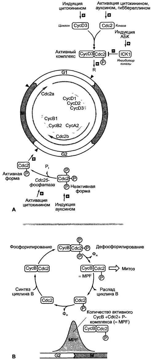
The main regulatory steps are engaged shortly before the critical transitions between the individual Phases of the cell cycle (Fig. 7.19):
✵ Before the G1->S transition (restriction point R, also known as START in yeast); here, the initiation of replication and thus the entry into a new cell division cycle is regulated; cell cycle arrest in the G1 phase precedes the onset of cell differentiation;
✵ Before the G2->M transition; here, the onset of mitosis is regulated; in the event of incomplete nuclear genome replication or DNA damage, the cell cycle arrests at this point;
✵ Before the M->G1 transition; here, actual cell division takes place; if chromosome alignment in the spindle apparatus is disrupted, the cell cycle arrests in metaphase (see 2.2.3.5).
The molecular processes of cell cycle control have been investigated primarily in mammalian cells and yeast. In higher plants (and presumably in all eukaryotes), these processes appear to operate similarly. A key role is played by cyclin-dependent protein kinases (Cdks). Baker's yeast possesses a single kinase (Cdc2, cell division cycle mutant), named after the yeast mutants that enabled the identification of the gene. In higher plants, however, cyclin-dependent protein kinases are represented by multiple similar proteins (in Arabidopsis thaliana, for example, There are two: Cdc2a and Cdc2b; see Fig. 7.19). Cyclins are proteins that appear at specific Stages of the cell cycle. Their Synthesis and degradation are strictly controlled by the ubiquitin/proteasome system (see 7.3.1.3). Cyclins act as Cdk activators. It is assumed that individual cell cycle transitions are controlled by various combinations of cyclins with cyclin-dependent kinases, which regulate The activity of various groups of target proteins essential for the respective transitions (e.g., transcription factors, histones, nuclear envelope proteins) via phosphorylation. The cell division-inducing factor MPF (maturation-promoting factor), for example, was identified as a complex of Cdc2 kinase with a G2->M B-type cyclin. Alongside their association with cyclins, kinases are further regulated by phosphorylation; in their hyperphosphorylated form they are inactive, but can be activated by specific Phosphatases such as Cdc25. Cdks decrease in activity upon association with inhibitory proteins (ICKs, inhibitors of cyclin-dependent kinases), meaning that a multitude of effective mechanisms exist to regulate cell cycle kinase activity. To date, over 50 different Cdc genes have been discovered in yeast alone.
External factors intervene in The regulation of the cell cycle: for instance, in yeast, the G1->S transition is controlled at the START point by nutrient availability, cell size, and pheromones, whereas the restriction point in animal cells is regulated by growth factors. It has recently been established that phytohormones participate in regulating cell cycle control in higher plants (see Fig. 7.19). Division-stimulating Cytokinins induce the Formation of the G1->S cyclin CycD3 and participate in activating cell cycle kinases at the G2->M transition. Their formation is under the control of Auxins (see 7.6.1). Abscisic acid (see 7.6.4) induces the formation of the kinase inhibitor ICK1 and thereby inhibits the G1->S transition.
In special cases, the reaction sequence can be interrupted at any point in the mitotic cycle (Fig. 7.20): DNA doubling without subsequent chromosome separation leads to polyteny; likewise, in many seeds, a resting period may be incorporated into the G2 phase following the DNA replication phase (S phase). However, if chromosome doubling occurs within an intact nuclear envelope without becoming outwardly apparent, and nuclear division fails to take place, endopolyploid cells arise (see Fig. 2.31). In cells possessing only a single plastid (many Algae, the moss Anthoceros, see 11.2) or even a single mitochondrion (the alga Micromonas), these organelles divide in strict synchrony with the cell nucleus. The mechanism underlying this coordination remains unknown.
Fig. 7.20. Processes comprising the cell cycle and their possible deviations

However, in the polyenergid cells of many algae and fungi, as well as in the multinucleate endosperm (see Fig. 2.33), multiple rounds of DNA replication, chromosome division, and nuclear division occur, while cell division is suppressed. During subsequent Cell wall formation in the multinucleate endosperm (e.g., in Haemanthus katharinae), cell walls arise between nuclei that are not sister nuclei, and consequently no mitotic spindle is present between them. Thus, cell wall formation here has lost its usual coupling to nuclear division. Cell divisions in which one of the daughter cells receives no nucleus generally do not occur in plants; enucleate cells, such as mature sieve tube elements, lose their nucleus only after division.
Very little is known about the Physiological aspects of mitosis. Cell divisions often occur rhythmically; partly, it appears, governed by diurnal periodicity (onion roots, zoospore formation in algae), although multiple cycles can also pass within 24 hours. In many algae, mitoses take place predominantly at night: Spirogyra, for instance, typically divides around midnight. In multinucleate cells, nuclear divisions often begin synchronously—likely facilitated by the cytoplasm—or progress in a wave-like manner from one end of the cell to the other (see 11.2, embryo sac). Like other physiological processes, cell division takes place only within certain species-specific temperature limits, often with a pronounced optimum (in pea, for example, between 0 and 45 °C, with an optimum at 28–30 °C). Seedlings may be adapted to lower temperatures than older plants.
Embryonic cells (such as apical cells or meristem initials, see 3.1.1) produce daughter cells that either continue to divide or, upon losing the capacity for division, immediately begin differentiation via extension growth. Cells entering the differentiation process leave the cell cycle in the G1 phase (see Figs. 7.19; 7.20) and enter a mitotically inactive state (G0 phase). This transition is subject, among other things, to hormonal control; however, many details remain poorly understood.
Under appropriate conditions, already differentiated cells can re-enter the cell cycle (G0 -> G1). This process is promoted, among other things, by phytohormones, a technique utilized in regenerating plants from cell cultures (see Figs. 7.1 and 7.6.2). Full-fledged plants with all species-specific traits are formed, which proves the totipotency of plant cells. Often, during in vitro Plant Regeneration from a single cell, a somatic embryo is formed first, resembling a zygotic embryo, whose apical meristems subsequently differentiate into a shoot and a root (example: Daucus carota, see Fig. 7.1). These regeneration processes are of economic importance: they are employed for the propagation of certain ornamental plants, such as orchids, which are difficult to grow from seeds. On an industrial scale, orchids are produced via micropropagation in in vitro cultures using mechanically isolated leaf mesophyll cells or meristematic tissue. Plant regeneration from cell or tissue cultures represents a crucial step in the generation of Transgenic Plants (see Box 7.3).
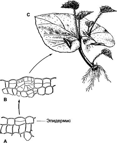
Following injury, roots develop on detached begonia leaves not only at the lower (basal) end of the petiole, but also at the base of the leaf blade. Adventitious buds readily form at the lower cut edge of isolated leaf Veins, which can subsequently develop into entire begonia plants. These adventitious shoots originate from a single epidermal cell that has regained an embryonic state (Fig. 7.21), whereas adventitious roots form from dividing cells near the phloem of the vascular bundle.
Fig. 7.21. Regeneration of shoots and roots on leaf cuttings of Begonia: A, B — formation of an adventitious shoot from an epidermal cell (fragments of a transverse leaf section, 150×): A — the epidermal cell has undergone a single division; B — the epidermal cell has given rise to a multicellular secondary meristem, which first produces an adventitious bud and subsequently a shoot from it (C)
The Formation of secondary meristems (such as the cork cambium and interfascicular cambium) serves as an example of the natural dedifferentiation (a sort of reversion to an embryonic state) of cells that had already differentiated during development, as well as the formation of wound callus resulting from tissue damage and tissue fusion during grafting.
In grafting, severed plant parts bearing buds (scions) are fused with the incised PARTS OF THE same or a compatible closely related species (rootstocks) with the help of callus tissue developing at the wound site. Phloem and xylem elements differentiate within the callus, connecting the corresponding tissues in the vascular bundles of the scion and rootstock upon successful grafting. Grafting is particularly important in horticulture and agricultural practice because it allows non-homozygous forms and varieties—which cannot be propagated by seeds—to be preserved and propagated on rootstocks (for example, in fruit growing, viticulture, and rose breeding).
Moreover, after fusion, each partner retains its genetic material unaltered. Through the metabolism taking place between the scion and rootstock, The properties of both graft components can be modified. This alteration of properties is particularly striking in grafts where adventitious shoots arise from the callus at the graft union, composed of fused tissues from both partners (chimeras). In sectoral chimeras, one sector of a shoot or leaf is formed by the scion, while the remaining sectors originate from the rootstock. Particularly noteworthy are periclinal chimeras, in which the epidermis and possibly some outer layers originate from one partner, while the inner tissues derive from the other partner (grafts in Cytisus species, grafts between Crataegus and Mespilus, etc.). Such "graft hybrids" may outwardly give the impression of true sexually produced hybrids, yet they are not identical to them: even in such intimate fusions, each individual cell or cell layer ultimately retains its own species-specific genetic material, despite the fact that the outward appearance clearly reveals the reciprocal Influence of the different tissue layers.
A central, yet insufficiently explained stage in developmental physiology concerns determination, i.e., the specification of The Fate of a differentiating cell, tissue, or organ. The consequence of determination is a reprogramming of gene activity that ensures The production of gene products required for the differentiation process. As mentioned earlier (see 7.1), cell-autonomous processes play only a limited role in this regard. Furthermore, the differentiation process is simultaneously under the inductive control of its environment. Inducing stimuli may originate from the organism itself (endogenous stimuli, e.g., phytohormones, see 7.6) and/or involve external influences. Such exogenous stimuli can be of biotic origin (i.e., originating from other living organisms, as in gall formation or root nodule formation — see Chapter 9) or of abiotic origin (physical or chemical stimuli, such as the light factor — see 7.7). A stimulus is defined as any physical or chemical signal that triggers a specific sequence of reactions in an organism, the Energy Requirements of which are met by the organism itself. Developmental processes that can be influenced by external factors are termed actionomic, whereas those unaffected by them are termed endonomic.1
1 The latter two terms are not in common use and were introduced primarily for natural-philosophical reasons (Greek aitios = causal, nomos = law, hence actionomic = requiring an [external] cause; Greek endos = internal, hence subject to internal laws). — Translator's Note.
Endonomic, for example, is the determination and the resulting development of phloem elements produced by cambial initials in Cephalotaxaceae, Taxodiaceae, and Cupressaceae (the "four-stroke cycle": sieve cell — phloem fiber — sieve cell — parenchyma cell, etc.).
Cellular differentiation is also endonomously determined in Volvox carteri (see 11.2). This alga consists of 2,000–4,000 somatic cells and 16 reproductive cells located at strictly defined positions within the cell aggregate. During embryogenesis, at the sixth cell division (and exclusively here!), i.e., during the transition from the 32- to the 64-cell stage, 16 cells of the 32-cell embryo undergo an unequal cell division: the resulting smaller cells (diameter < 6 µm) develop into somatic cells, while the larger ones (>9 µm) develop into gonidia. Exactly how cell size determines subsequent differentiation remains unknown.
Embryogenesis in multicellular plants is largely endonomously determined (see Figs. 3.1 and 7.4.1). An example of actionomic developmental processes, in which external factors alongside endogenous ones largely determine cell fate, is the transition of the shoot meristem into the floral meristem, controlled by Photoperiodism. This process has been well researched, particularly the mechanisms of organ formation, which are discussed in detail below (see 7.4.3).
Intracellular substance gradients lead to polarity; they can influence the differentiation of daughter cells and thereby establish the polarity of entire Organs. Furthermore, intercellular substance gradients are crucial for determination processes, in which a cell's fate is defined by its position within an organ or tissue; thus, substance gradients are also essential for patterning (see 7.4.2). Finally, substance gradients between organs are responsible for correlative developmental processes (see 7.5).
In biology, polarity refers to the physiological or morphological inequivalence of two poles or two surfaces within a living system, in the simplest case, within a cell. Morphological polarity is expressed, for example, in The structure of thallophytes and cormophytes: it is already noticeable in early embryogenesis (see Fig. 3.1) and is attributed to the biochemical (physiological) polarity already established in the zygote (see below). Unequal cell division, which (as in the Volvox example) represents a decisive stage of differentiation, likewise presupposes a physiological polarity manifested outwardly solely by the positions of the spindle apparatus and the newly formed cell wall. It is not the cell divisions themselves, but the underlying cellular polarization that determines the characteristic spatial body form of the plant.
When egg cells or spores in lower plants (e.g., egg cells of the brown algae Sargassum and Coccophora) leave the maternal plant, they are polarized by the maternal plant only in exceptional cases. As a rule, their polarization occurs only under the Influence of External stimuli (light, gravity); in egg cells, this is possible only after fertilization.
If spores of Equisetum or fertilized zygotes of Fucus or Pelvetia (Phaeophyceae) are illuminated unilaterally, an unequal distribution of substances within the cytoplasm is induced, ultimately resulting in unequal cell division, with the cell on the shaded side becoming the rhizoid pole and the other, larger cell acting as the initial cell for the remaining thallus (Fig. 7.22). In zygotes of Pelvetia or Fucus, the rhizoid forms outgrowths even before cell division, meaning that the division merely stabilizes the polarization that has already taken place within the cell (see below). As experiments involving the illumination of one half of a zygote demonstrate, the decisive factor for polarity induction is the reduction of overall illumination intensity within the cell, rather than the direction of light (Fig. 7.23, B).
Fig. 7.22. Polarization of an Equisetum spore: A — unpolarized spore; B — onset of polarization; C — segregation of the rhizoid and prothallial cells; D — early multicellular stage
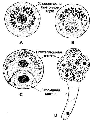
The duration of illumination required to induce polarization decreases as light intensity increases; consequently, the total photon dose is what matters. For maximum polarization in Equisetum spores, about 10 minutes are required at 2 W·m-2 of white light, 1–5 minutes at 20 W·m-2, and only 10-3 s when using a xenon flash lamp. The effective wavelengths for egg cells or zygotes of brown algae and spores of Equisetum typically lie in the blue and ultraviolet ranges. In brown algae, the putative photoreceptor is a retinal-containing protein showing similarity to the sensory rhodopsin of green algae. The first noticeable reaction in Equisetum spores polarized by unilateral illumination is the migration of plastids toward the light-facing side of the cell—into the prospective prothallial cell—and the Displacement of the cell nucleus in the opposite direction (Fig. 7.22, B). This movement is induced even when neither the plastids nor the nucleus are illuminated, but strictly the cytoplasm alone.
In the absence of an inducing unilateral light stimulus, gravity frequently exerts the inducing effect (with the rhizoid pole pointing toward the center of the Earth). If all other directional external factors are absent (achievable only experimentally), rhizoids develop in Pelvetia or Fucus zygotes at a random point of sperm entry, and in Equisetum spores at a specific site—the rhizoidal point—which typically remains unmanifested under directional induction by light or gravity. The Influence of neighboring cells on polarity induction has been demonstrated: if at least 10 Fucus zygotes are densely packed together, some of the inner cells fail to form rhizoids entirely, whereas rhizoids develop on the outer cells pointing toward the periphery of the group. Shortly after induction, polarity in Fucus zygotes can still be arrested or even reversed by applying an oppositely directed gradient (e.g., illumination).
The molecular processes underlying cellular polarization in Pelvetia and Fucus zygotes have been investigated in great detail (Fig. 7.23). Upon unilateral illumination, an F-Actin cytoskeletal "cap" first forms in the cortical cytoplasm on the side of the zygote opposite the light source (see 2.2.2.1). In the absence of any external influences, this structure forms at the site where the sperm entered the egg cell. The actin "cap" marks the prospective rhizoid pole of the cell and ensures that specific populations of Golgi vesicles are transported toward the rhizoid pole, where they fuse with the plasma membrane. These Golgi vesicles carry, on the one hand, specialized membrane proteins (such as Ca2+ channels and anchor proteins for microtubules) and, on the other hand, enzymes required, among other things, for cell wall remodeling during rhizoidal growth, as well as Structural components of The cell wall (including a specific sulfated fucan). The vesicle flow is enhanced and directed by a Ca2+ gradient that builds up toward the rhizoid pole. This Ca2+ gradient arises very soon after the formation of the F-actin "cap", likely as Ca2+ channels—initially distributed evenly throughout the plasma membrane—migrate to the newly formed rhizoid pole (toward the actin "cap"); later, fusing secretory Golgi vesicles deliver additional channel proteins. The rhizoid pole begins to grow in the region of the modified cell wall, with Golgi vesicles supplying the material required for cell wall construction.
Fig. 7.23. Establishment of cell polarity in a Fucus zygote: A — in the fertilized egg cell, the ESTABLISHMENT OF THE polarity axis begins at the site of sperm penetration (1); however, after the zygote cell wall forms and the zygote attaches to the substrate, this axis is replaced by a new polarity axis (2) under the influence of external factors, particularly light. At the emerging rhizoid pole, the plasma membrane and cell wall are specifically modified, and rhizoid growth begins (3, 4), with centrioles also aligning parallel to the polarity axis (4, 5). This establishes the plane of cell division and THE POSITION OF the new cell wall perpendicularly relative to the axis of cell polarization (5, 6). Both daughter cells differentiate into thallus or rhizoid under the restrictive influence of the cell wall. The modified cell wall region is located at the bottom. For further explanations, see text; B — rhizoid formation in a Fucus zygote at the darkest site
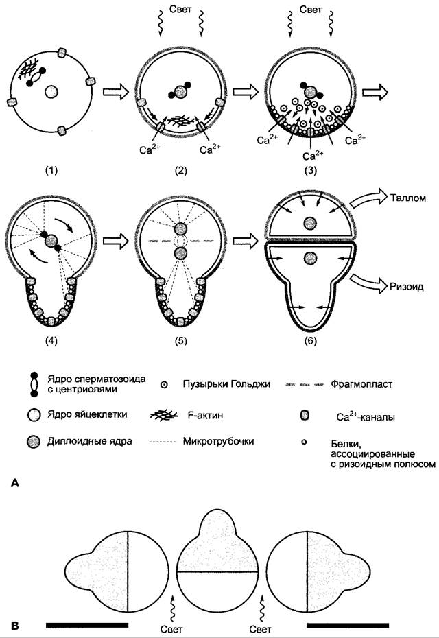
The cell, polarized along its axis, now undergoes its first unequal cell division, the plane of which is oriented strictly perpendicular to the axis of cellular polarization. The basal daughter cell differentiates into a rhizoid, whereas the apical daughter cell forms the thallus. For the Organization OF THE cell nucleus and the mitotic spindle, the rhizoid pole is also crucial because microtubules originating from one of the two centrosomes that form the prospective spindle pole (see Box 2.2) make contact with anchor proteins at the rhizoid pole via their free ends. Thus, the spindle poles are organized parallel to the cell's polarity axis (Fig. 7.23, A). Consequently, the plane of the phragmoplast (see 2.2.3.6) and therefore the plane of the future cell wall lie perpendicular to the longitudinal axis of the polarized zygote.
The subsequent fate of both daughter cells during differentiation is determined primarily by the different compositions of their cell walls: if protoplasts are isolated from these cells, further differentiation is disrupted. Conversely, if the two cells are separated, they retain their differentiation: independently of each other, the upper cell develops into a thallus, while the lower cell forms a rhizoid. If the protoplast of one daughter cell is combined with the cell wall of the other daughter cell, determination is altered: the protoplast of the rhizoid cell, when placed in contact with the cell wall derived from the thallus daughter cell, differentiates into thallus cells, and conversely, upon contact with the cell wall derived from the rhizoid cell, the protoplast of the thallus cell forms rhizoid cells.
It has been shown that similar processes occur during the polarization of other cell types. For instance, There are many parallels with the Cytology/cytology/16.html">Early stages of embryogenesis in angiosperms (see 7.4.1) and with the asymmetric growth of other cells driven by targeted local secretion of cell wall material, such as during yeast budding and, presumably, apical cell growth.
Once established, polarity in higher plants is particularly stable and generally irreversible. For example, when willow cuttings are kept in a humid atmosphere, buds sprout exclusively at the apical end, whereas only roots develop at the basal end, even though the latter possesses ample dormant bud primordia (Fig. 7.24). Root cuttings of plants such as snapdragon or chicory, when placed in moist soil, produce shoots on the proximal side, while roots emerge on the distal side (Fig. 7.25). Grafting experiments also reveal the polarity of the components, as union occurs only between correctly oriented tissues. This polarity is endogenously determined and cannot be reoriented by external factors, such as altered gravitational pull (see Figs. 7.24 and 7.25). Polarity is manifest in even the smallest fragments of shoots and roots, reminiscent of a permanent magnet, where even the smallest fragments retain two opposite poles. It thus seems justified to conclude that in higher plants, every individual cell is polarized, and the polarity of individual cells dictates the polarity of the organ as a whole.
Fig. 7.24. Polar regeneration and sprouting of willow stem cuttings suspended in a humid environment in their normal (A) and inverted (B) orientations

Fig. 7.25. Polar regeneration in root cuttings. Shoot buds invariably develop at the proximal end (closest to the root crown), regardless of spatial orientation

Only recently has it been discovered in animals, as well as in brown algal zygotes, that cell polarization also involves the asymmetric distribution of mRNA and, consequently, the asymmetric localization of translated proteins. This directed distribution of mRNA is likely mediated by specific mRNA-binding proteins that actively transport transcripts along cytoskeletal elements to the appropriate target region of the cell, consuming energy in the process.
Last update: 07/08/2026
Editorial and Educational Adaptation: This material has been compiled based on the primary/original source text. The project team performed an editorial review, corrected technical inaccuracies, structured sections, and adapted the content for an educational format.
What was processed:
- elimination of formatting defects (OCR errors, structural breaks, corrupted characters);
- editorial organization of content;
- standardization of terminology in accordance with academic sources;
- verification of factual statements against the original source text.
All mentions of the author, publication year, and origin of the primary text have been preserved in accordance with the source.