Biochemical Engineering Fundamentals, Part 1 - Bailey J., Ollis D. 1989
Chemical Foundations of Life
Amino Acids and Proteins
Proteinogenic Amino Acids and Polypeptides
The monomeric units of Polypeptides are α-Amino Acids with the general formula
Class="center">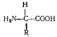
Thus, Amino acids differ in The Nature of the R group attached to the α-carbon atom (adjacent to the carboxyl group). Since, in general, all substituents on this carbon atom are different (except for Glycine, where R = H), this carbon atom is asymmetric.
Many biologically important Organic compounds, including sugars and amino acids, are optically active, meaning they possess at least one asymmetric carbon atom and can therefore exist in two forms, as shown below for amino acids:
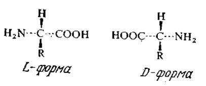
A solution of one isomer rotates the plane of polarized light to the right (dextrorotatory or d-form) or to the left (levorotatory or l-form). Optical isomerism is an extremely important phenomenon because, in the absence of Enzymes that interconvert isomers, living organisms can utilize only one of the isomers. As a rule, enzymes also catalyze the transformation of only one of the optical isomers. This property of enzymes is used industrially to resolve mixtures of racemic acylamino acids, where only one of the isomers is hydrolyzed, resulting in reaction products that contain two significantly different and therefore more easily separated substances. We will discuss this process in more detail in Chapter 4. Since direct physical Separation of optical isomers is expensive and inefficient, such microbiological and enzymatic (as well as chemical) resolution Methods can be much more advantageous.
Interestingly, only L-Amino acids are found in the vast majority of Proteins. D-Amino acids are rare in natural sources; they have been found in The Cell walls of certain microorganisms and in several Antibiotics.
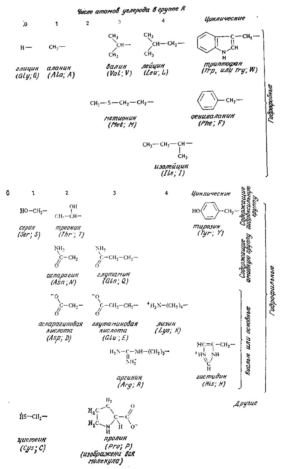
FIG. 2.15. 20 protein amino acids.
The acidic (—COOH) and basic (—NH2) groups of Amino acids can undergo ionization In aqueous solutions.
Table 2.7. pK (—log K) values of terminal amino and carboxyl groups, as well as groups in the R side chainsa
Amino acid |
pK1 α-COOH |
pK2 α-NHs |
pKR R group |
Glycine |
2.34 |
9.6 |
|
2.34 |
9.69 |
||
Leucine |
2.36 |
9.60 |
|
2.21 |
9.15 |
||
2.63 |
10.43 |
||
Glutamine |
2.17 |
9.13 |
|
Aspartic acid |
2.09 |
9.82 |
3.86 |
Glutamic acid |
2.19 |
9.67 |
4.25 |
1.82 |
9.17 |
6.0 |
|
1.71 |
10.78 |
8.33 |
|
2.20 |
9.11 |
10.07 |
|
2.18 |
8.95 |
10.53 |
|
2.17 |
9.04 |
12.48 |
a Reproduced from: Lehninger A. L., Biochemistry, 2nd ed., Table 4-2, Worth Publishers, Inc., New York, 1975; There is a Translation of an earlier edition: Lehninger A., Biochemistry.— M.: Mir, 1976.
An amino acid carries a positive charge (cation) at low pH values and a negative charge (anion) at high pH. At some intermediate pH value, The amino acid exists as a dipolar ion (zwitterion) with a net charge of zero. This pH value is called the isoelectric point and is determined by the Nature of the R substituent (Table 2.7). At the isoelectric point, the amino acid is unable to migrate to either the anode or the cathode under METABOLISM/18.html">The Influence of an electric field, and, furthermore, its solubility is minimal. Separation Methods such as Ion Exchange, electrodialysis, and Electrophoresis are based on these properties of amino acids (Ch. 11).
Figure 2.15 shows the structures of the 20 Amino Acids Commonly Found in Proteins. In addition to the carboxyl and amino (except for Proline) groups characteristic of all amino acids, some of them also contain ionizable groups in the R substituent. Some amino acids are characterized by nonpolar hydrophobic R groups, while in others, the R substituents possess hydrophilic properties. As we will see in the next section, the nature of these side chains is important for both protein function and Structure.
Simple proteins are polymers formed by the Condensation of amino acids alone. The condensation reaction underlying Protein Synthesis occurs between the amino group of one Amino Acid and the carboxyl group of another, leading to The formation of a peptide bond:

The peptide bond has partial double-bond character; therefore, the six atoms (shown inside the dashed rectangle in the diagram) lie in a single plane. Note that each amino acid is linked to the next by a peptide bond, so the entire protein chain can be built by a single enzyme; at the same time, The sequence of amino acids is determined by other mechanisms (Ch. 6).
The name of the fragment (amino acid residue) formed As a result of the peptide bond is derived from the name of the corresponding amino acid by adding the suffix -yl; for example, the residues of glycine and alanine are glycyl and alanyl, respectively. The residues in an oligopeptide are listed starting from the end bearing the free amino group.
Polypeptides are relatively short chains built from amino acid residues (Fig. 2.16). Clearly, as the chain length increases, the PHYSICOCHEMICAL PROPERTIES OF the polymer will be increasingly determined by the nature of the R groups of the amino acid residues, while The Role of the terminal amino and carboxyl groups will become less and less important. Relatively small polyamino acid chains are commonly referred to as polypeptides. Many polypeptides are of great biological importance; in particular, A number of Hormones, such as Insulin, Growth Hormone, and Somatostatin, are polypeptides.
Large polyamino acid chains are called proteins; the boundary between polypeptides and proteins is not strictly defined, but it is usually considered to lie between 50 and 100 amino acid residues. Since the average Molecular Weight of an amino acid residue is about 120, the molecular weight of proteins must exceed 10,000; some individual proteins are known to have molecular weights of over a million.

FIG. 2.16. A hypothetical polypeptide with various ionized groups. Note that the amino acid residues are listed starting from the N-terminus of the sequence. The numbers near each ionized group indicate the corresponding pK values.
The Amino acid composition of a protein or a protein mixture can be determined using an automatic amino acid analyzer. Complete Hydrolysis of the protein is carried out by heating in 6 N HCl for 10–24 hours at 100–120°C. This yields all amino acids (except Tryptophan, asparagine, and glutamine) quantitatively as hydrochlorides; they are then separated and quantified. Alkaline hydrolysis can be used to determine tryptophan content. The results of such a study on proteins from the bacterium E. coli are shown in Table 2.8. These and other data show that not all 20 amino acids (listed in Fig. 2.15) are present in every protein. In no known protein are amino acids present in equimolar amounts. For any specific protein, however, the relative amounts of the various amino acids are strictly defined values.
Amino acids are not the sole components of proteins. Many Conjugated Proteins contain other organic or even inorganic groups, called prosthetic groups. If a protein is composed only of amino acid residues, it is sometimes referred to as a simple protein, as mentioned above. A well-known example of conjugated proteins is Hemoglobin—the oxygen carrier in red Blood Cells—whose molecule contains four heme groups, which are iron-containing organometallic complexes. Myoglobin, which is similar in structure but has a lower molecular weight, contains a single heme. Each molecule of the enzyme L-Amino Acid Oxidase, which catalyzes the deamination of several L-amino acids, contains two flavin adenine dinucleotide (FAD) residues (see Fig. 2.9). As indicated above, RNA can be considered the prosthetic group of Ribosomes.
Table 2.8. Relative amounts of different amino acids in E. coli proteinsa
Amino acid |
Relative content (alanine content taken as 100) |
Amino acid |
Relative content (alanine content taken as 100) |
Alanine |
100 |
Threonine |
35 |
Glutamic |
Proline |
35 |
|
acid and glutamine |
Isoleucine |
34 |
|
83 |
29 |
||
Aspartic |
Phenylalanine |
25 |
|
acid and asparagine |
Tyrosine |
17 |
|
76 |
Cysteine |
14 |
|
Leucine |
60 |
Tryptophan |
8 |
Glycine |
60 |
Histidine |
5 |
Lysine |
54 |
||
Serine |
46 |
||
Valine |
46 |
||
Arginine |
41 |
a Reproduced from: Lehninger A. L., Biochemistry, 2nd ed., Table 5-3, Worth Publishers, Inc., New York, 1975; there is a translation of an earlier edition: Lehninger A., Biochemistry. — M.: Mir, 1976.
Last update: 06/08/2026
Editorial and Educational Adaptation: This material has been compiled based on the primary/original source text. The project team performed an editorial review, corrected technical inaccuracies, structured sections, and adapted the content for an educational format.
What was processed:
- elimination of formatting defects (OCR errors, structural breaks, corrupted characters);
- editorial organization of content;
- standardization of terminology in accordance with academic sources;
- verification of factual statements against the original source text.
All mentions of the author, publication year, and origin of the primary text have been preserved in accordance with the source.