Plant Physiology - Musiyenko, M. M. 2001
Physiology of Plant Growth and Development
Biogenesis of Cellular Structures and Plant Cell Ontogeny
The biogenesis of cellular structures is a multi-step process: DNA → RNA → Proteins → self-assembly of supramolecular functionally active blocks → functional activity of metabolic cycles → cellular Organelles.
Plants use Water, all nutrients, Organic compounds, mineral cations, and anions to synthesize A wide variety of substances that make up The Cell itself: Nucleic Acids, proteins, Lipids, secondary metabolites, phytohormones, etc. The fundamental principle of the biogenesis of secondary structures is the self-assembly of supramolecular complexes. Nucleic Acids and Proteins play a crucial role in these processes (Fig. 147). Every plant cell contains DNA and RNA. In eukaryotes, DNA is localized in The Nucleus and serves as the primary component of Chromosomes. Organelles (Mitochondria and METABOLISM/14.html">Chloroplasts) contain circular DNA. All Cells also possess Three types of RNA synthesized on a DNA template, a process known as Transcription. These include Messenger RNA (mRNA), ribosomal RNA (rRNA), and Transfer RNA (tRNA). Proteins are synthesized with the participation of all forms of RNA present within Polysomes. This process is called translation. The sedimentation coefficients of cytoplasmic, mitochondrial, and chloroplast RNA are 80S, 78S, and 70S (Svedberg), respectively. Information is stored in DNA as specific combinations of four NUCLEOTIDES. This information is encoded along the DNA helical chain (as a triplet code) in the form of a sequence of nitrogenous bases specific to each Organism. DNA information encodes the Amino Acid Sequence of the synthesized proteins. In turn, The amino acid sequence determines the specific Structure and function of the proteins. Information in proteins is encoded by a 20-amino-acid alphabet. The transfer of information from the DNA code to the protein code is mediated by RNA.
Class="center">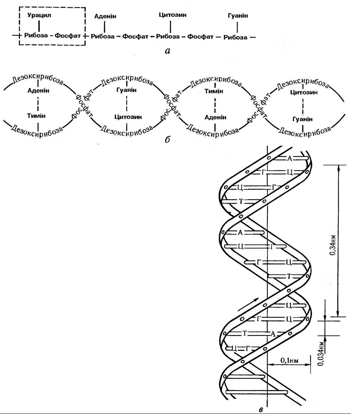
Fig. 147. Diagram of nucleic acid structure: a — RNA molecule (one nucleotide highlighted); b — DNA molecule; c — Watson-Crick model of the DNA molecule. Nitrogenous bases are indicated by letters (adenine, thymine, guanine, cytosine)
The DNA double helix and Histones together form nucleosomes. A nucleosome is defined as a complex consisting of 8 histone protein molecules around which DNA loops are wrapped, each containing 140–200 Base Pairs. Nucleosomes have a diameter of 10 nm. Located between nucleosomes are DNA segments of 30–50 base pairs (up to 10–20 nm in length) associated with histones. This Organization of DNA into Chromatin allows the molecule to fold into loops and higher-order structures. DNA within nucleosomes is less accessible to Nucleases, making internucleosomal regions more susceptible to Cleavage. Genes (cistrons) encoding functionally related proteins are distributed across different regions of chromosomes rather than being organized into operons, as observed in prokaryotes.
Most RNA molecules are synthesized as precursors (pro-RNA). Following The formation of the primary transcript, Processing (maturation) takes place,
which involves a series of reactions transforming primary transcription products into functional molecules. The half-life of most eukaryotic messenger RNA molecules ranges from 3 to 48 hours (Fig. 148).
In addition to unique nucleotide sequences, Eukaryotic DNA contains A large number of repetitive sequences, or repeats. Their copy number can reach 106 per genome. In higher plants, unique genomic regions account for only 20–30%. While a large number of repeats are required for the synthesis of rRNA and histones, the majority are utilized in the Introduction/30.html">Regulation of Gene Expression.
The majority of The Genome is occupied by introns — sequences within a structural gene that do not code for a protein product. Following gene transcription, sequences corresponding to introns are excised from the mRNA during splicing.
Splicing is the enzymatic removal of introns and the joining of two exon ends during mRNA synthesis. Exons are the regions of a structural gene that encode the amino acid sequence
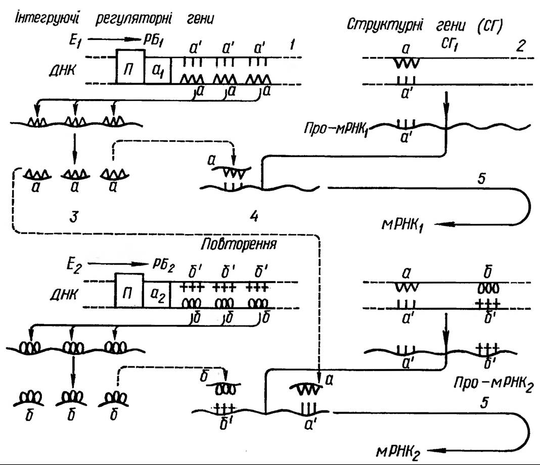
Fig. 148. Hypothetical scheme of mRNA Gene Expression regulation in eukaryotes: E1, E2 — effectors; RB1, RB2 — effector receptor proteins; a1, a2 — acceptor zones, P — promoter
of the protein product. Genes also contain spacers, which are either never read or are transcribed and subsequently degraded.
DNA Structure AND Synthesis. DNA is a polymer composed of four nucleotides: Purines (adenine and guanine) and Pyrimidines (cytosine and thymine), along with deoxyribose and a phosphoric acid residue. It is a double helix in which the nucleotides of each strand are covalently linked via phosphoric acid residues. The two strands interact through Hydrogen Bonds formed within the interior of the helix between purine and pyrimidine bases, where adenine always pairs with thymine (A–T, T–A) and guanine with cytosine (G–C, C–G) (see Fig. 147). This phenomenon of complementarity forms the basis for DNA Replication, as well as DNA-dependent synthesis of all RNAs and Protein Synthesis within the cell. Eukaryotic DNA molecules contain 109 to 1011 such base pairs.
This process occurs during a specific phase of the mitotic cycle in the presence of triose phosphates of all four deoxynucleotides, magnesium ions, a DNA primer, and DNA polymerase, and is termed replication (i.e., copying). It proceeds in a semi-conservative manner, meaning that the DNA molecule doubles by replicating each of its two strands. Consequently, each DNA double helix consists of one "old" and one "new" polynucleotide strand. During replication, the double-helical DNA structure locally unwinds at multiple sites simultaneously. From these origins, replication proceeds in both directions until adjacent replication forks meet. The new strand is synthesized by DNA polymerase, maintaining the polarity of polymer assembly across all regions.
DNA replication involves a interacting complex of factors, including DNA polymerase, initiation factors, and components responsible for the local unwinding of the DNA double strand.
Structure, Forms, and Synthesis of RNA. All RNAs are likewise built from nucleotides containing four bases: adenine, cytosine, guanine, and uracil. The pyrimidine uracil replaces DNA's thymine in RNA, and ribose is used as the sugar instead of deoxyribose. RNA sizes vary considerably. Messenger RNA accounts for up to 3% of the total cellular RNA and comprises 10,000 to 40,000 nucleotide sequences. Ribosomes contain Ribosomal RNAs (rRNAs) with sedimentation coefficients of 18S and 28S (synthesized in the nucleolus, along with 5.8S and 5S in certain
chromosomes). Transfer RNA consists of 75–80 nucleotides, with about 20 different forms of tRNA present in the cell (each specific to a particular amino acid).
RNA molecules lack a double helix, but different regions can pair with one another, leading to loop formation and The Development of a Secondary structure. For instance, tRNA molecules can adopt a cloverleaf secondary structure. Characteristically, tRNAs contain various methylated and other unusual nucleotides.
RNA Synthesis is carried out on a DNA template with the participation of RNA polymerases (Fig. 149).
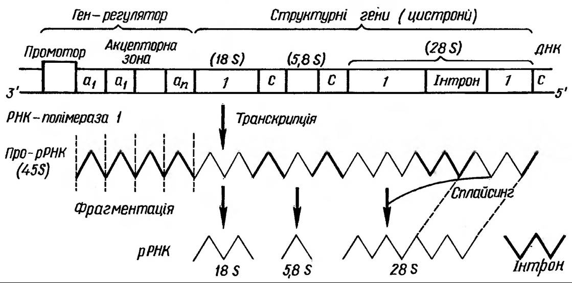
Fig. 149. Eukaryotic rRNA structural genes, their transcription, and processing: S — spacer, 1 — exon
Furthermore, eukaryotic nuclei contain three functional RNA polymerases:
·RNA polymerase responsible for the synthesis of ribosomal RNA, localized in the nucleolus;
·RNA polymerase responsible for the synthesis of messenger RNA;
·RNA polymerase responsible for the transcription of tRNA and 5S rRNA.
The latter two polymerases are localized in the chromatin and nucleoplasm.
RNA synthesis takes place on a DNA template. The Regulation of transcription occurs at various levels: through chromatin fragmentation via histone structure modification (methylation, phosphorylation, etc.), changes in RNA polymerase activity and localization, control over all processing stages, as well as the binding of messenger RNAs into ribonucleoprotein complexes (informosomes) and their subsequent release. A vital role in transcription regulation is played by specific non-histone proteins that act as activators and repressors, receptors for physiologically active substances, phytohormones, and Enzymes.
Protein Structure and Synthesis
Proteins form The basis of the Cytoplasm. They are composed of 20 standard Amino Acids, although significantly more non-protein Amino acids are known. Amino acids are linked within a protein by peptide bonds —NH—CO—. Polypeptides can occur in a free state within plant organisms, Glutathione being a prime example. In addition to peptide bonds, there is another type of covalent bond — Disulfide Bonds (-S-S-) — which connect separate regions of a molecule or polypeptide chains. Various weaker bonds, such as hydrogen and ionic bonds, are also found in protein molecules.
Primary and secondary protein structures are distinguished. The specific sequence of amino acids linked by covalent peptide bonds constitutes the Primary Structure of a protein. Hydrogen bonds between certain regions cause the molecule to fold, thereby forming an α-helical structure. These are the so-called α-structures, which may occur in specific segments of the molecule. A β-structure is also found; it is similarly driven by hydrogen bonds, but not within a single chain, rather between two adjacent chains. Both α- and β-structures define the Secondary structure of a protein. Various spatial arrangements of the helical protein molecule create its specific configuration, which constitutes the Tertiary Structure of the protein.
Proteins quite frequently consist of multiple subunits joined by non-covalent bonds. These complexes form The quaternary structure of proteins. Based on their shape, fibrous and globular proteins are distinguished. Since they contain amino acids with both carboxyl and amine groups, proteins are amphoteric compounds. In alkaline solutions, they dissociate as acids and carry a negative charge, whereas in an acidic medium, proteins dissociate as bases and thus carry a positive charge. The pH at which the equilibrium between positive and negative charges of a protein is established is called the isoelectric point. In this state, the protein is at its least
soluble and readily precipitates. Hydroxyl and amine groups are hydrophilic, enabling them to bind water molecules. Under certain conditions, proteins acquire a gel structure — this is associated with the formation of reticular structures from protein molecules, inside of which appropriately oriented water molecules are located. When such a reticular structure breaks down, the entire complex transitions into a more hydrated sol state. The gel–sol transitions play a crucial role in cytoplasmic activity.
information regarding protein structure is contained within messenger RNA in the form of codons, each consisting of three bases — triplets equivalent to a specific amino acid. Because RNA nucleotides comprise four nitrogenous bases, 64 codons are possible for amino acids, meaning that more than one codon corresponds to each amino acid. Protein synthesis takes place on an mRNA template in the cytoplasm during translation (Fig. 150).
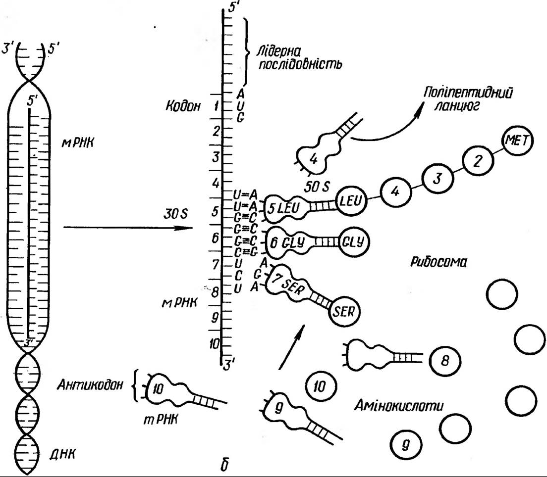
Fig. 150. Schematic representation of transcription (a) and translation (b) processes
The mechanisms of translation are considerably more complex than those of transcription. While transcription is driven by about two dozen proteins, translation requires up to 50 specialized proteins. In addition to initiation, the polypeptide synthesis process includes elongation and termination phases.
To be incorporated into a polypeptide, amino acids are first activated, converting into aminoacyl adenylates with the participation of ATP and aminoacyl-tRNA synthetase; under the action of the same enzymes, these adenylates then attach to their respective tRNAs (Fig. 151). For each amino acid, there is one specific tRNA and one aminoacyl-tRNA synthetase. Polypeptide chain synthesis within the ribosomal complex proceeds until the mRNA termination codon is reached. A protein termination factor binds to this codon, which not only recognizes the required codon but also ensures the release of the polypeptide from the tRNA. Following polypeptide cleavage, deacylated tRNAs and mRNA dissociate. The process concludes with the dissociation of the ribosome into subunits.
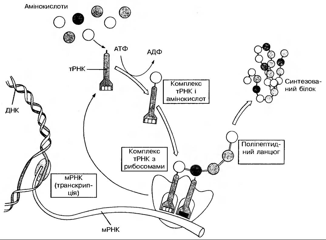
Fig. 151. Diagram of protein synthesis
Characteristically, each mRNA molecule can be read multiple times. It should be noted that a specific physicochemical environment (the
presence of magnesium, potassium, and manganese ions, pH, etc.) plays a vital role in protein synthesis. Within a cell, protein synthesis at any given time executes a specific physiological program; therefore, the activation and reading of numerous mRNAs encoding newly formed proteins must be strictly coordinated. When the physiological state of a cell changes, virtually the state of all components of The Translational Apparatus changes as well. A decrease in protein synthesis intensity, in turn, inhibits The transport of mRNA from the nucleus to the cytoplasm. Consequently, regulatory systems function within The plant cell to coordinate the transcription of various RNA classes and the translation of various proteins, while ensuring the close interaction of all these processes.
Self-Assembly and Biogenesis of Cellular Structures
Transcription and translation are the first stages in the formation of intracellular structures. The subsequent stages involve the assembly of supramolecular complexes and their delivery to specific cellular sites. All Stages of Protein molecule transformation, starting from the synthesis of its ribonucleic template to its incorporation into specific cellular components, are associated with self-assembly processes. These very processes form the basis of the formation and biogenesis of cellular structures.
Self-assembly is The process of spontaneous aggregation of homogenous or diverse molecules, leading to their ordering and the growth of multicomponent structures. If covalent bonds form immediately following self-assembly, such a process is termed polymerization. The mechanism itself relies on weak interactions: electrostatic forces (at a distance of 0.7 nm), hydrogen bonds (0.1 nm), Van der Waals forces, and hydrophobic interactions.
The selectivity of the self-assembly mechanism is ensured by the presence of recognition sites on biopolymer molecules for corresponding partner molecules. It should be borne in mind that molecular self-assembly can be a reversible process because it involves only weak bonds. The Nature of self-assembly is determined by the Features of the polymer's primary structure and also depends on the physicochemical conditions of the environment, the presence of specialized regulatory molecules, and other factors. For instance, self-assembly in biological systems is manifested in the bilayer arrangement of Phospholipids in membranes, the specific sequence of nitrogenous bases in nucleic acids, the enzyme-substrate interaction, the receptor-effector protein interaction (e.g., phytohormones), the formation of enzymatic complexes, and so forth.
It should be remembered that in an aqueous medium, and consequently in the cytoplasm, the aggregation of substances results in the formation of a liquid-crystalline state of the system's components. This state can be viewed as the fourth state of matter (liquid crystals). Proteins, nucleic acids, Polysaccharides, and lipids in water form structures that respond to various environmental influences — light, electric and magnetic fields, pH — thus exhibiting properties characteristic of living cells.
Synthesized proteins and lipids participate in the self-assembly of cell membranes, ribosomes, microtubules, microfilaments, Plastids, and other cellular components. A leading role in the biogenesis of cell membranes belongs to the Endoplasmic reticulum, which serves as the primary site for the synthesis of both Membrane Proteins and lipids, notably Phospholipids and Glycolipids. The Biosynthesis of Steroids and all Unsaturated Fatty acids also takes place here. It is precisely here that plant-specific fatty acids — linoleic, linolenic, and arachidonic — are synthesized. Derivatives of endoplasmic reticulum membranes include the membranes of vacuoles, spherosomes, Microbodies, and so on. The endoplasmic reticulum is directly connected to the nuclear envelope and participates in the Synthesis of the Plasmalemma. The transition of a membrane into various types of organelles is termed "membrane flow." THE CONCEPT OF the interaction of cellular membrane components is known as the "endomembrane system" concept, according to which membrane flow explains their functional continuity and Participation in the vital activity of all cell organelles.
Last update: 07/08/2026
Editorial and Educational Adaptation: This material has been compiled based on the primary/original source text. The project team performed an editorial review, corrected technical inaccuracies, structured sections, and adapted the content for an educational format.
What was processed:
- elimination of formatting defects (OCR errors, structural breaks, corrupted characters);
- editorial organization of content;
- standardization of terminology in accordance with academic sources;
- verification of factual statements against the original source text.
All mentions of the author, publication year, and origin of the primary text have been preserved in accordance with the source.