BIOCHEMISTRY AND MOLECULAR BIOLOGY - W. ELLIOTT - 2002
CHAPTER 19. DNA: STRUCTURE AND LOCALIZATION IN CELLS
In this chapter, we will explore The Structure of DNA and METABOLISM/2.html">THE CONCEPT OF the Gene, while the next two chapters will cover DNA Synthesis AND its role in protein expression. The primary function of DNA lies in its participation in Protein Synthesis, where the Amino Acid Sequence dictates the vast array of processes that have formed The basis of life over millions of years of evolution.
What are Nucleic Acids?
Deoxyribonucleic acid, or DNA, was first isolated from Cell nuclei, which is why it was named nucleic acid (from the Latin nucleus - core or kernel). Its acidic properties are due to the presence of phosphoric acid residues within the molecule. The carbohydrate component is 2-deoxy-D-ribose. Together, these features gave rise to the name deoxyribonucleic acid. Cells also contain another major Class of nucleic acids—Ribonucleic Acids—in which the carbohydrate is D-ribose, hence the name ribonucleic acids, or RNA (see Chapter 21). Unlike D-ribose, 2-deoxy-D-ribose lacks an oxygen atom at the second carbon position; it is commonly referred to simply as deoxyribose.
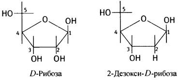
In Eukaryotic cells, the bulk of DNA is confined to The Nucleus (with minor amounts found in Mitochondria and Chloroplasts), whereas the majority of RNA resides in the Cytoplasm, although it is synthesized in the nucleus (except for Mitochondrial and Chloroplast RNAs). Although nucleotide synthesis was already discussed in Chapter 18, for the sake of convenience, we will revisit this class of compounds before turning our attention to DNA.
Introduction/19.html">Primary Structure of DNA
DNA is a polynucleotide. The structure of a nucleotide can be represented as follows: phosphate-sugar-base.
The structure of a deoxynucleotide, shown here in its un-ionized form for simplicity, appears as follows.
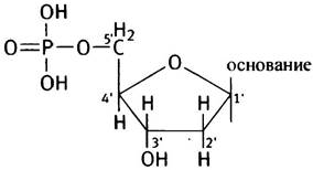
As noted above, the sugar is 2-deoxy-D-ribose (deoxyribose) in the furanose form, forming a five-membered ring. A prime symbol (') is added when numbering the atoms of the deoxyribose ring to distinguish them from the numbering of atoms in the bases. Therefore, the carbon atoms in the carbohydrate residue are designated as 1', 2', 3', 4', and 5' (pronounced "five prime", etc.), and these numbers are written on the outside of the ring.
Which bases are found in DNA?
Unlike Proteins, which are built from 20 different Amino Acids, DNA contains only 4 distinct bases, whose structures are scarcely more complex than those of amino acids. These bases are adenine, guanine, cytosine, and thymine, or abbreviated as A, G, C, and T, respectively. A and G are Purines, while C and T are Pyrimidines. The numbering of atoms within the bases is written inside the ring.
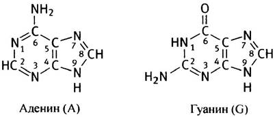
Note the methyl group in thymine! Hereafter, the names of the bases will be used in their abbreviated forms.
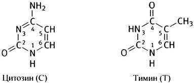
Bases can exist in tautomeric forms (keto-enol and amino-imino). At neutral pH, the bases in DNA exist predominantly in the keto and amino tautomeric forms:
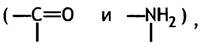
rather than in the enol and imino forms:
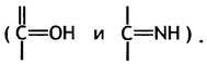
Linkage of Bases to Deoxyribose
The C1' carbon atom of the sugar is linked via a glycosidic bond to the nitrogen atom at position 9 of a purine base or position 1 of a pyrimidine base, respectively. This linkage is termed a β-glycosidic bond, a designation determined by the conformation of the sugar ring.
A compound consisting of a base linked to a carbohydrate is called a nucleoside; when the carbohydrate is deoxyribose, it is termed a deoxynucleoside. All deoxyribonucleosides have their own specific names.
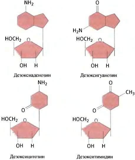
A nucleoside containing adenine as its base is called deoxyadenosine, and the guanine derivative is deoxyguanosine. Deoxycytidine and deoxythymidine are the deoxynucleosides of cytosine and thymine, respectively.
What are the Physical Properties of polynucleotide components?
In DNA, NUCLEOTIDES occur as 5'-phosphate derivatives — dAMP, dGMP, dCMP, and dTMP. As noted earlier, the latter can simply be designated as TMP, since its ribose counterpart is practically non-existent.
Both hydroxyl groups of phosphoric acid are ionized at physiological pH values, with a pKa of ~2 for one and ~7 for the other, which indicates their hydrophilic nature. Due to the phosphate groups, the DNA molecule carries a strong negative charge. The sugar with its hydroxyl groups also exhibits hydrophilic properties. The bases, conversely, are almost insoluble in Water and possess a pronounced hydrophobic character, although A number of atoms retain The ability to form Hydrogen Bonds.
Structure of a deoxyribopolynucleotide
Let us mentally remove a water molecule from two nucleotides. The resulting structure will represent a dinucleotide (the non-ionized form is shown):
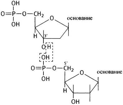
The formation of a dinucleotide is accompanied by a large positive change in Free energy ∆G°′; consequently, synthesis cannot proceed via the direct Condensation of two nucleotides.
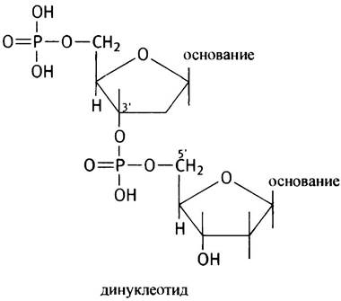
In a mononucleotide, the phosphate group is linked to deoxyribose by a simple ether bond. In a dinucleotide, a phosphodiester bond is formed. It is called a diester bond because the phosphate is linked to two carbohydrate residues:
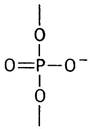
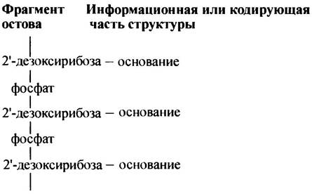
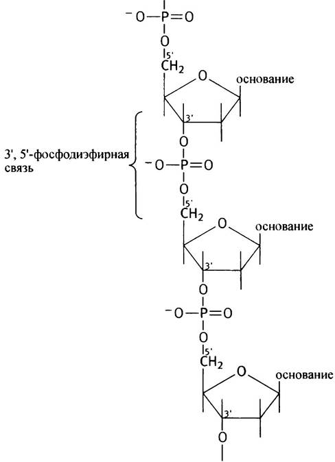
In the dinucleotide shown above, this is a 3',5'-phosphodiester bond, where the phosphate links the 3'-OH group of one nucleotide to the 5'-OH group of another. If nucleotides are further attached in a similar manner, we obtain a structure called a polynucleotide. The primary structure of DNA is a polynucleotide of enormous length. When discussing proteins, we noted the presence of a polypeptide backbone with amino acid residues protruding from it. A polynucleotide also has its own backbone, consisting of alternating sugar-phosphate-sugar groups, with the bases attached to each sugar residue at position 1'. The encoded information is written in The sequence of bases. A DNA chain can be schematically represented as follows.
The STRUCTURE OF THE chain can be depicted as follows.
Why deoxyribose?
Why not ribose?
The Cell contains a number of nucleotides (e.g., AMP) as well as ribonucleic acids containing ribose as their carbohydrate component. It is believed that ribonucleotides appeared earlier in evolution than deoxyribonucleotides, and consequently, RNA appeared before DNA. At the same time, cells expend considerable energy to convert ribonucleotides into deoxyribonucleotides, which are necessary for DNA synthesis. Why?
One of the most compelling reasons for storing Genetic information, which has accumulated over millions of years in the form of DNA molecules, is as follows: chemical molecules are always characterized by a certain degree of instability and a capacity for spontaneous degradation. DNA molecules, however, remain unchanged and largely intact across many generations. Furthermore, DNA is more stable than RNA. This is because the 2'-OH group of ribose readily undergoes nucleophilic attack in the presence of phosphate hydroxyl ions. As a result, the phosphodiester bond is cleaved to form 2',3'-Cyclic Nucleotides, whereas in DNA, where 2'-OH groups are absent, this cannot occur.
The OH- ion promotes The conversion of the 2'-OH group into a 2'-O- group, which is attacked by the phosphorus atom. This leads to the Cleavage of the polynucleotide chain and the formation of a 2',3'-cyclic nucleotide. Subsequent Hydrolysis of the cyclic nucleotide yields a mixture of 2'- and 3'-nucleotides, depending on the site of hydrolysis.
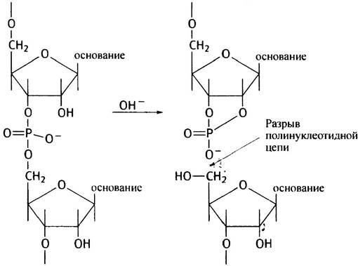
The difference in stability between RNA and DNA is further confirmed by the fact that a dilute NaOH solution completely destroys RNA at room Temperature while having no effect on DNA. Thus, DNA is a more stable and reliable information storage medium than RNA. Note that the fact that some Viruses use RNA as an information carrier does not contradict this statement.
The DNA double helix
Almost all DNA exists as a double strand, with the exception of the DNA of certain viruses. In other words, DNA consists of two polynucleotide molecules that form a pair. What holds them together? Base pair complementarity. This means that when A and T in two DNA strands are positioned opposite each other, 2 hydrogen bonds spontaneously form between them. Recall that hydrogen bonds have a short range of action, and their formation therefore requires the precise alignment of a pair of atoms. G and C also form a complementary pair, though 3 hydrogen bonds arise between them. Other base combinations cannot be complementary, so G does not pair with A or T, and so on. Only A-T and G-C pairs are found in DNA; they are known as Watson-Crick Base Pairs. Strikingly, the formation of hydrogen bonds is a completely spontaneous process that requires no Enzymatic Catalysis. After all, hydrogen
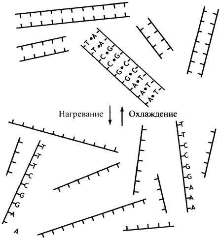
bonds are weak and easily broken, for instance, upon heating. Slight heating will be accompanied by the disruption of base pairing, and cooling by the reverse association of the molecules.
The geometry of base pairs is shown in Fig. 19.1.
Fig. 19.1. Hydrogen bonds in Watson-Crick base pairs
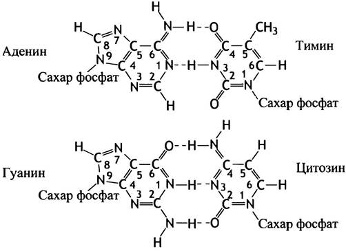
Note that a base pair always contains one purine (larger molecule) and one pyrimidine (smaller molecule); consequently, base pairs are always of uniform size. The spontaneous process of base pairing is called Hybridization. If a DNA molecule is cut into thousands of short double-stranded fragments ranging from 20 to 100 nucleotides in length and then heated to about 95° C, the paired strands of each DNA fragment will separate from one another, resulting in DNA melting. This occurs due to the heat-induced disruption of hydrogen bonds, yielding single-stranded DNA fragments with various base sequences. However, if the solution is cooled again, the fragments will slowly re-pair with their original complementary strands (Fig. 19.2). This hybridization technique, sometimes referred to as annealing, forms the basis of molecular gene biology (see Chapter 24). The thermodynamic driving force behind hybridization is the release of energy that accompanies the formation of Hydrogen bonds and other weak interactions. In the most stable state, free energy is at its minimum, which corresponds to the base pairing arrangement that maximizes the number of hydrogen bonds. Experimental evidence of hybridization demonstrates that G-C and A-T base pairs form spontaneously, representing a genuine physical phenomenon.
Fig. 19.2. Spontaneous hybridization of complementary DNA fragments. To illustrate the principles of hybridization, The base sequence is shown on only one DNA fragment
Because of base complementarity, The amount of G in a DNA molecule equals the amount of C, and the amount of A equals the amount of T. Different DNA molecules vary in their [A + T] and [G + C] pair content due to differences in nucleotide composition, which accounts for their distinct roles as carriers of genetic information.
As a first approximation, a DNA strand can be visualized as long continuous lines representing the sugar-phosphate backbone, with the hydrogen-bonded bases always oriented inward.
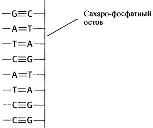
Note that in DNA regions rich in [G + C], the two strands are held together more strongly than in regions with a higher proportion of [A + T] pairs.
However, weak interactions significantly influence the conformation of large molecules, and DNA is no exception. Therefore, the straight-strand structure described above does not occur under normal conditions.
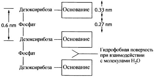
The length of the phosphodiester bond is 0.6 nm (1 nanometer = 10-9 m), and the thickness of the bases is about 0.33 nm, meaning gaps would remain between them in a straight, ladder-like structure. As already mentioned, the planes of the bases are hydrophobic. In a "straight" structure, they would be forced into an aqueous environment, rendering the molecule unstable. How can hydrophobic bases bond with each other while avoiding contact with H2O? They must stack upon one another by bending the phosphodiester bond; hydrophobic interactions between the base planes will then drive this stacking process (Fig. 19.3, a).
Each coplanar base pair is parallel to its immediate neighbors, forming what is known as a base stack. Furthermore, the bases are oriented in such a way that hydrogen bonds can form between opposing strands. Nevertheless, this "ladder" structure does not actually occur in DNA because it is stereochemically impossible. In reality, the two DNA strands form a helix in which the bases are located on the inside, while the hydrophilic sugar and phosphate groups are on the outside (Fig. 19.3, b, c).
Fig. 19.3. Nucleotide packing model in DNA: a - Base pair bonding type in the "slant ladder" structure; b - Backbone contour in the DNA double helix; c - Same as b, with base pairs shown in the center of the helix (each cylinder represents a base pair); d - Arrangement of two adjacent base pairs in The Double Helix, showing the displacement determined by the double helix structure. S - sugar, P - phosphate, B - base. A more detailed model corresponding to c is shown in Fig. 19.4
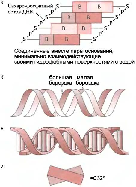
This structure ensures close interaction between base pairs and prevents contact with water, though the base stack is no longer strictly vertical. The base pairs are slightly displaced relative to one another; as shown in Fig. 19.3, d, there are about 10 pairs per turn.
The helix is right-handed; looking along its axis, the turns follow a clockwise direction (imagine driving a screw while holding a screwdriver in your right hand: the direction of rotation gives a sense of the helical twist). The structure of the double helix
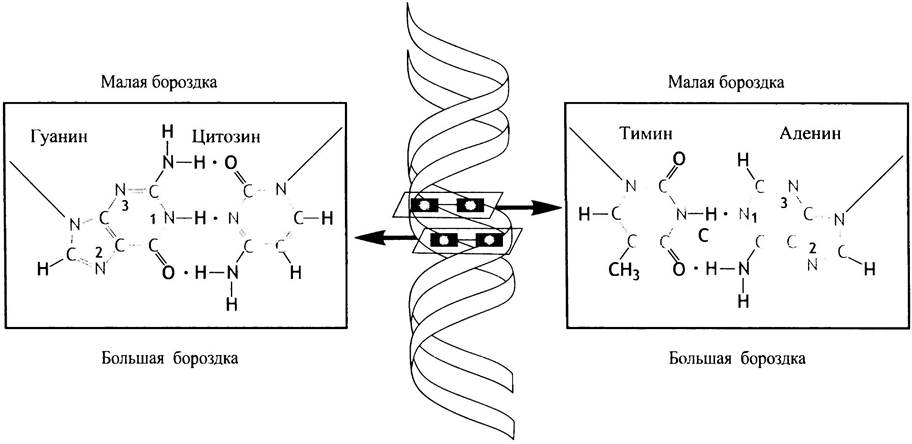
is such that it features two grooves—a major groove and a minor groove (see Fig. 19.3, b). Although the grooves are depicted flat on the page, they are difficult to visualize outside of three-dimensional space. If you have the opportunity to examine a 3D DNA model, it will help clarify the helical sense and reveal the two grooves, which is critically important. Portions of each base are exposed in both the Major and minor grooves, making them accessible for interactions with other molecules. The major groove provides easier access for proteins that "recognize" specific bases. Base pairs look quite distinct when viewed from the perspective of the two different grooves (Fig. 19.4); The Significance of this will be explained in Chapter 21, where Genetic regulation is discussed.
Fig. 19.4. Schematic diagram showing that the same DNA base pair looks different when viewed from the major and minor grooves
DNA-binding proteins designed to recognize specific sequences in DNA can bind to their target sites without unwinding the helix
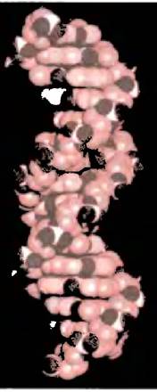
The DNA conformation described above is known as the B-form (Fig. 19.5): this is the standard form of DNA typically found in cells. However, DNA can alter its configuration depending on environmental conditions. Upon dehydration, the double helix adopts a more compressed shape with a larger base tilt angle, a conformation referred to as the A-form. This form of DNA is found in spores. Another known configuration is the Z-form, in which the sugar-phosphate backbone forms a zigzag path along the helix. The Z-form double helix is left-handed, whereas the B-form is right-handed. Evidence suggests that the Z-form occurs under high Ionic strength conditions in short synthetic DNA molecules with alternating purine and pyrimidine bases. The Biological Significance of the A- and Z-forms remains unclear, though adaptive configuration changes in short chromosomal regions cannot be ruled out.
Fig. 19.5. Model of the B-form of DNA. A three-dimensional model of a DNA fragment is shown, featuring two major grooves and one minor groove
An essential property of the double helix is its ability to bend. A DNA molecule can be a million times longer than the largest cells or nuclei; to fit inside them, the molecule must be flexible.
It should be noted that although DNA almost always exists in the double-helical form, single-stranded DNA does occur, for example, in certain bacterial viruses. In such cases, the molecule forms a complex folded structure to ensure thermodynamic stability. Crucially, even here, the viral life cycle includes a stage where DNA adopts the double-helix conformation (see Chapter 23); therefore, the fundamental principles of genetic information transfer, which rely on complementary base pairing, remain fully valid.
DNA strands are antiparallel: what does this mean?
By antiparallel, we mean a structural arrangement in which the two strands of the DNA double helix run in opposite directions. The two antiparallel DNA strands are illustrated in Fig. 19.6.
Fig. 19.6. Two antiparallel DNA strands. B — bases

What is meant by the polarity, or directionality, of a DNA strand? Each linear strand has two ends; at one end, There is a deoxyribose 5'-OH group (not linked to another nucleotide), which is sometimes phosphorylated. This is the 5'-end. At the other end lies a free 3'-OH group (also unlinked to another nucleotide and potentially esterified with a phosphate group). This is the 3'-end. Consequently, each end of the double helix always features one 5'-end and one 3'-end. You will never find two 5'-ends or two 3'-ends together! This is precisely what antiparallelism means. Even if a DNA fragment is circular (lacking free ends), the polarity of the individual strands is still preserved. This can be illustrated by the orientation of the sugar residues within the chains.
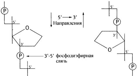
You can determine the direction by looking at the 5' and 3' positions. For instance, in the structure on the left, the strand runs in the 5’ —> 3' direction from top to bottom, whereas on the right, 5' —> 3' corresponds to the bottom-up direction (by convention, nucleotide sequences in DNA are always written in the 5' —> 3' direction).
Pay attention to a seeming contradiction! When moving along a DNA strand in the 5' —> 3' direction (from the 5'-end to the 3'-end), each phosphodiester bond you "pass" is formed between the 3'-OH group of the preceding deoxyribose and the 5'-OH group of the succeeding nucleotide. This means that—judging by THE POSITION OF the phosphate—you are moving from the 3'-end to the 5'-end of the strand. It is important to remember that for a linear DNA strand, the 5' —> 3' direction means moving from the terminal 5'-OH group to the terminal 3'-OH group, or from the 5' group of one deoxyribose residue to the 3' group of the adjacent one. This must be kept in mind, both because of The Importance of the topic itself and for understanding the material in subsequent chapters.
The antiparallel orientation of the strands imposes specific stereochemical constraints on the structure of the double helix.
There are established conventions for writing nucleotide sequences in DNA. A simple polynucleotide structure is typically depicted as a sequence of letters representing the corresponding nucleotide bases. Sometimes, the phosphodiester bond is explicitly indicated by the letter "p" placed between the bases (e.g., CpApTpGp, etc.).
Let us assume we have a double-stranded DNA fragment with the following base sequence:
5' CATGTA 3'
3' GTACAT 5'
Sometimes the sequence is written out for each strand, but in most cases there is no need to depict both strands since the second one is complementary
to the first. Therefore, despite a gene having two strands, its structure is often represented as a single base sequence. Provided it is written starting from the 5' end on the left, special designation of the 5' and 3' ends is not required. Thus, the structure shown above can be denoted simply as CATGTA.
What are the dimensions of a DNA molecule?
The sizes of DNA molecules vary greatly when measured by the number of base pairs they contain. Yet even the shortest of them are very large molecules, while DNA molecules with the longest chains are simply gigantic. Small viral DNAs consist of several thousand base pairs, whereas E. coli DNA contains about 4 million base pairs. The Human Genome (the complete set of chromosomal DNA) reaches 1–2 m in length and comprises 6 billion base pairs. Considering that The Human Body consists of 1013 cells, calculating the total length of all its DNA yields an astronomical figure comparable to the diameter of the solar system.
Clearly, the more information that needs to be stored, the greater the amount of DNA required. Consequently, the more complex the Organism, the more DNA its cells contain. However, this rule does not always hold true. For example, large bean cells contain more DNA than human cells, and amphibians have more than other vertebrates. The Emergence of anomalies (errors) in the DNA Structure leads to the loss of certain functional properties.
How is DNA packaged within the nucleus?
In biochemistry, one is frequently struck by the ingenious solutions evolution has found to various problems! DNA packaging is one of them.
Human cells contain 46 Chromosomes, and the total length of DNA confined within a single cell is 1–2 m, yet it is packed inside a nucleus millions of times smaller. This clearly demands a highly sophisticated packing system. In eukaryotic cells, DNA exists as part of Chromatin, a complex of DNA and proteins. These proteins are predominantly Histones—basic proteins rich in Arginine and Lysine. Due to their positive charge, they form ionic bonds with the negatively charged phosphate groups located on the outer surface of the DNA double helix. The amino acid sequence of eukaryotic histones is highly conserved. For instance, the primary structure of histones isolated from a plant (pea) and a mammal (cow) differs by only Two amino acids, and even these substitutions are relatively minor (valine for isoleucine, lysine for arginine). Such a pronounced constancy of structure suggests that this protein must interact precisely with an equally conserved structure or structures.
Let us begin by examining four histones: H2A, H2B, H3, and H4. They form an octameric protein complex called the nucleosome core (nucleosome core), around which the DNA molecule makes two turns (approximately 146 base pairs). This complex of histone proteins and DNA serves as the fundamental structural unit of chromatin and is called a nucleosome. Individual nucleosomes are connected by linear stretches of DNA known as linkers (Fig. 19.7, a, b). If chromatin in vitro is treated with an enzyme isolated from microorganisms that is capable of hydrolyzing DNA into nucleotides, the linear stretches of DNA are indeed hydrolyzed, whereas the approximately 146 base pairs "wrapped" around the nucleosome core remain protected from the enzyme's action (it is unable to hydrolyze this portion of the DNA). DNA extracted after such Treatment contains intact fragments. This observation formed the basis of our current understanding of nucleosome structure.
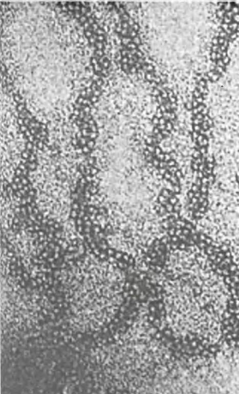
Nucleosomes are approximately 10 nm in diameter and form 10 nm thick fibers (see Fig. 19.7, b). Nevertheless, this level of DNA packaging is not sufficient; a fifth histone participates in its further Organization: it is not part of the nucleosome, and its exact role remains unclear. Next, the 10 nm fiber condenses to form a 30 nm fiber (Fig. 19.7, c). This nucleosome packing is often described as a solenoid; however, this is merely one of several models. According to an alternative model, the packing has a zig-zag conformation. Since the precise structure of the solenoid has not yet been established, the diagram of the 30 nm fiber is presented without packing details. An electron micrograph of such a fiber is shown in Fig. 19.8. At this stage, the DNA is condensed 100-fold—meaning its length is reduced by a factor of one hundred. The 30 nm fibers form long loops anchored to a central protein scaffold of the chromosomes (Fig. 19.7, d). Loop formation renders the structure even more compact, though it remains unclear whether this coiling involves additional supercoiling to achieve the 10,000-fold condensation of the original DNA molecule.
Fig. 19.7 Model of chromatin packaging in eukaryotes: a - Nucleosome; b - nucleosomes with linker DNA regions; c - 30 nm chromatin fiber; d - loops of this fiber attached to a central scaffolding protein. The loops further condense via supercoiling to form a highly compact metaphase chromosome. The final stage of condensation is not shown in the figure
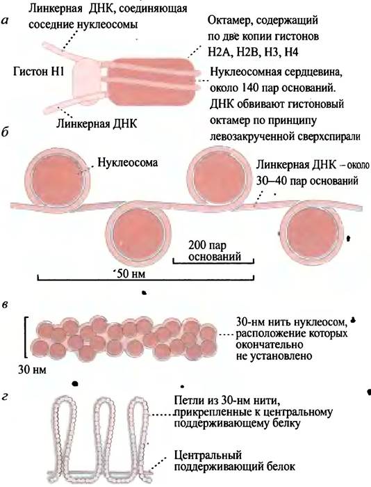
Fig. 19.8. Transmission electron micrograph of a 30 nm chromatin fiber
Thus, there are several levels of DNA organization: 1) wrapping around nucleosomes; 2) packaging of nucleosomes into a 30-nanometer fiber containing 3-5 nucleosomes per turn; 3) formation of loops thousands of nucleosomes long, attached to a central supporting structure; 4) formation of spirals and/or folds from the loop (see Fig. 19.7, a, d).
To perform its function, packaged DNA must remain accessible to the Enzymes involved in reading genetic information and Replication.
In E. coli cells, the DNA double helix is circular. It is about 1,000 times longer than the cell itself. Prokaryotes lack a nuclear membrane and do not possess ordered structures resembling eukaryotic nucleosomes. Nevertheless, histone-like proteins have been found in E. coli that presumably perform a similar function. Because proteins are bound less tightly to DNA in bacterial cells than eukaryotic histones, nucleosome-like complexes are not detected. The looped structure of DNA in E. coli is stabilized by proteins, but the details of this process remain incompletely understood.
How does DNA structure relate to the compact eukaryotic chromosomes visible under a Light Microscope?
Chromosomes in stained dividing eukaryotic cells appear under the light microscope as compact, dense entities similar to those shown in Fig. 19.9.
Fig. 19.9. A metaphase chromosome consisting of two sister chromatids
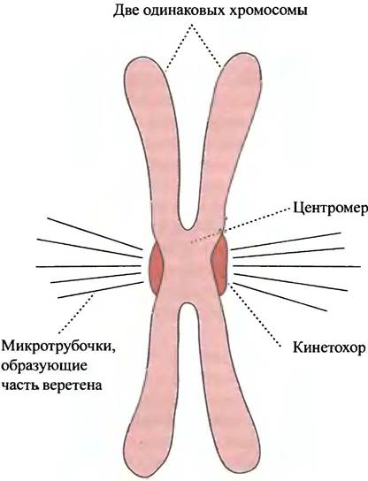
The compact structures in dividing cells represent so-called metaphase chromosomes. Here, DNA is in its most condensed state, which facilitates the proper segregation of chromosomes into each daughter cell following Cell Division.
The most densely packed region of DNA, consisting of repetitive sequences, corresponds to the centromere. It holds a pair of chromatids together, while kinetochore Protein Complexes are located on their outer surfaces. Microtubules of the mitotic spindle attach to the kinetochore to separate sister chromatids (see Chapter 29). After cell division, chromosomes become less condensed and are referred to as interphase chromosomes. The DNA of metaphase chromosomes is functionally inert, whereas interphase DNA is functionally active (although more condensed than naked DNA). The degree of DNA condensation in eukaryotic cells is critically important. Before the genetic information can be utilized, DNA must become accessible to enzymes and other proteins rather than remaining tightly packed. Certain regions of eukaryotic chromosomes contain DNA that presumably does not form part of any gene, meaning it does not carry the information required for protein synthesis. In these regions, even in interphase chromosomes, DNA remains condensed as so-called heterochromatin, whereas actively functioning regions—euchromatin—are less condensed. The exact function of heterochromatin is not yet fully understood.
In addition to histones, chromatin contains other non-histone proteins. They are involved in forming long DNA loops attached to axial supporting protein structures. Thus, chromatin is a complex structure. Depending on Gene Expression, both the degree of its packaging and the compaction of the DNA within chromatin change. The term gene expression refers to The process of protein synthesis controlled by a gene, and is also used when genes encode specific RNA molecules (see p. 283).
WHAT IS A gene in molecular terms?
A gene is the unit of heredity. It represents a segment of a giant DNA molecule and contains encoded information for the amino acid sequence of a single polypeptide chain (see p. 264 for a detailed description of a gene). Information is stored as a sequence of DNA bases, and one gene differs from another solely in its encoded information. For clarity, one can draw an analogy between a chromosome and a magnetic tape: the gene is then comparable to a segment of a musical recording, and the base sequence to the magnetic signals. The above applies to "standard" (typical) protein-coding genes, which constitute the overwhelming majority.
Some variations of the "standard" gene
Occasionally, multiple copies of the same gene exist. For example, genes for certain histone proteins are found in hundreds of tandemly repeated copies in sea urchins. This may result from Gene Duplication during evolution. Multiple copies of a gene can ensure accelerated protein synthesis. From an evolutionary standpoint, this allows one of the copies to adapt to perform some other (alternative) function. There are also pseudogenes—copies of functional genes that are capable of expression but synthesize a non-functional protein or protein fragment.
There is a group of genes that do not encode proteins: these are the genes for ribosomal and Transfer RNA. They encode RNA molecules essential for protein synthesis (see Chapter 22).
An ordinary "standard" gene is a DNA segment located at a specific site on a chromosome. DNA sequences are not always rigidly fixed; sometimes they contain fragments capable of moving from one site in the DNA to another. These are called mobile, or jumping genes (see p. 259). Chromosomes can also acquire new genes from Retroviruses (see p. 313).
Gene structure is not always constant. Under certain conditions, the DNA segment corresponding to a particular gene can undergo Amplification, resulting in a huge number of gene copies and a corresponding sharp increase (approximately 1,000-fold) in the Synthesis of the protein it encodes. This phenomenon is observed in experiments using mouse Cancer cell cultures treated with methotrexate, an "antifolate" drug used in the treatment of leukemias (see p. 226).
Repetitive DNA
In human cells, not all DNA is organized into genes. For instance, our chromosomes contain fairly long DNA stretches (several hundred bases long) that are repeated hundreds and thousands of times, interspersed throughout the chromosome. An example is provided by Alu sequences (Alu is an abbreviation derived from the restriction enzyme used for their cleavage). The function of Alu sequences remains undetermined.
Another type of repetitive DNA sequence is satellite DNA: it is frequently found in the centromeric region of chromosomes and forms part of heterochromatin. The term satellite arose because when giant eukaryotic DNA molecules were isolated using standard Methods, they fragmented. Upon density-gradient centrifugation, the bulk of the DNA sedimented as a single band, whereas repeated DNA tracts with a different density separated from the main band (appearing as satellites). Satellite DNA consists of short sequences repeated in tandem (one after another). The length of each fragment is approximately 105 bases. Other repetitive DNA sequences are dispersed throughout the chromosome.
Where are we now?
To keep from losing the main thread of our argument amid a wealth of detail, let us recall that we first dealt with Protein Structure before moving on to the questions related to its Biosynthesis. Now we turn to DNA biosynthesis. First, this is a logical continuation of our study of DNA structure; second, it will help us understand subsequent Stages of Protein Synthesis. Therefore, in the next chapter we will discuss DNA synthesis, and then proceed to examine the pathways of genetic information transfer directly linked to Protein Biosynthesis.
Questions for Chapter 19
1. Draw the structure of a dinucleotide.
2. Ribonucleic acid (RNA) appeared earlier than deoxyribonucleic acid. Why did DNA evolve?
3. The bases in DNA are hydrophobic. Explain how this affects the structure of double-stranded DNA.
4. List the key Features of the DNA double helix. Is it a left-handed or right-handed helix? How many base pairs make up one turn of the DNA helix?
5. Explain what is meant by the antiparallel orientation of DNA strands in the double helix.
6. Explain the Significance of the 5' —> 3' direction in a linear DNA molecule.
7. If you see a DNA sequence written as CATAGCCG, what does it represent in terms of double-helical structure and strand polarity? Explain your answer.
8. What is a nucleosome? What evidence Supports its existence?
Last update: 06/08/2026
Editorial and Educational Adaptation: This material has been compiled based on the primary/original source text. The project team performed an editorial review, corrected technical inaccuracies, structured sections, and adapted the content for an educational format.
What was processed:
- elimination of formatting defects (OCR errors, structural breaks, corrupted characters);
- editorial organization of content;
- standardization of terminology in accordance with academic sources;
- verification of factual statements against the original source text.
All mentions of the author, publication year, and origin of the primary text have been preserved in accordance with the source.