Fundamentals of Molecular Biology. Part 1: Molecular Biology of the Cell - A. N. Ogurtsov 2011
Fundamentals of Cell Theory
Mitochondria and Plastids
Cell/35.html">Mitochondria, present in all Eukaryotic Cells—both autotrophic (such as photosynthetic plants) and heterotrophic (such as animals and Fungi)—contain Enzymes and other protein-membrane structures responsible for oxidizing Organic compounds and synthesizing adenosine triphosphate (ATP), the primary energy source for intracellular biochemical transformations.
Mitochondria are among the largest cellular Organelles, comparable in size to an E. coli bacterium. In some eukaryotic cells, mitochondria can occupy up to 25% of the Cytosol.
The number of mitochondria in cells varies significantly depending on the energy demands of a given cell type. Some species of Algae and Protozoa contain only a single mitochondrion, spermatozoa of various animal species contain 20–70, and mammalian body cells contain anywhere from 100 to several thousand. Mitochondria are most abundant in Tissues that constantly require large amounts of energy to perform their Functions, such as Muscles, Liver, and Kidneys. Mitochondria can account for 2% to 48% of the total cell volume, with the average being around 18%.
A mitochondrion is formed by two structurally and functionally distinct membranes—the outer and inner membranes—separated by the intermembrane space (Figure 20).
Class="center">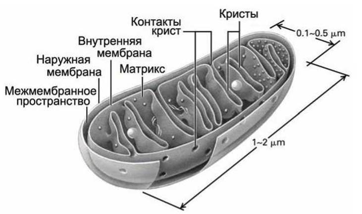
Figure 20 – Diagram of a mitochondrion
The inner membrane, which harbors the enzymes that catalyze oxidative reactions in the Respiratory Chain and synthesize ATP, forms numerous folds known as cristae that project inward toward the matrix, the central region of the mitochondrion.
Mitochondria contain their own DNA and Ribosomes, yet they receive most of their Proteins from the Cell Cytoplasm. To emphasize their role as the primary source of ATP, mitochondria are often referred to as the "powerhouses" of The Cell.
The outer mitochondrial membrane contains numerous porin proteins that allow ions and small metabolite molecules (with a Molecular Weight of up to 5,000 Da) to pass freely into the intermembrane space.
The main barrier regulating The entry of metabolites into the mitochondrial matrix is The inner mitochondrial membrane. The inner membrane is packed with Membrane Proteins (constituting up to 75% of its mass), some of which drive essential biochemical reactions, while others (transporters) shuttle metabolites across the membrane.
The inner membrane features an elevated concentration of cardiolipin (diphosphatidylglycerol), which reduces its permeability to protons and helps establish a significant proton-motive force across the inner membrane.
New mitochondria are formed exclusively through the division or budding of pre-existing mitochondria.
Plastids are found exclusively in photosynthetic eukaryotic organisms (higher plants, algae, and certain unicellular protozoa). Based on their pigment composition and associated functions, plastids are divided into three main types: METABOLISM/14.html">Chloroplasts (green plastids), chromoplasts (yellow or orange plastids), and leucoplasts (colorless plastids). Each individual cell contains plastids of only one type.
Chloroplasts contain the green pigment chlorophyll and carry out Photosynthesis in the presence of light, converting light energy into chemical energy that is then used to synthesize the compounds required by the plant. Chloroplasts are found in nearly all plant cells exposed to light, but are particularly abundant in leaf cells.
Leucoplasts occur in plant cells that are not exposed to sunlight. Their primary function is the accumulation and storage of nutrient reserves.
Chromoplasts accumulate yellow, orange, and red pigments (carotenoids). Much like pigmented vacuoles, chromoplasts impart vibrant colors to various plant parts. For instance, chromoplasts give color to the petals of buttercups, dandelions, and tulips, as well as to ripe tomatoes, rowan berries, watermelons, oranges, carrots, and beets.
Plastids are capable of interconverting. If the colorless underground part of a plant is exposed to light, leucoplasts will begin to accumulate chlorophyll under The Influence of light and transform into chloroplasts.
For this reason, a potato tuber left out in the sun turns green, for example. Conversely, as chloroplasts age, they lose their green coloration and turn into chromoplasts. This process occurs in leaves during autumn: the green chlorophyll in chloroplasts begins to break down, revealing the carotenoids that were present all along but masked by chlorophyll. As a result, green leaves turn yellow, orange, or red. These mutual transformations of plastids indicate that they represent different developmental stages (differentiation types) of a single organelle, which is further supported by the structural uniformity shared among different plastids.
The primary type of plastid is the chloroplast (Figure 21). They typically maintain a consistent shape of a prolate spheroid, with long and short semi-axes of 2–3 µm and 0.5–1.5 µm, respectively.

Figure 21 – Diagram of a chloroplast
Most plants have chloroplasts with a volume ranging from 30 µm3 to 60 µm3. (Smaller plastids, under 30 µm3, are found in poplars, aspens, ashes, and certain sedge species. Unusually large chloroplasts are characteristic of shade-tolerant plants, such as wild ginger at 227 µm3, stonecrop at 240 µm3, and violets at 100 µm3).
The number of chloroplasts per cell varies from 3 to 400, with an average of 10–30 chloroplasts per cell. (More than 400 plastids per
cell have been found in tobacco, rhubarb, and certain lily species. Low plastid counts are typical of cells in reeds (4–10), strawberries (8–12), and lemons (5–10)). The number of plastids in a cell correlates strongly with nuclear DNA content and much more weakly with cell volume. For estimation purposes, a standard statistical average of 14 chloroplasts per 10,000 µm3 of cell volume can be used.
Chloroplasts, much like mitochondria, are bounded by two membranes: an outer membrane containing porins that is permeable to metabolites, and an inner membrane containing transport proteins that facilitate metabolite exchange with the cell cytosol. Neither of these chloroplast membranes takes part in photosynthesis.
Unlike mitochondria, chloroplasts possess an additional, third membrane known as the thylakoid membrane.
In the mature chloroplasts of higher plants, Two Types of internal membranes are distinguished: (1) extended, flat folds formed by the inner envelope membrane that can span nearly the entire plastid, known as stroma lamellae, and (2) flat, closed, disk-like membrane sacs called thylakoids.
Thylakoids frequently form stacks resembling piles of coins, which are referred to as grana. The number of thylakoids per granum can range from a few to 50 or more.
The number of grana in chloroplasts can reach 40–60. Thylakoids within a granum are closely appressed so that the outer layers of their membranes fuse tightly. In addition to thylakoids, a granum typically incorporates regions of stroma lamellae, which thus interconnect the individual grana of the chloroplast.
All chloroplast thylakoids assembled into grana share a continuous thylakoid membrane that separates the thylakoid lumen—the internal volume of the thylakoids forming a single compartment—from the stroma, the other chloroplast compartment situated between the thylakoids and the inner chloroplast membrane.
Embedded within the thylakoid membrane are numerous integral membrane proteins containing specific prosthetic groups and light-absorbing pigments, such as chlorophyll.
Carbohydrate synthesis takes place in the stroma of the chloroplast. In photosynthetic Bacteria, folds of The Plasma Membrane form membranous structures that house the Photosynthetic Systems, which are likewise referred to as thylakoid membranes.
Molecules of plastid DNA, ribosomes, and various inclusions are found within the chloroplast stroma. Among these inclusions, starch granules and plastoglobules are the most prevalent.
The deposition of starch granules is closely linked to chloroplast function: a portion of the CARBOHYDRATES produced during photosynthesis is channeled into starch synthesis within the stroma. Starch granules feature an oval, spherical, or irregular shape characteristic of each plant species, exhibit a density of 1.5–1.6 g/cm3, and display birefringence. The diameter of starch granules ranges from 0.2 to 7 µm, particularly when the granules occupy nearly the entire volume of the plastid—typically a leucoplast, in which case it is termed an amyloplast. (Starch granules are largest in potatoes and smallest in rice and buckwheat. As a rule, the size of starch granules is variable).
Plastoglobules range in diameter from 0.2 to 1 µm depending on the plant species. Their number and size generally increase with the age of the chloroplasts, reaching a peak during the autumn senescence of leaves.
The rapid assembly of thylakoid membranes during the light-driven conversion of leucoplasts into chloroplasts is typically accompanied by a decrease in the number of plastoglobules. In this regard, plastoglobules are regarded as a reserve lipid pool for plastid membranes.
The Structure of Chloroplasts in lower photosynthetic plants (green, brown, and red algae), referred to as chromatophores, broadly resembles that of chloroplasts in higher plant cells.
Algae frequently contain a single large chromatophore per cell, the shape of which can vary widely, such as a long spiral ribbon or a branched network. Much like in higher plants, chromatophores are bounded by an envelope consisting of outer and inner membranes, the latter of which forms internal invaginations resembling parallel flat sacs.
However, grana are absent in algal chromatophores. In green algae, the chromatophore contains pyrenoids, which are specialized zones surrounded by small vesicles around which starch is deposited.
Just like new mitochondria, new plastids in a cell arise exclusively through the division or budding of pre-existing ones. An increase in plastid number typically occurs via the division of unstructured proplastids or initial particles, followed by their development and differentiation into chloroplasts or leucoplasts.
Nevertheless, in young, growing cells, not only proplastids divide, but also chloroplasts possessing a well-developed internal membrane system.
During chloroplast maturation, as A large accumulation of plastoglobules builds up, their capacity for division is lost. Nonetheless, throughout its life cycle, the cell generally maintains a pool of smaller chloroplasts that retain The ability to divide. In young leaves, such chloroplasts account for 5%, and in mature leaves, 1% of the total plastid population.
As a rule, Cell Growth and Division are accompanied by chloroplast division. Chloroplast division ceases when the cell reaches its maximum size. At the same time, cell growth and chloroplast division are relatively independent of one another.
Prokaryotic Cells lack internal membranes, yet the plasma membrane of photosynthetic bacteria contains mesosomes—intracytoplasmic vesicular and tubular membrane structures formed by the invagination of the plasma membrane into the cytoplasm.
Last update: 12/08/2026
Editorial and Educational Adaptation: This material has been compiled based on the primary/original source text. The project team performed an editorial review, corrected technical inaccuracies, structured sections, and adapted the content for an educational format.
What was processed:
- elimination of formatting defects (OCR errors, structural breaks, corrupted characters);
- editorial organization of content;
- standardization of terminology in accordance with academic sources;
- verification of factual statements against the original source text.
All mentions of the author, publication year, and origin of the primary text have been preserved in accordance with the source.