Molecular Biology of the Cell - Volume 3 - Alberts B., Bray D., Lewis J., Raff M., Roberts K., Watson J. 1994
From Cells to Multicellular Organisms
Features of Plant Cells
Cellular Basis of Plant Development
Despite the striking diversity of flowering plants, certain features of their shape and development remain remarkably constant. The presence of a rigid Cell wall forces plants to adopt reproductive, growth, and developmental strategies quite different from those of animals. This section explores several General Principles underlying plant development and discusses their cellular basis. Plant reproduction features are summarized in Figure 20-2. We begin by examining the fertilized zygote and the initial events of early Embryogenesis. Like animals, plants rely heavily on spatial regulation during Cell Differentiation. However, rather than cell migration and rearrangement—processes so crucial to animal embryo development (see Chapter 16)—plant morphogenesis depends fundamentally on coordinated Cell Division and tightly regulated cell growth. These processes are governed by environmental cues, such as light, gravity, and nutrient availability, as well as internal signals like phytohormones. The discovery of growth factors has made it possible to culture plant Cells and Tissues in vitro and to employ these cultures for various genetic manipulations.
Class="center">
Fig. 20-57. A highly simplified diagram showing the layout of the main meristematic zones and regions of most rapid and continuous cell division in higher plants. The activity of ROOT and SHOOT apical Meristems is responsible for longitudinal growth, whereas lateral meristems (such as the cambium) provide for the thickening of various plant parts.
20.5.1. Embryonic development begins with the ESTABLISHMENT OF THE root-shoot axis and arrests after seed formation [28]
At the earliest stages of higher plant development, the zygote divides asymmetrically, thereby establishing the polarity of the future embryo. One product of this division is a small, dense Cytoplasm-containing cell that gives rise to the embryo proper. The other, a large vacuolated cell, continues to divide to form the suspensor—a Structure somewhat analogous to the mammalian Placenta. The suspensor anchors the embryo to maternal tissue and facilitates the supply of nutrients from the surrounding endosperm (a triploid nutritive tissue formed separately during Fertilization; see Fig. 20-2).
At the next stage of development, the diploid embryonic cell divides to form a rounded cluster of cells in which the plant's first meristems are established. During growth, cell division occurs almost exclusively within specialized regions called meristems, which consist of self-renewing stem cells. Meristems are generally of two types: (1) apical meristems at the tips of growing shoots and roots, which primarily drive elongation of the plant or its parts, and (2) lateral meristems—cylindrical sheets of immature cells responsible for radial growth in thickness (Fig. 20-57). A lateral meristem of vital importance to all plants is the cambium, a layer of cells that gives rise to Vascular Tissues.
The spherical cluster of embryonic cells soon segregates into two groups of meristematic cells. One of these locates at the end of the embryo adjacent to the suspensor and forms the root apical meristem, while the other, situated at the opposite pole, gives rise to the shoot apical meristem. These two meristems establish the fundamental root-shoot axis of the future plant. Subsequently, as the embryo grows, the vascular cambium develops, ultimately generating the conducting tissues between root and shoot. A bit later in development, the shoot apical meristem forms the embryonic seed leaves, or cotyledons—one in monocots and two in dicots (see Fig. 20-12). At this point, further development typically arrests, and the embryo becomes packaged into a seed adapted for dispersal and survival under harsh conditions.
The embryo within the seed is stabilized by dehydration and can remain dormant for extremely long periods, even millennia, as demonstrated by cereal seeds recovered from Egyptian tombs. Upon contact with Water, the seeds imbibe moisture, and embryonic development resumes. This process is fueled by abundant nutritional reserves stored in the triploid endosperm tissue (see Fig. 20-2). During seed maturation, these nutrient reserves may be partially or fully transferred to the cotyledons. For instance, the seeds of monocots (such as wheat and maize) and certain dicots retain large reserves of endosperm, whereas the seeds of most dicots (including peas and beans) store nutrients within thick cotyledons.

Fig. 20-58. A simple example of the modular architecture of plants. Each module (color-coded) consists of a stem, a leaf, and a bud containing a potential meristem. Modules arise successively As a result of the continuous activity of the apical meristem.
20.5.2. Meristems continuously generate new Organs and new meristems, resulting in the iterative production of identical modules [29]
A vertebrate embryo represents a miniature version of the adult Organism and possesses most of the characteristic Organs of the adult animal. In contrast, a plant embryo housed within a seed bears little resemblance to the mature plant. However, each of its two meristems is capable of producing the complete Complement of required organs at the appropriate time. Upon rupture of the seed coat during germination, cells enlarge and the root emerges to anchor the seedling in the soil as quickly as possible. This is followed by robust cell division in the apical meristems; for example, in the root apical meristem of maize, cells divide every 12 hours, generating 5 × 10⁵ cells per day. Rapidly growing roots enhance their capacity to absorb water and inorganic ions from the soil, while shoots increase their photosynthetic activity (see Fig. 20-2).
The plant apical meristem produces Two Types of structures: (1) the primary root and shoot system, whose growth is sustained indefinitely by the meristems themselves, and (2) flowers and leaves, which have determinate growth. Structures of the first type, responsible for continuous development, persist throughout the plant's life—and in the case of grafted scions, such as old apple varieties, significantly longer. Structures of the second type have a short lifespan, undergoing programmed senescence. Contrast this annual turnover of leaves with the virtual absence of somatic cell turnover in the rest of the plant body!
As the shoot elongates, the apical meristems produce an ordered sequence of nodes and internodes. Thus, the continuous activity of meristems leads to the generation of an ever-increasing number of identical modules, each comprising a leaf, a stem, and a bud (Fig. 20-58). The modules are interconnected by ground and vascular tissues, with each successive module positioned in a strictly defined geometric relation to the preceding one, thereby producing the characteristic ordered architecture of the plant (Fig. 20-59).
Each individual module possesses a degree of autonomy, such that mature plants share certain features with colonial organisms like Sponges or corals. Accordingly, each module initiates as a Swelling of a characteristic size on the apical meristem, though the specific local environment in which the module develops can modify its final shape. For example, unusually large, dark-green leaves typically develop in shaded microclimates, whereas short, thick petioles tend to form in windy locations. Controlled experiments have proven that these local adaptations affect only the individual module without altering the plant as a whole. Furthermore, in the acute angle formed by the stem and the departing leaf (the axil), every module develops a bud containing a new apical meristem that has the potential to branch into another shoot. However, many of these new meristems remain dormant unless the original apical meristem is damaged or removed—a phenomenon well known to gardeners who pinch back shoot tips to stimulate lateral branching.

Fig. 20-59. The orderly arrangement of sequential modules derived from a single apical meristem results in regular, albeit complex, geometric patterns in leaf arrangement (phyllotaxy, A), floral structures (B), and fruit placement (C). (Courtesy of A. Davies.)
The modular nature of plants is strikingly demonstrated by meristem culture in vitro. Using an appropriate nutrient medium, isolated plant apical meristems can be induced to grow and regenerate into normal, whole plants. This technique is now widely applied in horticulture for the propagation of valuable varieties of roses, orchids, and agricultural crops such as strawberries and sugarcane.
20.5.3. Plant form depends on mechanisms that pattern the apical meristems [30]
The repeating pattern of shoot growth across internodes implies that cells within the apical meristem must "sense" their distance and orientation relative to the preceding node in order to differentiate appropriately. In plants, as in animals, mechanisms exist to regulate The formation of developmental primordia. These operate over distances of less than 1 mm and therefore encompass relatively small groups of cells (see Section 16.4.4). The precursors of each new node (the branching initiation points) appear as sequentially emerging swellings near the very tip of the meristem, with each swelling comprising roughly 100 cells (Fig. 20-60). According to the simplest models proposed to explain this process, each new node produces a diffusing substance—a morphogen—that inhibits the formation of a subsequent node until it becomes sufficiently diluted by internodal growth at a certain distance. However, virtually nothing is known about the molecular mechanisms underlying this fundamental morphogenetic process in the plant kingdom.

Fig. 20-60. Shoot apex of a young tobacco plant. (A) Scanning electron micrograph showing a shoot apex with two leaf primordia. The leaf primordia appear as lateral outgrowths on either side of the dome-shaped apical meristem. (B) Thin section of a similar apex, illustrating how young leaf primordia arise from a small cluster of cells (~100 cells) within the outer four or five cell layers. (C) Highly schematic diagram illustrating how leaf primordia emerge at short intervals during early shoot development. Subsequent elongation of the apex ultimately yields the internodes that spatially separate leaves along the stem (see Fig. 20-58). (A and B, from R.S. Poethig and I.M. Sussex, Planta 165:158–169, 1985.)
20.5.4. The generation of new structures depends on coordinated cell division, elongation, and differentiation [31]
Thus far, our Discussion of higher plant development has focused primarily on the Tissue and organ levels. What cellular changes underlie all these processes? Because plant cells are immobile due to rigid cell walls, plant morphogenesis must rely on regulated cell division coupled with strictly oriented cell expansion. For instance, most cells produced by the root apical meristem undergo three major developmental phases: division, elongation, and differentiation. These three stages overlap in time and space, establishing the characteristic structural zonation of the root tip. Although cell differentiation often begins while The Cell is still enlarging, it is relatively easy to distinguish the zone of cell division, the zone of elongation (where root lengthening occurs), and the zone of differentiation within the root tip (Fig. 20-61). Upon completing differentiation, some specialized cell types remain alive (e.g., phloem elements), whereas others undergo programmed cell death (e.g., xylem vessel elements).
Precise control over the plane of cell division is of paramount importance for plant morphogenesis and various developmental pathways, such as stomatal development or leaf initiation, which are triggered by shifts in the division plane. Continued cell division within the meristem frequently gives rise to files of daughter cells oriented parallel to the axis of growth (Fig. 20-61), and alterations in the division plane are intimately tied to morphogenetic events. In the epidermis, anticlinal divisions frequently occur, producing additional cells in the surface layer. These may accompany radial thickening driven by periclinal divisions (Fig. 20-62A). The formation of complex organs such as flowers, leaves, and lateral roots results from combinations of localized Changes in the rate and orientation of cell division planes (Fig. 20-62B).

Fig. 20-61. A. Structural and functional Organization of a growing root tip (showing a 2 mm region). The BOUNDARIES OF THE three zones—where cell division, elongation, and differentiation occur, respectively—are indicated only approximately. B. Apical meristem and root cap of a corn root, showing orderly rows of developing cells. (B - after P. H. Raven, R. F. Evert, S. E. Eichhorn, Biology of Plants, 4th ed., New York: Worth, 1986.)

Fig. 20-62. The relationship between the plane of division, cell expansion, and morphogenesis. A. Three planes of cell division in a typical plant organ. The shape of various morphogenetic structures in plants is determined by variations in the relative proportions of these planes, combined with directed cell expansion. B. Cytology/practical/54.html">Longitudinal section of a young periwinkle bud. Small domes formed by cells that will subsequently differentiate into floral parts arise through combinations of newly emerging cell division planes, which are determined by the reinforcement of Cellulose hoops in The cell wall. (After N. H. Boke, Am. J. Bot., 36, 535-547, 1949.)
We have already seen that turgor-driven plant cell expansion, often resulting in a fiftyfold or greater increase in volume, is governed by the orientation of cellulose microfibrils in the cell wall, which in turn depends on the orientation of cortical microtubules in the cytoplasm. The Cytoskeleton also plays a crucial role in determining the plane of cell division.
20.5.5. Cytoskeletal Architecture Determines the Plane of Cell Division [32]
For organized plant growth to occur, cells in specialized regions must divide in a specific plane and at a specific time; in other words, cell files must be properly oriented. Microtubules are involved from the very beginning in orienting the plane of cell division. Thus, the first visible signs that a Higher Plant Cell is preparing to divide in a particular plane appear immediately after interphase, when cortical microtubules disappear in preparation for mitosis. At this time, a narrow band of microtubules encircling the entire cell appears directly beneath The Plasma Membrane (Fig. 20-63). Because the microtubules making up this band appear in G2 before prophase begins, this region is called the preprophase band. Although it disappears before metaphase, the boundary of the division plane is somehow remembered: when a new cell plate forms later during cytokinesis, it begins to grow outward, fusing with the parental wall precisely at the site originally occupied by the preprophase band (Fig. 20-64). Even if the cell contents are displaced by centrifugation after the preprophase band has disappeared, the growing cell plate will tend to return to the plane defined by the previous preprophase band.
It is now known that the preprophase band contains numerous Actin filaments In addition to microtubules. The latter are not restricted to the cortical layer but also form radial, disk-shaped arrays of filaments that traverse the vacuole, interconnecting and supporting the centrally positioned dividing nucleus. After the microtubules in the preprophase band depolymerize, these radial actin filaments persist, providing the "memory" that dictates THE POSITION OF the division plane. During cytokinesis, as the phragmoplast expands centrifugally like ripples on water, the edges of the growing cell plate remain connected to the site of the preprophase band by actin filaments (Fig. 13-73).


Fig. 20-63. The preprophase band of microtubules in higher plant cells. A. Simplified diagram showing changes in microtubule distribution during the Cell Cycle in higher plant cells. Four types of orientation are shown. As cells transition from interphase to prophase, cortical microtubules (1) condense into a dense preprophase band (2) 1–3 µm wide, which often comprises over a hundred microtubules. This preprophase band determines the future plane of cell division. Next, the mitotic spindle forms (3), organizing and aligning Chromosomes along the metaphase plate (4). Finally, during cytokinesis, microtubules form the phragmoplast (5), which determines the new cell wall (6) (see Fig. 13-71). In daughter cells, microtubules polymerizing from amorphous centrosomal material on the nuclear envelope (7) re-establish the interphase orientation (8). B. Immunofluorescent staining of microtubules in the preprophase band of onion root tip cells. A single cell is shown on the left, while two focal planes on the right help illustrate the band's association with the Early stages of extranuclear spindle formation. C. Although the two cells shown here have the same shape, the preprophase band dictates that the upper cell will divide transversely and the lower cell longitudinally. (B and C courtesy of K. Goodbody and C. Lloyd.)

Fig. 20-64. This series of scanning ELECTRON MICROGRAPHS OF sugarcane leaf epidermal cells demonstrates the strict sequence of symmetric and asymmetric cell divisions involved in stomatal development. The plane of each division is predetermined by the preprophase band of microtubules. A. Middle photo: the upper cell is preparing for asymmetric mitosis, which will cleave off a subsidiary cell (the lower cell has already divided to form another subsidiary cell). The position of the preprophase band is indicated by brackets, with structural details at high magnification shown in the left and right photos. B. The upper cell enters metaphase; the preprophase band has disappeared. C. A cell plate forms in the upper cell, with its edges curving downward toward the site of the former preprophase band. At the same time, the large central cell—the guard cell mother cell—prepares for symmetric division, with the position of the preprophase band indicated by brackets. D. Cytokinesis is complete in the upper cell and nearing completion in the guard cell mother cell. This final division produces a pair of guard cells flanking the stomatal pore (see Fig. 20-11). (Courtesy of C. Busby.)
Regardless of whether a cell division is symmetric or asymmetric, transverse, periclinal, or anticlinal, the plant preprophase band invariably establishes the future site of cell division well before mitosis begins (see Fig. 20-63, B). Such spatial control is particularly critical in asymmetric cell divisions, which give rise to two daughter cells with distinct developmental fates; for instance, stomatal cells, root Hair cells, and generative cells of pollen grains all develop from the smaller daughter cell. During such divisions, The Nucleus migrates to the appropriate region of the cell prior to mitosis (Fig. 20-65). Although The Mechanism of nuclear migration remains unknown, evidence suggests that both microtubules and actin filaments are involved.
Such rigorous control over cell division planes is only possible if the plant tissue and its constituent cells possess a defined polarity structure, which can subsequently be either fixed or modified. Although the structural basis of this polarity in higher plants has not yet been elucidated, well-studied Examples in lower plants provide models that help shed light on the process.
20.5.6. Plant Cell Polarity Depends on the Asymmetric Distribution of Ion Channels and Carrier Proteins [33]
The polarity of PLANTS AND THEIR constituent cells exhibits striking constancy: small stem segments, even when inverted upside down, consistently regenerate roots and shoots at their original basal and apical ends, respectively. It remains unclear how such cellular polarity is established, maintained, and transmitted to daughter cells, but there is mounting evidence that asymmetrically localized ion channels and carrier proteins in the plasma membrane, capable of generating intracellular ion currents, may participate in this process.
A particularly well-studied organism in this regard is the ubiquitous marine brown alga *Fucus*. This large alga releases thousands of free-swimming sperm and eggs into the surrounding water. The large, round fertilized egg of *Fucus* is initially completely homogeneous and apolar. However, within 18 hours, the zygote polarizes and undergoes an asymmetric division, in which the resulting smaller basal cell develops into a structure that subsequently anchors the alga to a rock, while the larger apical cell forms the thallus (the photosynthesizing region). The key to understanding the polarization mechanism in the early developing zygote is a small electrical current passing through the cell. This current is generated and sustained, in part, by the passive influx of Ca2+ ions at the basal pole and active leakage of Ca2+ ions from another region, likely reflecting an asymmetric distribution of calcium channels and pumps in the plasma membrane. Such a transcellular current is capable of electrophoretically redistributing highly charged intracellular components; furthermore, evidence suggests that one consequence of this polarization is the accumulation of secretory vesicles at the basal pole, followed by the localized deposition of specific sulfated Polysaccharides in that region of the cell wall.
The apolar *Fucus* zygote becomes polarized in response to external cues, such as a gradient of light or gravity. These factors are thought to induce the initial uneven distribution of Ca2+ ions within the egg (Fig. 20-66). Even subtle shifts in the homogeneous distribution of Ca2+ ions apparently have profound consequences, as they are rapidly amplified and propagated through a feedback mechanism. Although the mechanism stabilizing the zygote axis remains elusive, it is known that both Actin filaments and the cell wall are required for this process. Cytoskeletal elements likely participate in linking calcium channels to cell wall fibrils by forming transmembrane bridges at the future basal pole. Intracellular Ca2+ gradients are also believed to exist in many other plant cell types that exhibit tip growth. For example, electrical currents, partially driven by Ca2+ ions, have been shown to pass through the growing tips of A wide variety of polarized structures, including root hairs, pollen tubes, and entire roots, while being absent in non-growing regions. Thus, the same mechanisms that determine *Fucus* egg polarity may govern cell polarity in other plants as well.

Fig. 20-65. Asymmetric division in an elongating root epidermal cell. During interphase (A), cortical microtubules are arranged along the cell wall. However, during preprophase (B), they assemble into a discrete band encircling the cell. This preprophase microtubule band precisely predicts the site where the new cell wall will join the old one when the cell divides. C. This epidermal cell divides asymmetrically to produce a large daughter cell, which remains epidermal, and a small daughter cell, which becomes a root hair cell.

Fig. 20-66. Diagram illustrating how the fertilized egg of the brown marine alga *Fucus* becomes polarized (1). In response to one of the aforementioned asymmetric environmental cues, Ca2+ channels and pumps appear (or are activated) in the plasma membrane, establishing a transcellular Ca2+ ion current (2). This initially labile axis is stabilized through the involvement of actin, and negatively charged cell wall polysaccharides are deposited at the prospective basal end (3). The initial structural Asymmetry is locked in During the first cell division, which produces a large apical cell and a small basal cell. The polarity of higher plant cells may be determined by a similar asymmetric distribution of ion channels and pumps in the plasma membrane.
20.5.7. Plant GROWTH AND DEVELOPMENT Respond to Environmental Signals [34]
Environmental conditions often exert a much more pronounced influence on plant development than on animal development. Plants have evolved specialized mechanisms for perceiving gravity, Temperature, as well as light intensity and duration. Responses to these signals are often complex and can be either rapid and transient (such as light-induced chloroplast movement; see Section 20.4.10) or slow and sustained (such as the long photoperiod required to induce flowering in some plants, or the prolonged chilling period needed for the germination of many seeds).
Responses to light exposure are among the most complex reactions, falling into two main categories. Long-term responses typically involve changes in Gene Expression, whereas short-term responses generally do not engage this level of regulation. Although plants possess several types of photoreceptor molecules, the most thoroughly studied is Phytochrome, a high-molecular-weight protein (124,000 daltons). This compound contains a light-sensitive chromophore and can exist in two interconvertible forms: the inactive phytochrome form is produced by far-red light irradiation (between the visible red and Infrared Spectra), whereas its active form is generated by red light. Phytochrome is known to participate in numerous light-activated plant responses, including plastid differentiation, seed germination, stem elongation, initiation of leaf growth, and flowering.
Some insights into how activated phytochrome triggers a response in competent cells have been gained from the fern Onoclea. Spore germination in this plant requires red light, whereas far-red light inhibits it. Initially, phytochrome appears to trigger a transient influx of Ca2+ from the extracellular environment, thereby increasing cytosolic Ca2+ levels. Ca2+ ionophores accelerate germination even in the dark. The exact role of phytochrome in light-induced responses that alter gene METABOLISM/31.html">Transcription remains unclear. It is likely that protein Kinases sensitive to the Ca2+ influx undergo phosphorylation, thereby activating transcription factors specific to photoregulated genes.
Phytochrome can function across a fairly broad range of light intensities, from single photons to bright sunlight, with each process reaching saturation at different light levels. Nevertheless, in almost all such processes, the action of light is coupled with other environmental factors, and furthermore, molecules of one or more plant growth regulators are involved throughout.
20.5.8. Plant Growth and Development Are Regulated by Chemical Messengers [35]
The coordinated processes of cell division, growth, and differentiation are controlled by multiple factors. Among these, a group of signaling molecules known as phytohormones (or plant growth regulators) stands out, as they specifically regulate plant growth and play a pivotal role in development. Five classes of such compounds are recognized: Auxins, Gibberellins, Cytokinins, Abscisic acid, and the gas Ethylene. As shown in Fig. 20-67, all of these are small molecules capable of easily diffusing through the cell wall. These substances are synthesized in PLANT CELLS AND either act locally or are transported along specific pathways to target cells. For instance, the net flux of auxins in shoots is directed from the apex to the base (at a rate of about 1 cm/h). Despite the relatively small number of Hormones, plants manage their regulatory tasks through the versatile utilization of each hormone: their cells typically respond to specific combinations of these substances. Thus, auxin on its own promotes root formation, in combination with gibberellin it induces stem elongation, together with cytokinin it controls lateral bud growth, and with ethylene it stimulates lateral root growth.
An important general principle of hormonal regulation in both plants and animals is that each cell, guided by its own genetic program, responds specifically to particular signals (see Section 12.1.3). Moreover, the same cell at different Selection/3.html">Stages of development will respond differently to the identical signals. For example, fruit ripening is initiated by ethylene (see Section 20.1.8); however, cells acquire sensitivity to ethylene only at late stages of fruit development. Similarly, in barley seeds, gibberellic acid secreted by the embryo induces Enzymes that mobilize starch reserves in the endosperm, yet the responding cells in the seed coat become receptive to gibberellic acid only after the dehydration process necessary for seed maturation.
It remains unknown how growth regulators exert their effects on target cells, and the corresponding receptor proteins have not yet been isolated and characterized. However, much like animals, plants appear to possess two classes of receptors: 1) Plasma Membrane Receptors that activate ion channels (and possibly other, as yet unknown pathways), and 2) intracellular receptors that act on the nucleus to regulate gene expression.
The availability of purified growth regulators has made it possible to culture Plant Tissues and cells in vitro, greatly facilitating The Study of various aspects of plant growth and development.

Fig. 20-67. Structural formulas of plant growth regulators. One naturally occurring representative is shown for each of the five groups of growth regulators.

Fig. 20-68. Wounding response in the common houseplant Coleus (longitudinal stem section, light micrograph). Vascular bundles in the wound area were severed. Seven days later, stimulated by cell division and redifferentiation of neighboring cortical cells, numerous new xylem and phloem cells formed, restoring the continuity of conducting structures across the injury site. Auxin normally flows toward the Base of the plant from the apex. It is believed that tissue damage triggers a novel directed auxin flux, which in turn induces the vascular regeneration shown in this micrograph. (Courtesy of N. P. Thompson.)
20.5.9. Cell and Tissue Culture: A Powerful Tool for Studying Plant Development [36]
Under certain conditions, mature plant cells retain the capacity to divide and, in some cases, may even embark on a new developmental pathway. Such plasticity enables the plant to recover from injury. For example, if a stem is incised, nearby cells begin to divide and seal the wound, while other cells undergo redifferentiation to form xylem, restoring the continuity of the vascular tissue around the wound (Fig. 20-68).
This plasticity is not restricted to the intact plant: when plant tissue is cultured in a medium containing essential nutrients and growth regulators, many cells begin to divide. Capable of unlimited proliferation, they eventually form a mass of relatively undifferentiated cells called a callus. With the proper Balance of Nutrients and growth regulators, shoots and subsequently roots begin to form within the callus; in many species, an entirely new plant can be successfully regenerated in this manner (Fig. 20-69).
A callus can be mechanically dissociated into individual cells and subsequently grown in suspension culture. In such cultures, the cells are remarkably uniform: all possess a thin primary cell wall and large vacuoles traversed by delicate strands of cytoplasm (Fig. 20-70). In several instances, whole adult plants have been successfully grown from single cells isolated from Suspensions. Successful results have been achieved with potato, tobacco, petunia, and carrot cells (Fig. 20-69). This ability of a single somatic cell to give rise to a complete plant clearly illustrates the totipotency of many plant cells.

Fig. 20-69. Regeneration of an intact plant from callus cells. Callus cultures of Freesia (A) were induced to develop first shoots (B) and then roots (C) by altering the auxin-to-cytokinin ratio in the medium. A higher cytokinin-to-auxin ratio in the medium promotes shoot and leaf formation. (Courtesy of G. Hussey.)
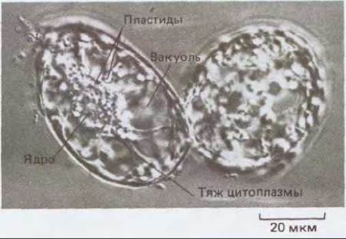
Fig. 20-70. Callus cultures can be maintained in liquid medium as suspensions of single cells. This micrograph shows two such cells from a sycamore callus. They are highly vacuolated, with cytoplasmic strands extending from the nucleus. These cells feature a primary cell wall.
Plants regenerated from cultured cells exhibit genetic instability (somaclonal variation). The same phenomenon occurs when an intact plant is subjected to stress. It appears that various chromosomal rearrangements take place in plant cells under stress conditions, which include tissue culture environments (see Section 10.5.10). The resulting mutant plants may confer advantages in altered or unfavorable environments. Today, plant breeders utilize somaclonal variants as starting material to develop improved agricultural crop varieties.
20.5.10. Application of Molecular Genetics to Plant Development
The capacity of plant somatic cells for regeneration in culture makes it possible to perform diverse genetic manipulations on these cells and generate Transgenic Plants. To facilitate The entry of foreign DNA into plant cells, their rigid walls are removed. This is achieved by treating the cells with enzymes that hydrolyze the bonds in cell wall polysaccharides. Such Treatment converts the cells into protoplasts (Fig. 20-71). Following the Introduction of foreign DNA, protoplasts can be induced to resynthesize a cell wall, resume division, and ultimately regenerate a new plant.
To introduce foreign DNA into intact plant cells, a vector derived from the Agrobacterium T-DNA discussed earlier is employed (see Section 20.3.3). For this purpose, the genes responsible for crown gall tumor formation on the plant are excised and replaced with genes of interest. After transferring the modified T-DNA into an appropriate Agrobacterium Ti plasmid, the Bacteria are co-cultivated with leaf explants. This system enables the T-DNA to integrate into the plant chromosome, stably incorporating the new gene into the plant genome (Fig. 20-72). Unfortunately, this method has been successfully applied only to certain families of dicotyledonous plants1).
1) Recently, the range of transformable plant species has expanded significantly, and the technique has become applicable to certain monocotyledonous plants as well, such as maize and rice. — Transl. note.

Fig. 20-71. Light micrograph of protoplasts isolated from green tobacco leaf cells. Deprived of their cell walls, the cells assume a spherical shape and can be maintained only in an isotonic solution. Numerous Chloroplasts are clearly visible within each protoplast. (Courtesy of F. Burgess.)

Fig. 20-72. Scheme for producing a transgenic plant. The gene of interest encodes a bacterial protein toxic to insects. To ensure expression in plant cells, the 5'-end of the gene is joined to a plant promoter, and its 3'-end to a polyadenylation site. This modified toxin gene is then inserted into a plasmid that also contains a selectable marker gene (such as a kanamycin resistance gene) for selection purposes. The plasmid is engineered so that both the toxin gene and the marker gene are flanked by characteristic 25-base-pair repeats that naturally bound the T-DNA. The plasmid is transferred from E. coli cells into Agrobacterium, which harbors virulence genes on a separate plasmid. When such Agrobacteria are co-cultivated with leaf discs, the virulence gene products recognize the T-DNA repeats and transfer the DNA containing the marker and toxin genes into the plant chromosome. The leaf disc cells are then induced to divide by placing the explants on a suitable nutrient medium; however, only those cells containing the selectable marker gene retain the capacity to divide and form callus tissue. Transgenic plants are subsequently regenerated from the callus, expressing the bacterial insecticidal toxin gene and thereby exhibiting exceptional insect resistance.
Applying Genetic Engineering techniques to plant cells has greatly advanced our understanding in many areas, facilitating the ISOLATION OF PURE growth regulators and shedding light on the pathways of morphogenesis and gene expression mechanisms. Furthermore, it has opened up new horizons in agriculture by creating the theoretical possibility of breeding plants with tailored traits, such as resistance to pests or The ability to thrive in extreme environments (e.g., saline or waterlogged soils).
It is well established that breakthroughs in understanding animal developmental mechanisms have been achieved largely through The Use of invertebrate models that are amenable to genetic analysis and experimentation. For many years, the fruit fly Drosophila (see Section 16.5) and the nematode Caenorhabditis (see Section 16.3) have served as classic model organisms. Plant developmental biology has progressed at a much slower pace, primarily due to the long life cycles and large genomes characteristic of plants. In recent years, however, the small herbaceous plant Arabidopsis thaliana has gained widespread acceptance as a model organism for Molecular and genetic research. First, it is so small (Fig. 20-73) that it can be easily cultivated in test tubes under laboratory conditions. Second, its generation time is a mere 5 weeks. Finally, Arabidopsis possesses the smallest genome among higher plants (7 x 10⁷ Base Pairs), comparable in size to the genomes of Yeast (2 x 10⁷ base pairs), C. elegans (8 x 10⁷ base pairs), and Drosophila (10 x 107 base pairs). A wide array of interesting Arabidopsis thaliana mutants has now been identified, and a complete genomic library of the plant has been established (see Section 5.6.3). It is anticipated that in the near future Arabidopsis will play a role in plant developmental biology analogous to that of Drosophila in animal developmental biology.

Fig. 20-73. Arabidopsis thaliana, a small herbaceous plant belonging to the mustard family (Brassicaceae). While holding no direct agricultural value, it is exceptionally well-suited for molecular and genetic experiments involving plants. The compact genome of Arabidopsis (7 x 10⁷ base pairs) is comparable in size to that of Drosophila (10 x 107 base pairs). (Courtesy of Ch. Sommerville.)
In the earliest stages of higher plant development, embryonic cells undergo rapid and intensive division. As the embryo continues to grow, however, mitotic activity becomes restricted to specific regions known as meristems. An adult plant can thus be viewed as a series of repeating modules generated by the morphogenetic activity of these meristems.
Plant morphogenesis relies on the coordinated division, elongation, and differentiation of immotile cells. The control of cell division planes and directed cell elongation is mediated, in part, by microtubules associated with the inner surface of the plasma membrane. Plant growth and cell division are influenced by environmental cues such as light, gravity, and temperature, as well as by specialized low-molecular-weight growth regulators like auxins and cytokinins.
Many aspects of plant cell growth and development have been investigated using cultured cells, both as single cells and within callus tissues. Plant somatic cells exhibit a degree of developmental plasticity unknown in animal cells. The most striking manifestation of this somatic totipotency is the capacity of many plant cells to regenerate an entire, intact plant.
Protoplasts are plant cells stripped of their rigid cell walls. They can be studied in vitro using techniques similar to those applied to animal cells, with the added advantage that whole plants can be regenerated directly from them.
Genetic engineering techniques make it possible to introduce desired new genes into the chromosomes of both protoplasts and intact cells, frequently enabling the subsequent recovery of transgenic plants.
General
Cutter E.G. Plant Anatomy, 2nd, Part 1, Cells and Tissues; Part 2 Organs. London, Arnold, 1978.
Esau K. Anatomy of Seed Plants, 2nd ed. New York, Wiley, 1977.
Grierson D., Covey S.N. Plant Molecular Biology, 2nd ed. New York, Methuen, 1988.
Gunning B. E. S., Steer M. W. Ultrastructure and the Biology of Plant Cells. London, Arnold, 1975.
Raven P. H., Evert R. F., Eichhorn S. E. Biology of Plants, 4th ed., New York, Worth, 1986.
Roberts K., Johnson A. W.B., Lloyd C. W., Shaw PJ., Woolhouse H. W., eds. The Cell Surface in Plant Growth and Development. J. Cell. Sci., Suppl. 2. Cambridge, U. K., Company of Biologists Ltd, 1985.
Salisbury F.B., Ross C. W. Plant Physiology, 3rd ed. Belmont, CA, Wadsworth, 1985.
Cited
1. Bacic A., Harris P.J., Stone B.A. Structure and function of Plant Cell Walls. In Biochemistry of Plants: A Comprehensive Treatise (J. Preiss, ed.), Vol. 14, CARBOHYDRATES, pp. 287-371. San Diego, CA, Academic Press, 1988.
Brett С. Т., Hillman J. R., eds. Biochemistry of Plant Cell Walls. SEB Seminar Series, 28. New York, Cambridge University Press, 1985.
McNeil M., Darvill A. G., Fry S. C., Albersheim P. Structure and function of the primary cell walls of plants. Annu Rev. Biochem., 53, 625 663, 1984. Rees D.A. Polysaccharide Shapes. London, Chapman and Hall, 1977.
2. Cassab G. I., Varner J. E. Cell wall proteins. Annu. Rev. Plant Physiol., 39, 321 -353, 1988. Fry S. Cross-linking of matrix polymers in the growing cell walls of angiosperms. Annu. Rev. Plant Physiol., 37, 165-186, 1986.
Selvendran R. R. Developments in the chemistry and biochemistry of pectic and hemicellulosic polymers. J. Cell Sci. Suppl., 2, 51-88, 1985.
3. Carpita N., Sabularse D., Montezinos D., Delmer D. P. Determination of the pore size of cell walls of living plant cells. Science, 205, 1144 1147, 1979.
Milburn J. A. Water Flow in Planls. London, Longman, 1979.
4. Street H. E., Öpik H. The physiology of Flowering Plants, 3rd ed. London, U. K., Edward Arnold, 1984.
5. Fry S. C. The Growing Plant Cell Wall: Chemical and Metabolic Analysis. New York, Wiley, 1988.
Masuda Y., Yamamoto R. Cell-wall changes during auxin-induced cell extension. Mechanical properties and constituent polysaccharides of the cell wall. In Biochemistry of Plant Cell Walls; (C.T. Brett, J. R. Hillman, eds.), pp. 269 300. SEB Seminar Series 28, New York, Cambridge University Press, 1985.
6. Baker D. A., Hall J. L. Ion Transport in Plant Cells and Tissues. Amsterdam, North-Holland, 1975. Cheeseman J. M. Mechanisms of salinity tolerance in plants. Plant Physiol., 87, 547 550, 1988.
Morgan J. M. Osmoregulation and water stress in higher plants. Annu. Rev. Plant Physiol., 35, 299-319, 1984.
Zimmerman U. Cell turgor pressure regulation and turgor pressure-mediated transport processes. Symp. Soc. Exp. Biol., 31, 117-154, 1977.
7. Esau K. Anatomy of Seed Plants, 2nd ed. New York, Wiley, 1977.
Tanner W., Loewus F .A., eds. Encyclopedia of Plant Physiology, New Series Vol. 13B, Plant Carbohydrates II, Extracellular Carbohydrates. Heidelberg, Springer-Verlag, 1982.
8. Dugger W. M., Bartnicki-Garcia S., eds. Structure, Function, and Biosynthesis of Plant Cell Walls. Proc. 7th Annu. Symp. Botany Rockville, M.
D., American Society of Plant Physiologists, 1984. (Several relevant papers appear in this collection.)
Fry S. The Growing Plant Cell Wall: Chemical and Metabolic Analysis. New York, Wiley, 1988.
9. Gunning B. E. S., Overall R. L. Plasmodesmata and cell-to-cell transport in plants. Bioscience., 33, 260-265, 1983.
Gunning B. E. S., Robards A. W., eds. Intercellular Communication in Plants: Studies on Plasmodesmata. New York, Springer-Verlag, 1976.
10. Baron-Epel O., Hernandez D., Jiang L.-W., Meiners S., Schindler M. Dynamic continuity of cytoplasmic and membrane compartments between plant cells. J. Cell Biol., 106, 715 721, 1988.
Gunning B. E. S., Hughes J. E. Quantitative assessment of symplastic transport of pre-nectar into the trichomes of Abutilon nectaries. Aust. J. Plant Physiol., 3, 619-637, 1976.
Terry B. R., Robards A. W. Hydrodynamic radius alone governs the mobility of molecules through plasmodesmata. Planta, 171, 145-157, 1987. Zaitlin M., Hull R. Plant virus-host interactions. Annu. Rev. Plant Physiol., 38, 291-315, 1987.
11. Aloni R. Differentiation of vascular tissue. Annu. Rev. Plant Physiol., 38, 179 204, 1987.
Moorby J. Transport Systems in Plants. New York, Longman, 1981.
12. Baker D. A. Transport Phenomena in Plants. London, U.K., Chapman and Hall, 1978.
Clarkson D. T. Factors affecting mineral nutrient acquisition by plants. Annu. Rev. Plant Physiol., 36, 77-115, 1985.
Milburn J. A. Water Flow in Plants. London, Longman, 1979.
Passioura J. B. Water transport in and to roots. Annu. Rev. Plant Physiol. Plant Mol. Biol., 39, 245-265, 1988.
13. Cronshaw J. Phloem structure and function. Annu. Rev. Plant Physiol., 32, 465-484, 1981.
Cronshaw J., Lucas W. J., Giaquinta R. T., eds. Phloem Transport. New York, Liss, 1986.
Gunning B. E. S. Transfer cells and their roles in transport of solutes in plants. Sci. Prog. (Oxford), 64, 539-568, 1977.
Ho L. C. Metabolism and compartmentation of imported sugars in sink organs in relation of sink strength. Annu. Rev. Plant Physiol. Plant Mol. Biol., 39, 355-378, 1988.
14. Gianinazzi-Pearson V., Gianinazzi S., eds. Physiological and Genetical Aspects of Mycorrhizae. Paris, INRA, 1986.
Smith S. E., Gianinazzi-Pearson V. Physiological interactions between symbionts in vesicular-arbuscular mycorrhizal plants. Annu. Rev. Plant Physiol. Plant Mol. Biol., 39, 221-244, 1988.
15. Downie J. A., Johnson A. W. B. Nodulation of legumes by Rhizobium: the recognized root? Cell, 47, 153-154, 1986.
Peters N. K., Frost J. W., Long S. R. A plant flavone, luteolin induces expression of Rhizobium meliloti nodulation genes. Science, 233, 977-980, 1986.
Rolfe B. G., Gresshoff P. M. Genetic analysis of legume nodule initiation. Annu. Rev. Plant Physiol. Plant Mol. Biol., 39, 297-319, 1988.
16. Buchanan-Wollaston V., Passiatore J. E., Cannon F. The mob and oriT mobilization Functions of a bacterial plasmid promote its transfer to plants. Nature, 328, 172-175, 1987.
Klee H., Horsch R., Rogers S. Agrobacterium-mediated plant transformation and its further Applications to plant biology. Annu. Rev. Plant Physiol., 38, 467-486, 1987.
Stachel S. E., Messens E., Van Montagu M., Zambryski P. Identification of the signal molecules produced by wounded plant cells that activate T-DNA transfer in Agrobacterium tumefaciens. Nature, 318, 624-629, 1985.
17. Ayers A. R., Ebel J., Finelli F., Berger N., Albersheim P. Host pathogen interactions. Plant Physiol., 57, 751-759, 1976. (This should be read in conjunction with three related papers that follow it.)
Darvill A. G., Albersheim P. Phytoalexins and their elicitors - a defence against microbial infection in plants. Annu. Rev. Plant Physiol., 35, 243-275, 1984.
Ralton V. E., Smart M. G., Clarke A. E. Recognition and infection process in plant pathogen interactions. In Plant-Microbe Interactions. (T. Kosuge, E. W. Nestor, eds.), Vol. 2, pp. 217-252, New York, Macmillan, 1987.
Ryan C. A. Oligosaccharide signalling in plants. Annu. Rev. Cell Biol., 3, 295-317, 1987.
18. McDougall G. J., Fry S. C. Inhibition of auxin-stimulated growth of pea stem segments by a specific monosaccharide of xyloglucan. Planta, 175, 412-416, 1988.
Thanh Van K. T., et al. Manipulation of the morphogenetic pathways of tobacco explants by oligosaccharins. Nature, 314, 615-617, 1985.
19. Anderson J. M. Photoregulation of the composition, function, and structure of thylakoid membranes. Annu. Rev. Plant Physiol., 37, 93-136, 1986.
Mullet J. E. Chloroplast development and gene expression. Annu. Rev. Plant Physiol. Plant Mol. Biol., 39, 475-502, 1988.
Thomson W. W., Watley J. M. Development of nongreen Plastids. Annu. Rev. Plant Physiol., 31, 375-394, 1980.
20. Boller T., Kende H. Hydrolytic enzymes in the central vacuole of plant cells. Plant Physiol., 63, 1123-1132, 1979.
Boiler Т., Wiemken A. Dynamics of vacuolar compartmentation. Annu. Rev. Plant Physiol., 37, 137-164, 1986.
Marin В., ed. Plant Vacuoles: Their Importance in Solute Compartmentations in Cells and Their Applications in Plant Biotechnology. NATO ASI Series, Vol. 134. New York, Plenum, 1987.
Motile P. Biochemistry and function of vacuoles. Annu. Rev. Plant Physiol., 29, 193-213, 1978.
21. Mollenhauer H. H., Morre D. J. The Golgi apparatus. In The Biochemistry of Plants-A Comprehensive Treatise. (N. E. Tolbert, ed.), Vol. 1, pp.
437-488. New York, Academic Press, 1980.
Moore P. J., Staehelin L. A. Immunogold localization of the cell-wall-matrix polysaccharides rhamnogalacturonan I and xyloglucan during cell expansion and cytokinesis in Trifolium pratense L.; implications for secretory pathways. Planta., 174, 433 445, 1988.
Northcote D. H. Macromolecular aspects of cell wall differentiation. In Encyclopedia of Plant Physiology, New Series. Vol. 14A. Nucleic Acids and Proteins in Plants I (D. Boutler, B. Parthier, eds.), pp. 637-655. Berlin, Springer-Verlag, 1982.
22. Coleman J., Evans D., Hawes C., Horsley D., Cole L. Structure and Molecular organization of higher plant coated vesicles. J. Cell Sci., 88, 3545, 1987.
Robinson D. G., Depta H. Coated vesicles. Annu. Rev. Plant Physiol. Plant Mol. Biol., 39, 53-99, 1988.
Tanchak M. A., Griffing L. R., Mersey B. G., Fowke L. C. Endocytosis of cationized ferritin by coated vesicles of soybean protoplasts. Planta., 162, 481-486, 1984.
23. Brown R. M. Cellulose microfibril assembly and orientation: recent developments. J. Cell Sci., Suppl., 2, 13-32, 1985.
Delmer D.P. Cellulose biosynthesis. Annu. Rev. Plant Physiol., 38, 259-290, 1987.
Schneider В., Herth W. Distribution of plasma membrane rosettes and kinetics of cellulose formation in xyleme development of higher plants. Protoplasma., 131, 142 152, 1986.
24. Green P. B. Organogenesis-a biophysical view. Annu. Rev. Plant Physiol., 31, 51 82, 1980.
Herth W. Plant Cell wall formation. In Botanical Microscopy (A. W. Robards, ed.), pp. 285-310. New York. Oxford University Press, 1985.
25. Lloyd C. W., ed. The Cytoskeleton in Plant Growth and Development. New York, Academic Press, 1982. (Chapters 5-8 are particulary relevant.)
26. Kersey Y. M., Helper P. K., Palevitz B. A., Wessels N. K. Polarity of actin filaments in characean Algae. Proc. Natl. Acad. Sci. USA., 73, 165-167, 1976.
Parthasarathy M. V. F-actin architecture in coleoptile epidermal cells. Eur. J. Cell Biol., 39, 1-12, 1985.
Sheet: M. P., Spudich J. A. Movement of Myosin-coated fluorescent beads on actin cables in vitro. Nature, 303, 31-35, 1983.
Williamson R. E. Organelle movements along actin filaments and microtubules. Plant Physiol., 82, 631 634, 1986.
27. Haupt W. Light-mediated movement of chloroplasts. Annu. Rev. Plant Physiol., 33, 205-233, 1982.
Roberts I. N., Lloyd C. W., Roberts K. Ethylene-induced microtubule reorientations: mediation by helical arrays. Planta, 164, 439-447, 1985. Virgin H. I. Light and chloroplast movements. Symp. Soc. Exp. Biol., 22, 329 352, 1968.
Wagner G., Klein K. Mechanism of chloroplast movement in Mougeotia. Protoplasma, 109, 169-185, 1981.
28. Cutter E.G. Plant Anatomy, 2nd ed., Part 2, Organs. London, Arnold, 1978. Johri B. M. Embryology of Angiosperms. Berlin. Springer-Verlag, 1984.
Raven P. H., Evert R. F., Eichhorn S. E. Biology of Plants, 4th ed. New York. Worth, 1986. (Chapters 19-22 provide a good general outline of plant development.)
29. Harper J. L., ed. Growth and Form of Modular Oganisms. London, Royal Society, 1986. (There are several relevant papers in this collection.)
Walbot V. On the life strategies of plants and animals. Trends Genet., 1, 165-169, 1985.
30. Green P. B. A theory for influorescence development and Flower Formation based on morphological and biophysical analysis in Echeveria. Planta, 175, 153 169, 1988.
McDaniel C. N., Poething R. S. Cell Lineage patterns in the shoot apical meristem of the germinating corn embryo. Planta, 175, 13-22, 1988. Sachs T. Controls of cell patterns in plants. In Pattern Formation (G. M. Malacinski, S. V. Bryant, eds.). New York. Macmillan, 1984.
Sleeves T. A., Sussex I. M. Patterns in Plant Development, 2nd ed. New York, Cambridge University Press, 1988.
31. Gunning B.E.S. Microtubules and cytomorphogenesis in a developing organ: the root primordium of Azolla pinnata. In Cytomorphogenesis in Plants. (O. Kiermayer, ed.), pp. 301-325, New York, Springer, 1981.
Poethig R. S. Clonal analysis of cell lineage patterns in plant development. Am. J. Bot., 74, 581-594, 1987.
32. Gunning B. E. S., Wick S. M. Preprophase bands, phragmoplasts and spatial control of cytokinesis. J. Cell Sci. Suppl., 2, 157-179, 1985.
Lloyd C. Actin in plants. J. Cell Sci., 90, 185-188, 1988. Lloyd C. W. The plant Cytoskeleton: The impact of Fluorescence Microscopy. Annu. Rev. Plant Physiol., 38, 119-139, 1987.
Pickett-Heaps J. D., Northcote D. H. Organization of microtubules and Endoplasmic reticulum during mitosis and cytokinesis in wheat meristems. J. Cell Sci., 1, 109-120, 1966.
Wick S. M., Seagull R. W., Osborn M., Weber K., Gunning B. E. S. Immunofluorescence microscopy of organized microtubule arrays in structurally stabilized meristematic plant cells. J. Cell Biol., 89, 685-690, 1981.
33. Hepler P. K., Wayne R. 0. Calcium and plant development. Annu. Rev. Plant Physiol., 36, 397-439, 1985.
Kropf D. L., Kloareg B. Quatrano R. S. Cell wall is required for fixation of the embryonic axis in Fucus zygotes. Science, 239, 187-190, 1988. Quatrano R.S., Griffing L. R., Huber-Walchli V., Doubet R. S. Cytological and biochemical requirements for the establishments of a polar cell. J. Cell Sci., Suppl., 2, 129-141, 1985.
Schnepf E. Cellular polarity. Annu. Rev. Plant Physiol., 37, 23-47, 1986.
34. Jordan B. R., Partis M. D., Thomas B. The biology and molecular biology of plytochrome. Ox. Sur. Plant. Mol. Cell Biol., 3, 315-362, 1986. Kuhlemeier C., Green P. J., Chua N. Regulation of Gene Expression in higher plants. Annu. Rev. Plant Physiol., 38, 221-257, 1987.
Tobin E. M., Silverthorne J. Light regulation of GENE EXPRESSION IN higher plants. Annu. Rev. Plant Physiol., 36, 569-593, 1985.
Verma D.P.S., Goldberg R. B. Temporal and Spatial Regulation of Plant Genes. New York, Springer-Verlag, 1988.
von Wettstein D., Chua N.-H., eds. Plant Molecular Biology. NATO AS1 Series A, Vol. 140. New York, Plenum, 1987. (Several relevant papers in this collection.)
35. Hoad G. V., Lenton J. R., Jackson M. В., Atkin R. K. Hormone Action in Plant Development: A Critical Appraisal. London, Butterworth, 1987. Salisbury F. Б., Ross C. W. Plant Physiology, 3rd ed. Belmonl, CA, Wadsworth, 1985. (Chapters 16 and 17 are relevant.)
Theologis A. Rapid gene regulation by auxin. Annu. Rev. Plant Physiol., 37, 407-438, 1986.
Trewavas A. J. Growth substance sensitivity: the limiting factor in plant development. Physiol. Plant., 35, 60-72, 1982.
36. APPLICATION OF PLANT Cell and Tissue Culture. CIBA Foundation Symposium 137. New York, Wiley, 1988.
Lee M., Phillips R. L. The Chromosomal Basis of somaclonal variation. Annu. Rev. Plant Physiol. Plant Mol. Biol., 39, 413 437, 1988.
Vasil I. K., ed. Perspectives in PLANT CELL AND Tissue Culture. Int. Rev. Cytol., Suppl. 11A and B, 1980.
37. Bevan M. Binary Agrobacterium Vectors for Plant transformation. Nucl. Acids Res., 12, 8711 -8721, 1984.
De Block M., Herrera-Estrella L., Van Montague M., Schell J., Zambryski P. Expression of foreign genes in regenerated plants and in their progeny. EMBO J., 3, 1681-1689, 1986.
Finkelstein R., Estelle M., Martinez-Zapater J., Somerville C. Arabidopsis as a tool for the identification of genes involved in plant development. In Temporal and Spatial Regulation of Plant Genes. (D. P. S. Verma, and R. B. Goldberg eds.), pp. 1-25. New York, SpringerVerlag. 1988.
Myerowitz E. M. Arabidopsis thaliana. Annu. Rev. Genet. 21, 93-111, 1987.
Vaeck M., et al. Transgenic plants protected from insect attack. Nature, 328, 33 37. 1987.
Last update: 12/08/2026
Editorial and Educational Adaptation: This material has been compiled based on the primary/original source text. The project team performed an editorial review, corrected technical inaccuracies, structured sections, and adapted the content for an educational format.
What was processed:
- elimination of formatting defects (OCR errors, structural breaks, corrupted characters);
- editorial organization of content;
- standardization of terminology in accordance with academic sources;
- verification of factual statements against the original source text.
All mentions of the author, publication year, and origin of the primary text have been preserved in accordance with the source.