Molecular Biology of the Cell - Volume 3 - Alberts B., Bray D., Lewis J., Raff M., Roberts K., Watson J. 1994
From Cells to Multicellular Organisms
Features of Plant Cells
Features of the Internal Organization of a Plant Cell
Cells of higher plants contain the same intracellular compartments previously described for animal cells: the Cytosol, Golgi apparatus, Endoplasmic reticulum, Nucleus, Cell/35.html">Mitochondria, Peroxisomes, and Lysosomes. In addition, plant cells possess a Cytoskeleton consisting of Actin filaments, microtubules, and Intermediate filaments comparable to those found in animal cells. Nevertheless, plant cells are easily distinguished from animal cells by the presence of two distinct Types of Membrane-enclosed compartments—vacuoles and Plastids. The presence of these Organelles is closely related to the stationary lifestyle of plant cells. These and other Features of the internal architecture of plant cells will be discussed in this section.
Class="center">
Fig. 20-35. Electron micrograph of a typical proplastid from a bean ROOT tip cell. The proplastid envelope consists of two membranes, and the internal system of membrane structures is poorly developed. (From B. Gunning, M. Steer, Ultrastructure and the Biology of Plant Cells. London: Arnold, 1975.)
20.4.1. METABOLISM/14.html">Chloroplasts are members of the family of organelles known as plastids, which are unique to plant cells [19]
Plants manufacture all the organic molecules they require through Photosynthesis, which takes place in chloroplasts. The products of photosynthesis can be used directly by cells for various biosynthetic processes, stored as an osmotically inert polysaccharide (typically starch), or converted into relatively low molecular weight sugars (such as sucrose) that are transported to other Tissues of the plant.
Chloroplasts represent just one type of a closely related family of organelles called plastids. Plastids are present in all living plant cells, with each cell type containing its own characteristic set of these organelles. All plastids share A number of common features. They possess a small genome, identical in all individuals of the same species, and are enclosed by an envelope consisting of two concentric membranes.

Fig. 20-36. Electron micrograph of two forms of plastids in oat seedlings. A. An etioplast from a dark-grown seedling. The lattice-like Structure formed by the internal membranes contains protochlorophyll. B. Upon illumination, the etioplast develops into a chloroplast. Shown is a portion of a young, greening chloroplast. Reorganization of the etioplast membrane system is underway: it already contains chlorophyll and is beginning to form small grana. (Courtesy of B. Gunning.)
Since the Structure and function of chloroplasts were discussed in detail in Chapter 7, we will focus our attention on other members of this same group of plant organelles.
All plastids, including chloroplasts, develop from proplastids—relatively small organelles present in meristematic cells (Fig. 20-35). The Fate of proplastids is dictated by the needs of differentiated cells. For example, if a leaf develops in the dark, its proplastids enlarge and differentiate into etioplasts. The internal membranes of etioplasts form a crystalline lattice-like structure containing protochlorophyll (a yellow chlorophyll precursor) instead of chlorophyll (Fig. 20-36A). Upon exposure to light, etioplasts develop into chloroplasts through The conversion of protochlorophyll to chlorophyll and the synthesis of new membranes, pigments, photosynthetic Enzymes, and Electron Transport Chain components (Fig. 20-36B).
It is not surprising that many processes occurring within plants are regulated by light. Photoreceptors, including Phytochrome (see Section 20.5.7), control the Transcription of numerous genes involved in chloroplast development, and these genes are located not only within the chloroplast itself but also in The Nucleus. The Mechanism of this control remains unclear. Approximately one-fifth of the 120 genes comprising the Chloroplast Genome are regulated by light (see Section 7.5.4).
Chromoplasts represent another form of plastid (Fig. 20-37A). They accumulate carotenoid pigments responsible for the yellow-orange colors of flower petals (such as daffodils) and fruits (such as tomatoes). Another variety of plastids is leukoplasts, which, aside from their larger size, differ little from proplastids and are found in many epidermal and internal tissues that do not become green and are incapable of photosynthesis. A widespread form of leukoplasts is amyloplasts (Fig. 20-37B), which function as starch storage depots in storage tissues. In some plants, such as potatoes, amyloplasts can enlarge to the size of an average animal cell.
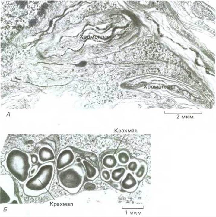
Fig. 20-37. Diverse forms of plastids. A. Chromoplasts from cells of orange-yellow daffodil petals. These plastids have a convoluted outline; their internal membranes, which are arranged quite randomly, contain the pigment (β-carotene) that imparts the characteristic color to the petals. B. Three amyloplasts (starch-storing plastids) in a soybean root tip cell. (From B. Gunning, M. Steer, Ultrastructure and the Biology of Plant Cells, London: Arnold, 1975.)
Although the exact Factors Determining the differentiation of a proplastid into a particular plastid type remain unknown, it is evident that the nuclear genome plays a crucial role in regulating this process. Nuclear Mutations can switch development from chromoplasts to chloroplasts, or block development altogether, resulting in Various Forms of leucoplasts or immature, abnormally pigmented chloroplasts characteristic of many ornamental plants.
Plastids within the Cytoplasm can grow and divide, thereby multiplying (see Section 7.5.1). The only cell type that has lost plastids is the sperm cell in certain higher plants; such plants (e.g., maize) inherit plastids exclusively from the egg cell from which they developed. In this case, plastids, much like animal mitochondria, are maternally inherited (see Section 7.5.10).
It is important to appreciate that plant plastids are organelles involved in much more than just photosynthesis. They are the site of intermediate stages of numerous metabolic pathways. Plastids generate sources of chemical energy and reducing equivalents (ATP and NADPH) consumed by the plant in biosynthetic reactions. Furthermore, the Synthesis of Purines and Pyrimidines, Most Amino Acids, and all Fatty acids takes place in plastids in plants, whereas in animals these processes occur in the cytosol.
20.4.2. Plant vacuoles are organelles with remarkably diverse Functions [20]
The most prominent compartment in most plant cells is a very large fluid-filled vesicle called the vacuole (Fig. 20-38). A single cell may contain several vacuoles, each separated from the cytoplasm by a single membrane known as the tonoplast. Vacuoles typically occupy more than 30% of the total cell volume, though this figure varies widely—from 5% to 90% depending on The Cell type. Most biologists consider this organelle not to be part of the cytoplasm, which is why plant cells are commonly described as containing a nucleus, a vacuole, and cytoplasm, with the cytoplasm housing all other membrane-bounded organelles, including plastids. Vacuoles originate in young dividing cells, likely through the fusion of increasing numbers of vesicles budding off from the Golgi apparatus. Structurally and functionally, they are similar to the lysosomes of animal cells; in particular, they also contain a multitude of various hydrolytic enzymes. At the same time, the functions of vacuoles in plant cells are astonishingly diverse. They can serve for nutrient storage, waste containment, cell enlargement, and The regulation of turgor pressure. Vacuoles with different functions (e.g., acting simultaneously as lysosomes and storage compartments) are frequently present within the same cell. The Role of vacuoles as space-filling compartments during cell expansion was discussed earlier (see Fig. 20-10). The following section describes the involvement of vacuoles in storage functions.
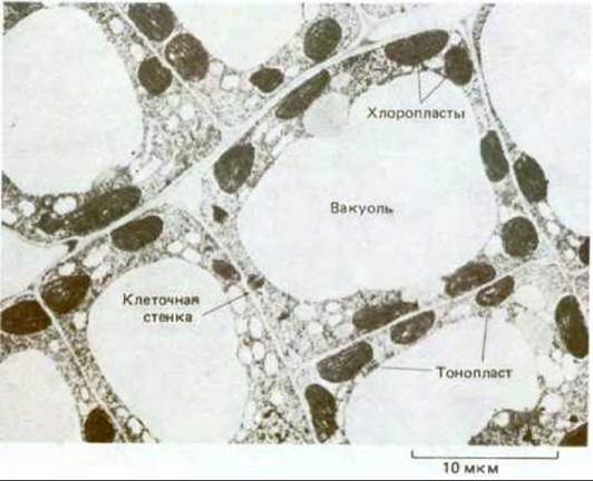
Fig. 20-38. Electron micrograph of cells in a young tobacco leaf. In these highly vacuolated cells, the cytoplasm, which is rich in chloroplasts, is confined to a thin peripheral layer along The Cell wall. (Courtesy of J. Burgess.)
20.4.3. Vacuoles can serve as storage organelles [20]
Vacuoles can accumulate and store A wide variety of molecules, including essential cellular substances that would be potentially hazardous if present in the cytosol in large amounts. For instance, in specialized cells of certain plants, vacuoles accumulate well-known products such as rubber (in Hevea brasiliensis) and opium (in Papaver somniferum). Even ubiquitous Na+ ions can be selectively concentrated in these organelles, where their osmotic activity helps maintain turgor pressure. Studies on giant algal cells of Nitella have shown that sodium pumps in the tonoplast maintain a relatively low concentration of Na+ in the cytosol by generating a 4-to-5-fold excess of these ions within the vacuoles. Because the vacuole occupies a much greater volume in a Nitella cell than the cytoplasm, the bulk of the cellular sodium pool is concentrated in the vacuole.
The vacuole plays a vital role in the Homeostasis of plant cells, which are exposed to diverse environmental fluctuations. If, for example, the extracellular pH drops, the influx of H+ ions into the cytoplasm is buffered, at least in part, by enhanced transport of H+ ions into the vacuole. Similarly, many plant cells maintain turgor pressure at a remarkably constant level despite significant environmental changes. This is achieved by adjusting the osmotic pressure in the cytosol and vacuole through the controlled breakdown and resynthesis of polymers such as polyphosphates within the vacuole. Turgor pressure can also be kept constant by modulating The rate of Transport Across the Plasma Membrane and tonoplast. The permeability of these two membranes depends on a specific set of transport Proteins that facilitate the movement of specific sugars, amino acids, and other metabolites across each lipid bilayer (see Chapter 6).

Fig. 20-39. Light micrograph of a cell from a developing pea seed. The initial Stages of Protein deposition can be seen at the margins of the well-developed vacuolar system. The seeds of legume plants accumulate large quantities of storage protein in their cell vacuoles, which is utilized by the embryo during seed germination. (Courtesy of S. Craig.)
The substances contained within the vacuole differ both qualitatively and quantitatively from those present in the cytoplasm. However, because the tonoplast has limited mechanical strength, the hydrostatic pressure in the vacuole and the cytoplasm must be approximately equal, and both compartments must contribute jointly to the osmotic equilibrium required to maintain turgor pressure.
Among the products stored in vacuoles, various metabolites occupy an important place. For example, succulent plants open their Stomata at night, absorb carbon dioxide from the air (thereby reducing transpirational Water loss), and store it in their vacuoles as malate until the following day, when it is converted into sugar using solar energy while the stomata remain closed. Organic molecules can also be stored in vacuoles for much longer periods, as is the case with storage proteins in the cells of many seeds, particularly peas and beans. Upon seed imbibition, these proteins are hydrolyzed, and the Amino acids are used to nourish the developing embryo (Fig. 20-39).
A number of substances accumulated in vacuoles are involved in plant interactions with animals or other plants. For instance, anthocyanins impart color to the petals of certain flowers, which helps attract insect pollinators. Other substances perform defensive functions. Plants cannot move to escape destruction by herbivores; instead, they synthesize a myriad of toxic compounds that are released from vacuoles upon cell damage. These include highly toxic Alkaloids as well as unpalatable substances that adversely affect Digestion. Trypsin inhibitors commonly found in seeds, as well as protease inhibitors produced in leaf cells in response to wounding, accumulate in the vacuole and likely affect herbivore digestion. Throughout their evolutionary history, plants—much like animals—have continually diversified their chemical warfare toolkit. The balance has shifted back and forth when, for example, a potent new repellent against herbivores emerged in the plant world or, conversely, when an insect evolved The ability to neutralize or degrade a plant's toxic metabolite, thereby gaining the ability to feed on the synthesizing plant. In such cases, the toxin itself could transition from a repellent to an attractant.
20.4.4. Golgi Vesicles Deliver Cell Wall Material to Specific Regions of The Plasma Membrane [21]
Most of the matrix Components of the cell wall are transported in Golgi vesicles to the plasma membrane, where they are subsequently discharged from the cell via exocytosis. However, unlike animal cells, in which the Golgi apparatus secretes Glycoproteins, the plant Golgi apparatus is primarily involved in the production and secretion of a wide range of extracellular Polysaccharides (Fig. 20-40). The details of the synthesis of these polysaccharides remain largely unknown, which is hardly surprising given that: 1) each cell wall polysaccharide is formed from two or more sugars; 2) at least 12 different polysaccharides are utilized; 3) the molecules of most of these polysaccharides are branched; and 4) after synthesis, the polysaccharides undergo numerous covalent modifications. It has been established that several hundred different enzymes participate in assembling the polysaccharide components required to form a typical primary cell wall. Most of these enzymes are found in the Endoplasmic reticulum and the Golgi apparatus, whereas some enzymes associated with later covalent modifications of polysaccharides are present within the cell wall itself, with a fraction of them being covalently bound to it.

Fig. 20-40. Electron micrograph of a Hair cell from the green alga Bulbochaete. Distinct stacks of Golgi cisternae and associated transport vesicles containing secreted polysaccharides are clearly visible. A polar Organization is readily apparent: extending from The endoplasmic reticulum through the cis, intermediate, and trans Golgi cisternae to the transport vesicles. In plant cells (unlike animal cells), the Golgi cisternae belonging to different stacks are not interconnected. (Courtesy of T. Frazer, after V. Gunning and M. Steer, Ultrastructure and the Biology of Plant Cells. London: Arnold, 1975.)
Because the chemical Composition and Structure of the cell wall vary across different Regions of the cell surface, vesicles carrying the appropriate matrices must be selectively targeted to specific domains of the plasma membrane. This directional transport is mediated, at least in part, by elements of the cytoskeleton; one striking example is the de novo Formation of the primary cell wall that separates two daughter cells following mitosis (Fig. 20-41). At the end of telophase, a bundle of microtubules arranged parallel to the spindle axis persists between the two daughter nuclei. This bundle consists of two sets of polar spindle microtubules with opposite polarities; the ends of microtubules from the different sets overlap in a disk-shaped region called the phragmoplast, which lies in the equatorial plane of the former division spindle (Fig. 20-42). Transport vesicles containing various cell wall precursors, particularly pectin, move along these oriented microtubules toward the equator and, upon reaching the central disk, fuse with one another to form the cell plate. The cell plate expands through the incorporation of more and more vesicles directed to the area by new microtubules forming at the periphery of the phragmoplast as the more centrally located microtubules depolymerize. Eventually, the growing plate fuses with the maternal cell wall, resulting in two separate daughter cells (see Figs. 20-41 and 13-71). It remains unclear which phragmoplast components—microtubules, actin filaments, or perhaps both—are responsible for driving the movement of Golgi-derived vesicles.

Fig. 20-41. A time-lapse series of light micrographs of a dividing staminal hair cell. Numbers indicate the time in minutes elapsed since THE START OF recording. The vesicles whose fusion leads to The formation of the cell plate become distinguishable by the 42nd minute. The plate gradually expands and ultimately fuses with the parent cell wall. (Courtesy of P. Herler.)
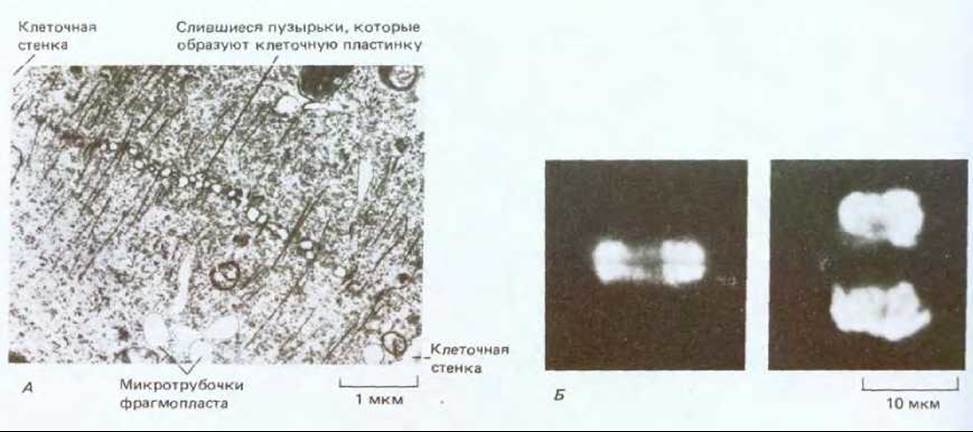
Fig. 20-42. A. Electron micrograph of a phragmoplast in a dividing plant cell. Microtubules direct the movement of vesicles containing cell wall precursors toward the growing cell plate. For details, see Fig. 13-71. B. Fluorescence micrographs of cytokinesis in onion root-tip cells. Staining with fluorescent Antibodies reveals the developing cell plate and the two sets of phragmoplast microtubules flanking this plate (left); fluorescent DNA staining (right) shows the positions of the two daughter nuclei that will be separated by the new cell wall. (A, courtesy of J. Pickett-Heaps.)

Fig. 20-43. Electron micrograph of a root-cap cell from timothy grass. In such cells, the Golgi apparatus is primarily engaged in producing and secreting slime, which is exuded onto the root tip surface to facilitate its passage through the soil. Material identical in appearance to the Contents of the Golgi cisternae can also be seen in vesicles and near the outer surface of the plasma membrane. (V. Gunning, M. Steer, Ultrastructure and the Biology of Plant Cells. London: Arnold, 1975.)
Not all polysaccharides produced and secreted by the Golgi apparatus are intended for Cell wall formation. For example, fructose-rich slime secreted by root-tip cells traverses the cell wall and acts as a lubricant on the outer surface of the root as it pushes through the soil (Fig. 20-43).
20.4.5. Fluid-Phase Endocytosis Involves Rapid Membrane Recycling [22]
What happens to the massive amount of new membrane material added to the existing plasma membrane during repeated fusion events with vesicles? In some actively secreting plant cells, the rate of exocytosis driven by Golgi transport vesicles is such that the entire plasma membrane surface area would double every 20 minutes. It is obvious, however, that the plasma membrane maintains a constant surface area and, consequently, that some mechanism of membrane turnover must exist. Numerous coated pits are present in the plasma membrane of plant cells (Fig. 20-44A). They are thought to participate in membrane recycling, analogous to the process in animal cells (see Section 6.5.4). A similar fluid-phase endocytic pathway has recently been identified in plant cells through the analysis of protoplast uptake of electron-dense markers such as ferritin or colloidal gold. When introduced into protoplasts, these markers are rapidly delivered to a complex network of membrane tubules termed the partially coated reticulum (Fig. 20-44B and C). This organelle is believed to be functionally equivalent to the endosomal compartment of animal cells (see Section 6.5.4). From here, the marker is transferred to large vacuoles and eventually ends up in the vacuole. Thus, the major intracellular pathways of metabolite synthesis, sorting, packaging, secretion, and endocytosis are remarkably similar in PLANT AND ANIMAL cells.

Fig. 20-44. Endocytosis in soybean protoplasts as visualized by Electron Microscopy. A. Cytoplasmic face of the plasma membrane. Protoplasts were attached to an Electron microscope grid, lysed, washed, and negatively stained. In addition to numerous membrane-associated pits involved in endocytosis, cortical microtubules are visible. B. A "partially coated reticulum" seen in thin section. It consists of a complex system of membrane-bound tubules with numerous clathrin-coated vesicles budding from them. This structure is thought to be the equivalent of the animal cell endosomal compartment. C. Endocytosed material—in this case, the electron-dense marker ferritin—first appears within the partially coated reticulum. (Courtesy of L. Fowke.)
20.4.6. Cellulose Synthesis Takes Place at The surface of Plant Cells [23]
Cell wall polysaccharides are typically synthesized in the Golgi apparatus and secreted via exocytosis. However, there is one major exception to this rule: in most plants, cellulose is synthesized on the outer surface of cells by a membrane-bound enzyme complex (cellulose synthase) that utilizes a sugar-nucleotide conjugate, presumably UDP-glucose, as a substrate. The newly formed cellulose chains spontaneously assemble into microfibrils that form a layer On the surface of the plasma membrane (a lamella) in which all microfibrils share a roughly parallel orientation. Because cellulose is synthesized right at the plasma membrane, each new lamella is laid down beneath the previous one. As a result, the cell wall consists of concentrically arranged lamellae, with the oldest one located on the outside.
Despite numerous attempts, cellulose synthesis has not yet been successfully reconstituted in vitro. This is likely due, in part, to the requirement for a functionally intact plasma membrane. Freeze-fracture Cytology/cytology/93.html">ELECTRON MICROGRAPHS OF higher plant cells reveal "rosettes" composed of intramembrane particles localized on the cytoplasmic fracture face. These rosettes most likely represent cellulose synthase complexes, as they are found in regions where new cellulose microfibrils are being deposited, such as in the plasma membrane overlying patterned wall thickenings in young xylem vessel cells (Fig. 20-45; see also Section 20.1.7).
20.4.7. The Shape of a Growing Plant Cell Is Determined by the Organization of Cellulose Microfibrils [24]
The final shape of every growing plant cell, and ultimately the form of the entire plant, is determined by cell expansion, which is subject to strict regulation. As noted above (see Section 20.1.5), cell expansion is the result of turgor pressure and wall plasticity. However, the question of how such an isotropic (nondirectional) pressure can produce directed cell elongation has not yet been addressed. In the lateral walls of an enlarging cell, the most recently deposited microfibrils are typically perpendicular to the axis of elongation, encasing the cell in a lamella where the microfibrils follow a helical path. Although the orientation of previously laid-down microfibrils in the outer lamella may differ, it is primarily the orientation of the cellulose microfibrils within this innermost lamella that governs the direction of cell growth.
If cellulose microfibrils in a newly formed lamella are transversely oriented, they will restrict cell expansion; however, because adjacent cellulose microfibrils within the lamella can separate from one another under turgor pressure, the cell will elongate (Fig. 20-46). For the existing cell wall to maintain its strength and thickness as the cell grows, new matrix components and microfibrils must be continually added to the existing wall. Newly secreted matrix components presumably penetrate the thickness of the wall, while additional microfibrils are deposited in a new lamella on the outer surface of the plasma membrane.
Thus, the future Morphology of plant cells is predetermined by The pathway of primary cell wall formation. It is still unclear precisely how the Fine Structure of all cell wall components is determined; however, cellulose deposition most likely depends on the arrangement of cortical microtubules.

Fig. 20-45. Electron micrograph of the plasma membrane of a developing xylem vessel element in a pepper root (prepared by freeze-fracture). Hexagonal Protein Assemblies (rosettes) are concentrated at sites where the cell wall thickens (A) and are less frequent in the interspace regions (B). These rosettes are thought to represent cellulose synthase complexes. Each of these complexes apparently synthesizes 60 to 70 cellulose chains that make up each microfibril. (Courtesy of W. Herth.)

Fig. 20-46. This diagram illustrates how the orientation of cellulose microfibrils in the cell wall influences the direction of cell elongation. The cells depicted in A and B initially have the same shape but different orientations of cellulose microfibrils. Due to cell wall loosening and turgor pressure, each cell expands in a direction perpendicular to the microfibril orientation. In turn, the final shape of an organ such as a SHOOT strongly depends on the direction in which its constituent cells expand. Although the fibrils appear to form hoop-like structures, numerous studies have shown that they are wound in a helix, as shown in the figure.
20.4.8. The orientation of cellulose microfibrils deposited on the cell surface is determined by microtubules in the cortical layer [25]
The orientation of cellulose microfibrils plays a crucial role in determining plant cell shape. But what dictates their arrangement? A clue to this question came from the observation that most microtubules in the cortical cytoplasm are oriented in the same direction as the cellulose microfibrils currently being formed in that region of the cell.
Cortical microtubules lie near the inner surface of the plasma membrane and are generally directed perpendicularly to the long axis of the cell (Fig. 20-47). Immunofluorescence microscopy of this region reveals that a system of overlapping microtubules surrounds the cell interior in a continuous, ordered layer (Fig. 20-48). The microtubules are attached to the plasma membrane by proteins whose nature is still poorly understood.
The coordinated orientation of microtubules located at the inner surface of the plasma membrane and cellulose microfibrils formed on its outer side is characteristic of many cells of various types and shapes. This phenomenon is observed during the formation of both Primary and secondary cell walls (Fig. 20-49), particularly during the local deposition of additional layers, such as when thickenings form in specific regions of xylem cell surfaces (see Fig. 20-15).
What happens if all microtubules of the cortical system are depolymerized by treating plant tissue with colchicine? The Effect of such Treatment on subsequent cellulose deposition is not as straightforward as one might expect. Colchicine does not inhibit the formation of new cellulose microfibrils, and in some cases cells can continue to deposit microfibrils oriented in their previous direction. However, any changes in microfibril arrangement during development that normally occur with the deposition of successive lamellas become impossible. Furthermore, a cell that would normally produce an ordered wall thickening for xylem vessel formation instead deposits wall material completely chaotically in the presence of the microtubule-depolymerizing agent (Fig. 20-50). Thus, pre-existing microfibril orientation can be maintained without microtubules, but any stage of cell development involving the deposition of differently oriented microfibrils requires the presence of intact microtubules to specify this new orientation.

Fig. 20-47. Electron micrograph of the contact zone between two adjacent cells in a wheat root tip, showing numerous cortical microtubules typical of interphase cells.

Fig. 20-48. Arrangement of cortical microtubules. A. Tangential section of a cell from a timothy grass root tip, showing cortical microtubules lying directly beneath the plasma membrane at right angles to the longitudinal axis of the cell. B. An isolated onion root tip cell. C. The same cell stained with fluorescent antibodies to reveal the microtubule pattern. (A — courtesy of B. Gunning; B and C — courtesy of K. Goodbody.)
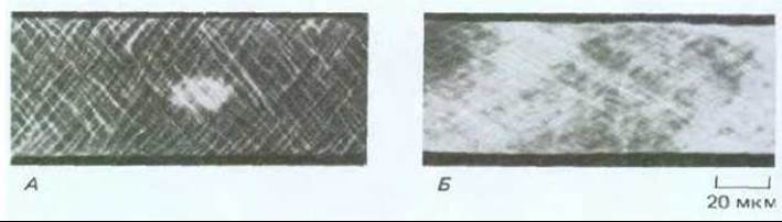
Fig. 20-49. Microtubules and cellulose microfibrils in developing cotton fibers. Microtubules (A) are stained with fluorescent antibodies and arranged in a spiral. The cell is flattened so that PARTS OF THE spiral are visible on both the front and back walls. Newly deposited cellulose microfibrils in a similar cell (B) are stained with the fluorescent dye Calcofluor White, which binds to growing cellulose molecules. Like the reinforcing cord of a garden hose, the microfibrils make the cotton fiber walls extremely strong. (Courtesy of R. Seagull.)
It is believed that the cellulose-synthesizing complexes located in the plasma membrane rotate around the long cellulose molecules. As Biosynthesis and self-assembly proceed concurrently, the distal end of the microfibril presumably forms cross-links with the previously deposited wall layer. Consequently, at the growing proximal end, the synthase complexes must move along the membrane in the direction of synthesis. Two mechanisms can be proposed for how microtubules influence the direction of this movement and, consequently, the orientation of the microfibrils. The cytoplasmic domain of the cellulose synthase complex may be directly or indirectly linked to cortical microtubules. According to another hypothesis, microtubules act like riverbanks, guiding the movement of synthase complexes parallel to a specific axis (Fig. 20-51). In this case, the rate of cellulose synthesis does not depend on microtubules; they merely define the membrane corridors within which the enzyme complex can advance. It remains unknown how the arrangement of cortical microtubules is controlled. Notably, the animal cell cytoskeleton also determines the orientation of Extracellular matrix components, with proteins such as Collagen and Fibronectin being deposited by cells in close association with their plasma membrane (see Section 14.2.18).

Fig. 20-50. The regular pattern of cell wall thickenings established during normal xylem Cell Differentiation (A) is determined by the presence of ordered arrays of cortical microtubules. In the presence of colchicine, cortical microtubules depolymerize, leading to disordered wall thickening (B).
20.4.9. In large plant cells, various Materials are transported by directed cytoplasmic streaming [26]
Cellular metabolism requires that substrates, intermediates, Cofactors, signaling molecules, and enzymes can move from one part of the cell to another. In small cells, such as Bacteria or even most animal cells, diffusion allows low-molecular-weight solutes to traverse distances comparable to the cell size in a fraction of a second. However, plant cells can grow quite large due to their cell walls, vacuoles, and turgor pressure: typically exceeding 100 µm in length, and in some cases measuring millimeters or even centimeters. Diffusion is relatively inefficient here, as the time required for a molecule to reach its destination by diffusion alone is proportional to the square of the distance (see Section 3.1.3). Meanwhile, some cells in a mature plant may be located quite far from sources of oxygen and nutrients. Therefore, it is unsurprising that large plant cells feature vigorous cytoplasmic streaming that mixes cell components and ensures their rapid Circulation.
Studies of living plant cells have shown that the larger the cell, the more active its cytoplasmic movement. Small cells typically exhibit saltatory organelle movements (from the Latin *saltare* — to dance, jump). As in animal cells, cytoplasmic particles here move as though they occasionally receive a strong push in a specific direction. In larger plant cells, this movement becomes partially directed, and in cells where the cytoplasm forms only a thin layer around a giant central vacuole, one can often observe a nearly continuous circular movement of cytoplasm at a rate of several micrometers per second. In giant Cells of the green alga *Nitella*, the polarity of cortical actin filaments is such that the movement of Myosin filaments along them could generate a directed cytoplasmic stream. This streaming likely facilitates not only intracellular organelle transport but also intercellular transport of solutes toward the pores of plasmodesmata connecting neighboring cells.
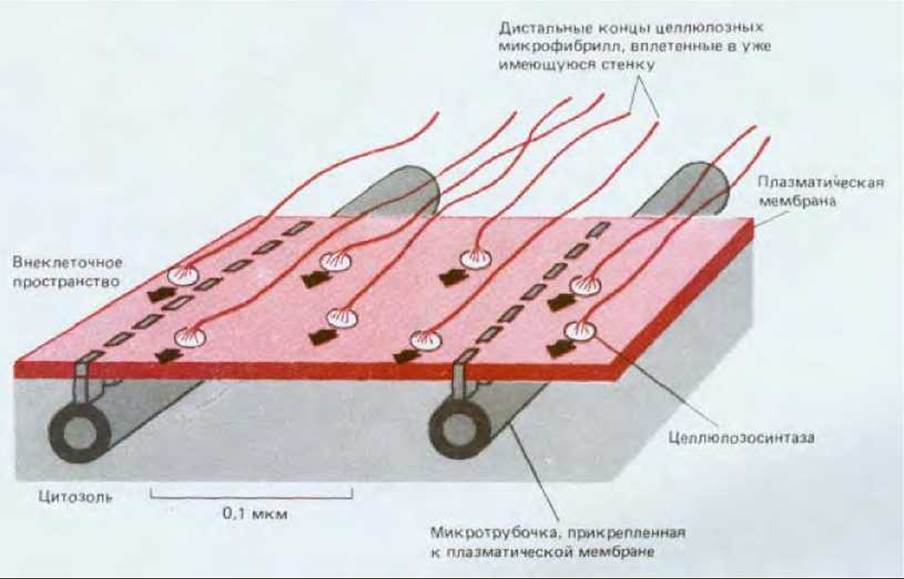
Fig. 20-51. A simple diagram illustrating how the orientation of cortical microtubules can determine the orientation of newly formed microfibrils. Large cellulose synthase complexes are integral Membrane Proteins that assemble microfibrils on the outer surface of the plasma membrane. Because the distal ends of rigid microfibrils are embedded in the wall, their elongation at the proximal end pushes the synthase complex along the membrane plane. Since microtubules are attached to the plasma membrane in a way that restricts the entire complex to specific membrane channels, microtubule orientation can determine the axis along which microfibrils are deposited.
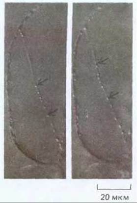
Fig. 20-52. The stamens of tradescantia flowers are covered with long, thin hairs, each consisting of a single row of large cells. Light micrographs taken at 5-second intervals reveal rapid streaming along cytoplasmic strands crossing the vacuole. Organelles move along these strands at speeds of up to 5 µm/s.
The thin, transparent cells forming hairs on the surface of plants provide an excellent model system for observing cytoplasmic streaming. Such cells contain a large central vacuole traversed by thin cytoplasmic strands about 1 µm in diameter (Fig. 20-52). Individual particles, such as mitochondria, can be seen "swimming" along these strands. These strands, which contain bundles of actin filaments but apparently lack microtubules, appear to originate in the region adjacent to the Cell Nucleus (Fig. 20-53). They can be observed constantly changing their shape and arrangement—vanishing, branching, fusing, and reforming.
20.4.10. The plant cell Cytoskeleton Responds to Extracellular Signals [27]
For plant cells enclosed within a rigid cell wall, subtle responses to environmental changes, particularly light, are of vital importance. As noted above, Changes in the direction of plant cell growth often depend on the cytoskeleton; thus, it is hardly surprising that both Actin filaments and microtubules react in highly complex ways to external stimuli.
Many plant cells can respond to changes in light intensity and direction by repositioning their chloroplasts. Under low-light conditions, chloroplasts tend to arrange themselves in a single layer perpendicular to the light source, thereby maximizing Light absorption. High light levels induce chloroplasts to migrate and align along cell walls parallel to the incident rays, which minimizes photodamage (Fig. 20-54). The Molecular Mechanism of this movement has been studied in a rather unusual alga. Mougeotia is a green alga in which each cylindrical cell contains a single flat plate-like chloroplast. When lighting conditions change, the chloroplast rotates and reorients itself to lie either parallel or perpendicular to the incident light, depending on the light intensity. Photoreceptors that induce this response include phytochrome (see Section 20.5.7) and a blue-light receptor located either on or immediately adjacent to the plasma membrane. Irradiation of these receptors with microbeams of light triggers an influx of Ca2+ ions (the exact mechanism remains unclear), which then binds to calmodulin. Calmodulin, in turn, activates a network of actin filaments attached to both the outer chloroplast envelope membrane and the plasma membrane, driving the Rotation of the plastid. If only a small part of the cell is illuminated with a directed microbeam, only the irradiated portion of the chloroplast bends. Consequently, different regions of the active cytoskeleton within the same cell can respond independently of one another (Fig. 20-55).
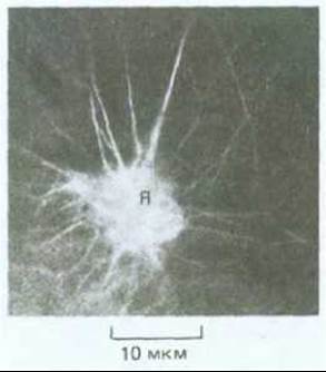
Fig. 20-53. Fluorescence micrograph of a small portion of a large vacuolated stem cell. Bundles of actin filaments are visible within the strands crossing the vacuole. The cell was stained with rhodamine-phalloidin, a conjugate that binds tightly and specifically to actin filaments. Actin bundles radiating from the nuclear region (N) are thought to be involved in the rapid cytoplasmic streaming characteristic of large vacuolated cells in higher plants. Myosin likely plays some role in this process as well.
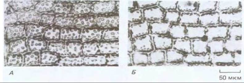
Fig. 20-54. Light micrograph of moss leaf cells showing chloroplast translocation in response to changing light conditions. The light beam is directed perpendicularly to the plane of the specimen. (A) Under dim light, disk-shaped chloroplasts position themselves to maximize light absorption. (B) After a 30-minute exposure of the same leaf area to bright light, the chloroplasts have migrated and now lie adjacent to the cell walls parallel to the incident rays.
The arrangement of cortical microtubules also changes rapidly in response to external stimuli. As discussed earlier, plant cell shape (and consequently plant morphology) depends on the orderly deposition of oriented cellulose layers, with the orientation of the innermost layer being the most critical (see Section 20.4.7). The outer cellulose layers of the cell wall often differ in orientation from the more recently deposited inner layers. There are at least two mechanisms by which old and new layers can be oriented differently, and both appear to function in plant cells: (1) cellulose microfibrils in older wall layers may slide past one another and reorient as they are displaced outward, accompanied by the breaking and reforming of cross-linking wall polysaccharides; and (2) new layers with an orientation distinct from the outer ones can be laid down directly into the wall at the plasma membrane.
Because the orientation of newly deposited cell wall material is tied to the arrangement of cortical microtubules, any change in one structure leads to a corresponding change in the other. Thus, the operation of the second mechanism requires A change in microtubule orientation. Such dynamic shifts in the helical arrangement of microtubules can be observed directly. For example, plant growth regulators such as Ethylene and gibberellic acid have opposing effects on microtubule orientation (and hence on the direction of cell growth) in the epidermal cells of young pea shoots. Gibberellic acid forces cortical microtubules to align perpendicularly to the long axis of the cell. As a result, cellulose is deposited in a way that restricts expansion to elongation, producing tall, slender shoots. However, if these shoots are treated with ethylene, the microtubules reorient within an hour to lie parallel to the long axis of the cell. Cellulose deposition along this axis causes the cells to expand laterally, yielding short, thick shoots (Fig. 20-56). The molecular mechanisms governing such dramatic cytoskeletal reorganizations remain poorly understood.

Fig. 20-55. Diagram illustrating chloroplast movement in the green alga Mougeotia. The cylindrical cells of this alga contain a single flat chloroplast that rotates in response to lighting conditions, thereby regulating The amount of light absorbed (A). A region of the chloroplast subjected to very bright light can alter its orientation independently of the rest of the organelle, pointing to a localized response mechanism (B).

Fig. 20-56. Ethylene and gibberellic acid (plant growth regulators) exert opposing effects on the orientation of cortical microtubules in young pea shoot cells. Most cells treated with ethylene (B) exhibit an exclusively longitudinal microtubule orientation, whereas most cells treated with gibberellic acid (C) show an exclusively transverse orientation. New cellulose microfibrils are deposited parallel to the microtubules. Because this dictates the direction of cell expansion, gibberellic acid and ethylene stimulate growth in opposite directions: ethylene-treated seedlings develop short, thick shoots (A), whereas gibberellic acid-treated seedlings form long, thin shoots (D).
Summary
Two Types of organelles—plastids and vacuoles—are unique to plant cells. Plastids comprise a diverse group of organelles that share a common genome, the most prominent being the photosynthetic chloroplasts found in all green tissues. The vacuole is a large intracellular compartment filled with an aqueous solution of variable composition and bounded by a membrane known as the tonoplast. Plant cells use vacuoles for a variety of purposes, such as an energetically inexpensive way to fill intracellular space during growth, storing nutrient reserves, or sequestering Metabolic waste products. Although plant cells themselves are incapable of locomotion, their cytoplasm—particularly in cells with large vacuoles—is constantly stirred by sustained directed streaming. In some cases, this cytoplasmic streaming has been shown to depend on the function of cytoplasmic actin filaments.
The internal ORGANIZATION OF THE PLANT CELL AND its cytoskeleton play a critical role in shaping the cell wall, which in turn determines cell growth direction and overall morphology. Cell wall matrix components are synthesized and exported by the Golgi apparatus, whereas cellulose microfibrils are synthesized directly at the cell surface. Both the sites of deposition for various wall components and the orientation of cellulose microfibrils are guided by cortical microtubules. Cytoskeletal elements can respond rapidly to various external stimuli, which can, for instance, drive light-induced chloroplast movements.
Last update: 12/08/2026
Editorial and Educational Adaptation: This material has been compiled based on the primary/original source text. The project team performed an editorial review, corrected technical inaccuracies, structured sections, and adapted the content for an educational format.
What was processed:
- elimination of formatting defects (OCR errors, structural breaks, corrupted characters);
- editorial organization of content;
- standardization of terminology in accordance with academic sources;
- verification of factual statements against the original source text.
All mentions of the author, publication year, and origin of the primary text have been preserved in accordance with the source.