Cytology, General Histology and Embryology - V. K. Napkhanyuk 2002
Fundamentals of General Embryology
Histo- and Organogenesis
Histo- and Organogenesis is The process of initiation and formation of tissues, Organs, and Organ Systems during embryonic development, resulting from a sequence of successive stages: induction, determination, proliferation, migration, Cell growth, intercellular interaction, and cell death.
Induction is METABOLISM/18.html">The Influence of organizing factors (Inducers) from certain Regions of the embryo on others, which drives the subsequent development of tissues and organs.
An organizing factor (inducer) is a specific region (site) of the embryo that affects other embryonic areas and determines the future course of their development.
Organizing factors (inducers) can be of the first (primary) and second (secondary) order.
A first-order organizer is an inducer capable of triggering primary differentiation in any area of the germ layer. Within this primarily differentiated region, a new organizer emerges that induces the next stage of differentiation—this is a second-order organizer.
Determination is a complex process that establishes the future developmental pathway of Cells on a genetic basis through the repression of specific genome components.
Determination serves as the foundation for differentiation processes, namely cell specialization.
Several types of differentiation are distinguished:
— ootypic — differentiation of presumptive cytoplasmic regions of the zygote;
— blastomeric — differentiation of individual regions of the blastula;
— embryonic — differentiation resulting in The formation of distinct regions of the germ layers (early gastrula stage);
— histogenetic — characterized by the appearance of various tissues within a single germ layer of the embryo.
Cell proliferation is the process of generating new cells similar to the parental cell. This process is driven by the cellular metabolic apparatus, which can operate in two modes:
— autosynthetic (cell metabolism is geared toward enhancing reproduction processes — increasing cell numbers);
— heterosynthetic (metabolic activity is focused on forming specific structures or synthesizing and secreting specific products).
Depending On the Relationship between autosynthetic and heterosynthetic cellular activity, two major directions of reproduction and differentiation processes are defined:
— the first — all cell reproduction processes are concentrated during Embryogenesis when the primary reserve of cells is established (e.g., Nervous Tissue);
— the second — all processes are directed toward the generation of poorly differentiated proliferating cells that subsequently ensure the continuous formation of new cells.
Based on these characteristics, all tissues in the body are classified into 3 types:
— stationary — tissue systems in which the age of the cells matches the age of the Organism (neurocytes);
— renewable — tissue systems in which a constant quantitative cell composition is maintained through a balanced ratio of dying and newly formed cells;
— growing — tissues characterized by continuous cell growth.
Migration is the process of active, mass morphogenetic translocation of cells from one part of the embryo to another, resulting in the formation of tissues and organs.
Growth is the process of formation, development, and Organization of animal or human cells driven by complex transformations occurring from one Cell Division to the next.
Cellular interaction is the process whereby cells of one primordium influence The Development of cells in another or the same primordium. Such interactions can be temporary or permanent.
Cell death is the process of irreversible cessation of all cellular Functions and their interaction with the environment.
Provisional (extraembryonic) organs are temporary structures that develop during embryogenesis outside the embryo's body and perform functions essential to ensure the GROWTH AND DEVELOPMENT of the embryo itself.
Provisional organs include: the yolk sac, amnion, serosa, allantois, chorion, and Placenta.
Class="center">
Fig. 27. Spermatozoon. Electron micrograph. x 25,000:
a — spermatozoon; b — cross-section of the tail; 1 — nucleus (occupies the entire HEAD of the spermatozoon); 2 — neck; 3 — proximal centriole; 4 — nuclear ring of the distal centriole; 5 — axial filament; 6 — Mitochondria; 7 — cell membrane; 8 — tail; 9 — connecting piece (after Fawcett)
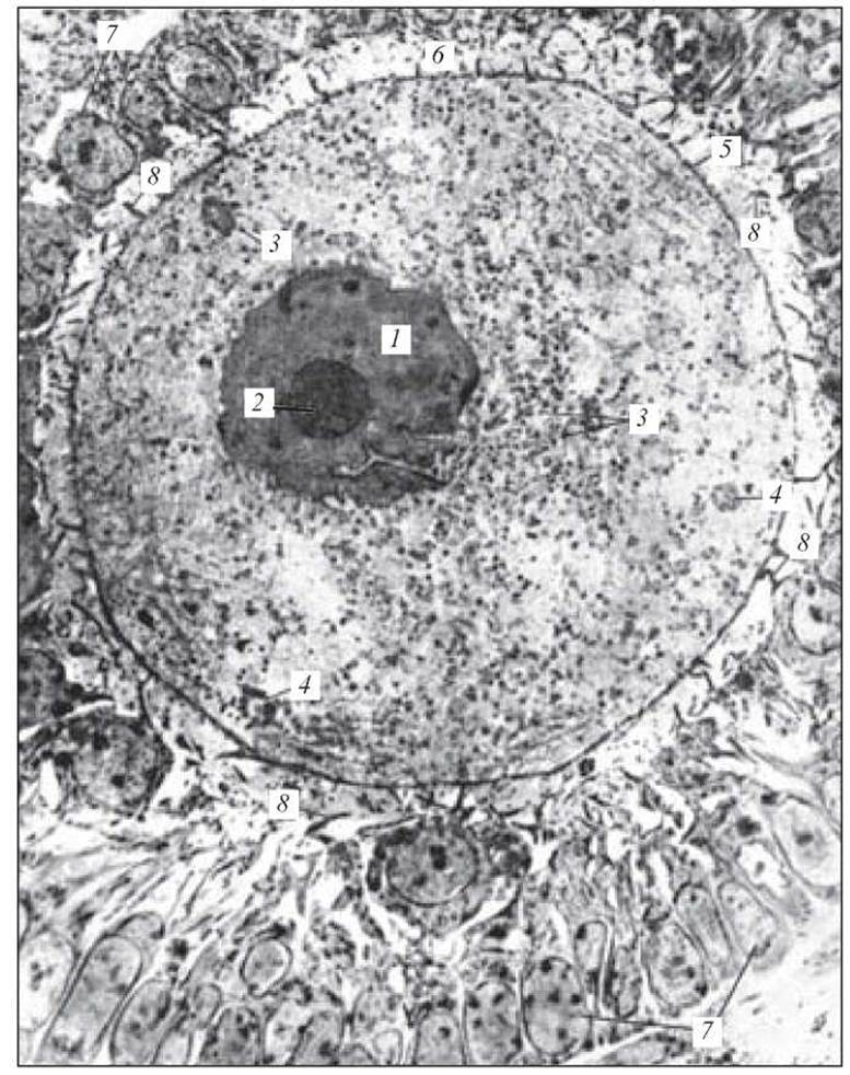
Fig. 28. Mammalian oocyte. Electron micrograph. x 3,000:
1 — nucleus; 2 — nucleolus; 3 — yolk granules in the Cytoplasm; 4 — multivesicular bodies; 5 — cell membrane (ovolemma) with microvilli; 6 — zona pellucida; 7 — follicular Cells of the granular layer; 8 — processes of follicular cells (from Rodin's atlas)
Control Questions
1. Embryology, its role and significance in medical training.
2. Ontogeny. Periods of ontogeny.
3. Progenesis.
4. Male Germ Cells. Structure. Function.
5. Female Germ Cells. Classification. Structure. Function.
6. Embryogenesis. CHARACTERISTICS OF THE stages of embryogenesis.
7. Fertilization. Phases of fertilization. Mechanism.
8. Cleavage. Types of cleavage. Characteristics of cleavage.
9. Gastrulation. Types of gastrulation.
10. Gastrulation in humans.
11. Histo- and organogenesis. Characteristics of the stages.
12. Induction. Concept of organizing factors (inducers).
13. Determination. Differentiation. Types of differentiation.
14. Cell reproduction.
15. Migration.
16. Intercellular interactions. Cell death.
17. Provisional organs. STRUCTURE AND FUNCTIONS.
Situational Problems
1. An electron micrograph (Fig. 27) shows a cross-section of a spermatozoon, revealing the axial filament surrounded by mitochondria. Through which part of the spermatozoon was the section made?
2. The ovum contains a moderate amount of yolk distributed unevenly. Determine the type of ovum, the cleavage pattern, and the type of blastula of the future embryo.
3. A histological specimen shows a blastula with a single-layered blastoderm and a blastocoel in the center. Determine what type of cleavage leads to the formation of such a blastula, and what the mechanisms of gastrulation will be.
4. In a chick embryo, the segmental pedicles (somitic stalks) are destroyed by a micromanipulator. What disorders will this experimental intervention cause?
5. Examine the electron micrograph of the oocyte (Fig. 28) and name its constituent parts.
Sample Examination Questions
1. Characteristics of ontogenesis periods. Progenesis.
2. Structure and function of spermatozoa.
3. Ovum. Classification. Structure.
4. Embryogenesis. Characteristics of embryogenesis stages.
5. Fertilization. Characteristics of fertilization phases.
6. Gastrulation. Types of gastrulation.
7. Main processes underlying histo- and organogenesis.
Last update: 10/08/2026
Editorial and Educational Adaptation: This material has been compiled based on the primary/original source text. The project team performed an editorial review, corrected technical inaccuracies, structured sections, and adapted the content for an educational format.
What was processed:
- elimination of formatting defects (OCR errors, structural breaks, corrupted characters);
- editorial organization of content;
- standardization of terminology in accordance with academic sources;
- verification of factual statements against the original source text.
All mentions of the author, publication year, and origin of the primary text have been preserved in accordance with the source.