BIOCHEMISTRY AND MOLECULAR BIOLOGY - W. ELLIOTT - 2002
CHAPTER 26. CHEMICAL SIGNALING IN THE ORGANISM
In Chapter 12, we discussed the REGULATION OF METABOLISM by external factors that ensure communication between Cells. The vital activity of every individual Cell is subordinated to the needs of the Organism as a whole. An animal cannot exist without a complex system of intercellular communications mediated through a diverse network of commands.
It is now apparent that external control over cells is far more complex than previously imagined. While the mechanisms underlying specific types of regulation are known, it remains completely unclear how all regulatory factors integrate into a unified system within the whole organism, or even within an individual cell. Intercellular Communication is mediated by signaling molecules. These are released from certain cells and migrate to others equipped with receptors capable of perceiving these signals—known as target cells. The binding of a signaling molecule to its receptor triggers a biochemical response in The Cell.
Signals can be transmitted via direct contact between two cells. An example is the Activation of a helper T cell (see Chapter 25) by an antigen-presenting cell. The presence of Gap Junctions or regulated pores between neighboring cells allows molecules to migrate from one cell to another, providing direct communication between cytoplasms and coordinating cellular Functions. In this chapter, we will examine the following questions.
✵ What types of cellular activity are controlled by external signals?
✵ What types of signaling molecules are involved in chemical signaling?
✵ Which cells secrete signaling molecules? How is their release controlled, and how do they reach their target cells?
✵ How do target cells recognize signals?
✵ How does a cell respond to a received signal?
What types of cellular activity are regulated by external signals?
For convenience, they can be divided into two groups.
Regulation not involving Gene Expression. Examples include: changes in The activity of Enzymes involved in fat and Carbohydrate Metabolism
(see Chapter 12); activation of voluntary skeletal Muscle contraction by acetylcholine (see Chapter 28); and the opening of Ligand-gated pores in nerve cell membranes during the generation of a Nerve Impulse (Fig. 26.24).
Regulation involving gene expression. Most external control factors act through this mechanism. Because Introduction/30.html">Regulation of Gene Expression affects virtually all processes occurring within cells, the list of such processes is virtually endless. In animals, Control of Gene Expression is executed primarily at the level of gene Transcription initiation, which in turn depends on the activity of specific transcription factors (see p. 267).
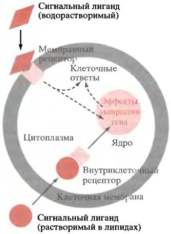
Thus, the main part of this chapter focuses on the mechanisms by which external control factors influence the transcription of specific genes. Most external signaling molecules do not enter the cell to deliver their messages; instead, they interact with the extracellular domains of membrane-bound receptors. This raises several questions: how do signals perceived outside The cell membrane lead to specific events inside the cell (Transmembrane Signal Transduction)? How is a signal generated on the cytoplasmic side of the membrane transmitted to the controlled locus? In the case of gene control, this means transmitting the signal from The Plasma Membrane to the Cell Nucleus (Fig. 26.1). Additionally, a small number of lipid-soluble signaling molecules can pass through The Lipid Bilayer into the cell, where they interact with intracellular receptors involved in gene regulation (see Fig. 26.1).
Class="center">Fig. 26.1. Receptor-mediated signaling. Water-soluble signaling molecules cannot cross the lipid bilayer. They bind to the extracellular domains of receptors. Lipid-soluble signaling molecules, such as Steroids and thyroxine, enter the cell directly and bind to intracellular receptors. Some intracellular receptors reside in The Nucleus; steroid receptors migrate to the nucleus upon ligand binding. In the case of nitric oxide, the intracellular receptor generates cGМР, which can produce direct metabolic effects
What are signaling molecules?
From a chemical perspective, these include Proteins, large and small Peptides, steroids, Eicosanoids, catecholamines, thyroxine, and nitric oxide. The first six groups of substances comprise: Insulin, Glucagon, vasopressin, Sex Hormones, Prostaglandins, and adrenaline.
Biological Classification includes the following groups of regulators:
1) hormones;
2) growth factors and cytokines;
Hormones
These are "classic" signaling molecules, most of which have been known for a long time, probably because they are present in the body in relatively large amounts. Hormones are produced by cells specialized in their synthesis and grouped into glands that secrete hormones directly into the bloodstream rather than into ducts communicating with the external environment (as occurs with the secretion of pancreatic digestive enzymes into the intestine). For this reason, Endocrine glands are also referred to as ductless glands. Hormones "find" their target cells via the Circulatory system, reaching Tissues located at a considerable distance from the hormone-secreting gland. A large number of hormones are known, and their biological effects have been described in detail. The main hormones and their actions are summarized in Table 26.1.
Growth factors
A recently published article suggested that this class of signaling molecules is more aptly named developmental regulatory factors, since their role becomes clearer when viewed as regulators of development rather than factors that merely stimulate cell growth.
Growth factors are regulatory proteins secreted by Cells of the same tissue to which they belong, for example, hepatocytes, lymphocytes, etc. Growth factors interact with cell-surface receptors. The first to be discovered was platelet-derived growth factor (PDGF), which was subsequently found to be produced by many other cell types as well. Another classic example is epidermal growth factor (EGF). A wide variety of such factors have now been discovered. About 20 of them are involved in regulating The Development of hematopoietic cells in the Bone Marrow. Many growth factors are colony-stimulating factors (CSFs) or interleukins. They got their name because they stimulate the growth of colonies of specific white Blood Cells under experimental conditions. They are called interleukins "in honor" of leukocytes that influence other leukocytes via secretion. Most growth factors were discovered due to their mitogenic (growth-promoting) effect on cells, but their action is much more complex. Under certain circumstances, the same factor can stimulate or inhibit Cell Differentiation, exerting completely different effects on the same cell. This is why the term developmental regulation factor seems more appropriate for describing these regulators, though we will stick to the universally accepted term—growth factors.
The term cytokine is used by immunologists for growth factors involved in the Immune Response.
Despite The Diversity of their biological effects, all growth factors interact with cell-surface receptors and indirectly influence the initiation of transcription of specific genes.
Aside from the characteristics mentioned above, how does a growth factor differ from a hormone? As already noted, hormones are released into the circulatory system and distributed throughout the body to reach their target cells. Most growth factors are paracrine in their action: they diffuse over a short distance and act locally only on nearby cells. However, some act in an autocrine manner, stimulating the cells that secrete them (for example, interleukin-2 stimulates T-cell proliferation); in other ways, the release of insulin and glucagon from the Pancreas depends directly on blood glucose levels.
Table 26.1 Main hormones and their functions
Secreting organ |
Hormone |
Target tissue |
Function |
Releasing factors |
Anterior pituitary |
Stimulation of hormone secretion |
|
Somatostatin (also from the pancreas) |
Anterior pituitary |
Inhibits Growth Hormone release |
|
Anterior pituitary |
Thyroid-stimulating hormone (TSH) |
Stimulates the release of thyroxine (T4) and triiodothyronine (T3) |
|
Adrenocorticotropic hormone (ACTH) |
Adrenal cortex |
Stimulates the release of adrenocorticosteroids |
|
Gonadotropins (luteinizing hormone, LH, and follicle-stimulating hormone, FSH) |
Stimulates sex hormone release and cell development |
||
Growth hormone (somatotropin) |
Stimulates the synthesis of Insulin-like Growth Factors IGFI and IGFII |
||
Prolactin |
Mammary gland |
Essential for Lactation |
|
Posterior pituitary |
Antidiuretic hormone (ADH), or vasopressin |
Renal tubules |
Promotes water reabsorption |
Oxytocin |
Smooth muscle |
Stimulates uterine contraction |
|
Thyroid gland |
Thyroxine (T4) and triiodothyronine (T3) |
Liver, Muscles |
Stimulates metabolic processes |
Parathyroid gland |
Parathyroid hormone |
Bone tissue, Kidneys, intestine |
Maintains blood Ca2+ levels, stimulates reabsorption and uptake of dietary Ca2+ |
Calcitonin (also from The Thyroid Gland) |
Bone tissue, kidneys |
Inhibits Ca2+ reabsorption |
|
Adrenal cortex |
Glucocorticoids (cortisol) |
Many tissues |
Promotes Gluconeogenesis |
Mineralocorticoids (aldosterone) |
Kidneys, blood |
Maintains water-electrolyte balance |
|
Adrenal medulla |
Catecholamines (adrenaline, noradrenaline) |
Liver, muscles, Heart |
Mobilize Fatty acids and glucose into the bloodstream |
Sex hormones (testosterone from the testes, estradiol and progesterone from the ovaries) |
Reproductive Organs, secondary sex characteristics |
Promote body maturation and reproductive organ function |
|
Liver |
Somatomedins (insulin-like growth factors: IGFI, IGFII) |
Liver, bone tissue |
Stimulate growth |
Pancreas |
Insulin |
Liver, muscles |
Stimulates Glycogen synthesis, Lipogenesis, and Protein Synthesis |
Glucagon |
Many tissues |
Stimulates Glycogenolysis and lipolysis |
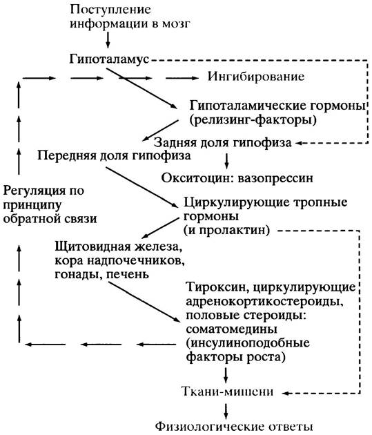
Fig. 26.3. Hypothalamic Regulation of Hormone release
Regulation of growth factor release
Relatively little is known about this, and it is quite possible that the release of these factors is controlled by multiple mechanisms. Damage to the epithelial lining of Blood Vessels leads to platelet rupture and the release of PDGF, which stimulates Cell Division and repair; however, PDGF is also produced by many other cells in the body. In The Immune System, the activation of B AND T cells triggers the release of growth factors (cytokines) that stimulate their proliferation (see Chapter 25). Growth factors can stimulate cells to secrete other growth factors. In short, growth factors are involved in developmental and repair processes as well as defense mechanisms, such as the immune response, which involves extensive control over cell proliferation and differentiation.
Regulation of neurotransmitter release
A nerve impulse traveling along the axon of a nerve cell triggers the exocytotic release of neurotransmitters from vesicles at nerve terminals. The Mechanism of this release will be described later.
Removal of signaling molecules
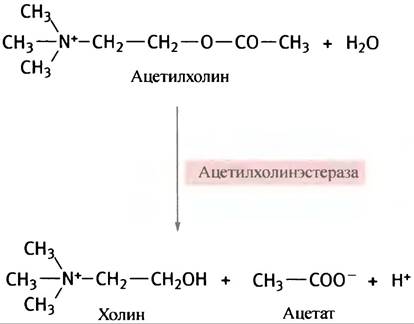
The Essence of control lies in its reversibility. Released signaling molecules must be removed; otherwise, the initial signal would persist indefinitely. A classic example of signal molecule removal is The breakdown of acetylcholine by acetylcholinesterase present in nerve synapses. Signal removal is necessary to prepare the synapse for receiving the next nerve impulse. For acetylcholine, this occurs via the following reaction:
Catecholamines released by nerve endings are taken up by neighboring cells or removed via metabolic degradation.
How are signals recognized by target cells?
As already noted, target cells possess receptors on their outer surface or inside the cell for each specific signal. Signaling molecules bind to these receptors, inducing Conformational Changes in the receptor protein(s) that lead to a specific cellular response. Several hormones (steroids, thyroxine, and nitric
oxide) are lipid-soluble and enter the cell directly across the lipid bilayer, binding to intracellular receptors (see Fig. 26.1). The structures of THYROID HORMONES and two Steroid Hormones are shown in Fig. 26.4. The remaining signaling molecules are water-soluble and bind to receptors on the cell membrane surface (see Fig. 26.1).
The essence of regulation is Specificity: a signaling molecule (referred to as a ligand or agonist) interacts with its receptor with extreme precision—much like a substrate with its enzyme. This is why a hormone distributed by the bloodstream throughout the entire body affects only its target cells. If a cell lacks the specific receptor, it is "blind" to that signal. Similarly, a television receiver without the appropriate antenna cannot pick up a satellite TV signal.
Fig. 26.4. Hormone structures: a - Structures of Tyrosine and thyroid hormones; b - Structures of two steroid hormones

How does ligand binding lead to a cellular response?
Intracellular receptor-mediated responses
Steroid hormones regulate the expression of specific target cell genes at the level of transcription initiation. These include glucocorticoids, estrogen, and progesterone (see Table 26.1). These lipid-soluble hormones readily cross the cell's lipid bilayer to reach their receptors.
Estrogen and progesterone receptors are localized in the nucleus, whereas the glucocorticoid receptor resides in the Cytoplasm. All of them contain "zinc fingers" (see p. 279) as their DNA-binding domain and exist in a complex with heat Shock proteins (Hsp). As we recall, heat shock proteins function as chaperones (see p. 298), participating in the folding and Maintenance of the specific conformation of various proteins.
The "zinc finger" of the glucocorticoid receptor is the best studied, so we will describe it as an example. In the cytoplasm, the receptor is bound to a complex of heat shock proteins that shield the nuclear localization signal (a specific peptide sequence).
When the glucocorticoid hormone binds to a specific site on the receptor, the heat shock protein complex dissociates from it, exposing the signal tag, and the receptor is transported into the nucleus. There, it assembles as a dimer and binds to the glucocorticoid-responsive element on the DNA (Fig. 26.5). The latter is a palindrome, with each half interacting with two "zinc fingers" (see Fig. 21.22) of the receptor dimer molecule.
Fig. 26.5. Gene activation by steroid hormones. The glucocorticoid receptor, belonging to the steroid and thyroxine receptor superfamily, is shown as an example. These receptors form complexes with heat shock proteins (Hsp) and bind to DNA regions via "zinc fingers"

In the case of other steroid Hormone Receptors, the nuclear localization signal is not masked by a heat shock protein complex, so the entire Structure is transported into the nucleus. However, DNA binding does not occur until Hsp is released upon hormone attachment.
Membrane receptor-mediated responses
Numerous intercellular communications in the organism are mediated through Membrane Receptors. Figure 26.6 illustrates Two Types of such receptors. In the first case, ligand binding activates the internal domain of the receptor; In the second case, ligand binding induces receptor dimerization, which is accompanied by the activation of internal domains. How the signal generated at the cytoplasmic face of the membrane is transmitted to the control site is a complex question; therefore, we will first examine membrane receptor-mediated responses and then explore individual signaling pathways leading to cellular responses.
Fig. 26.6. Transmembrane signal transduction by receptors. a - Ligand binding to the receptor causes an allosteric change in the cytosolic domain of the receptor protein molecule; b - ligand binding leads to the dimerization of membrane receptors. This mechanism applies to receptors with tyrosine kinase activity (see p. 353). The association of two cytoplasmic domains results in a biochemical response

Summary of membrane receptor-mediated responses
The main subject of Discussion in this section is protein phosphorylation mediated by ATP-utilizing protein Kinases. It is difficult to overstate The Importance of protein phosphorylation in cellular regulation: in animals, phosphorylation is involved in hormonal control, growth factor regulation, Cell Cycle phase regulation by cyclins, and the control of smooth muscle contraction. Figure 26.7 illustrates the Basic principles of regulation via phosphorylation. It is worth revisiting Figure 12.8, which illustrates The Central Role of phosphorylation in transmitting external signals.
Fig. 26.7. The fundamental regulatory principle for most external signals. Protein phosphorylation alters its properties, leading to a specific cellular response. Note that phosphorylation can either activate or deactivate various proteins. Ultimately, external signals affect the activity of protein kinases and protein Phosphatases
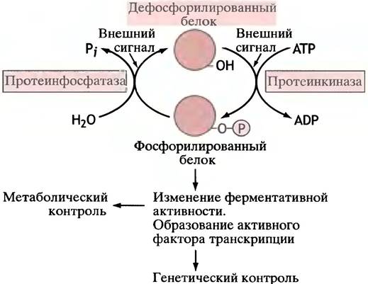
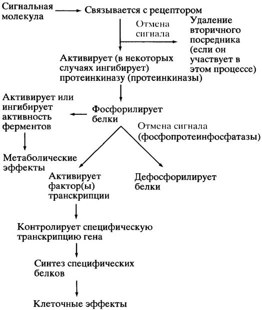
It clearly follows from the foregoing that, in most cases, receptor-mediated signaling involves the activation of protein kinases. The dephosphorylation of Phosphoproteins is catalyzed by protein phosphatases, and this step can also be subject to control. Figure 26.8 summarizes the relationships among regulatory systems mediated by membrane receptors. The diagram also illustrates the reversibility of the signal action through the degradation of the second messenger.
Fig. 26.8. Simplified depiction of key events in membrane receptor-mediated signaling. Note that a single arrow may represent multiple steps in the given process
Modulation of the receptor response to a signal
Repeated exposure of a target cell to a signal often results in a weaker response. In the case of one class of adrenergic receptors, feedback regulation
is accompanied by phosphorylation of the receptor protein and a decrease in the receptor's sensitivity to the ligand (a process known as desensitization). To avoid confusion, let us note that for Other types of receptors, which will be described later, phosphorylation is an integral part of the activation process. Another type of response modulation involves controlling the number of functional receptors in the membrane. In the case of the insulin receptor, the insulin-receptor complex is internalized into the cell via endocytosis. The recycling of the receptor back to the membrane is regulated by the number of receptors present in it. This can contribute to diminishing the cell's response to the given signal and thus attenuate the regulatory system; the latter is known as downregulation.
Description of individual Intracellular Signaling pathways linking receptor activation to cellular response
cAMP-mediated pathways
Since in Chapter 12 (see p. 166) we already examined Metabolic Regulation through changes in protein kinase activity mediated by cAMP, in this section we will discuss how intracellular cAMP levels are modulated and how cAMP regulates gene expression.
Many hormones as primary messengers, including adrenocorticotropic hormone (ACTH), antidiuretic hormone (ADH), gonadotropins, thyroid-stimulating hormone (TSH), parathyroid hormone, glucagon, catecholamines (epinephrine-norepinephrine), and somatostatin (see Table 26.1), utilize cAMP as a secondary messenger. This list is far from exhaustive and is presented merely to illustrate the remarkably diverse effects of cAMP on various cells. The system functions because the cAMP response in a given cell corresponds to the signal that the cell is equipped to detect via its specific receptors. Thus, in cell A, signal X increases the cAMP level, which triggers cellular responses specific to signal X. Cell B lacks receptors for signal X but possesses receptors for signal Y; these also elevate cAMP levels, but in cell B this elicits responses appropriate to signal Y.
We will examine two aspects of these effects: 1) how the binding of a signaling molecule to its receptor controls intracellular cAMP levels; and 2) how cAMP influences gene transcription.
Control of cAMP Levels in Cells
cAMP is produced within the cell from ATP through the action of the enzyme adenylyl cyclase, which is localized on the inner surface of the cell membrane (Fig. 26.9).
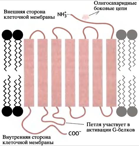
Let us consider a typical example of adenylyl cyclase activation by epinephrine, which results in cAMP generation in The Liver and Skeletal Muscle (see p. 167). The receptor (in this case, the β2-adrenergic receptor) is a protein whose polypeptide chain spans the membrane 7 times (Fig. 26.10). Each of the 7 transmembrane segments consists of approximately 19 hydrophobic Amino Acids. These form an α-helical structure sufficient to span the hydrophobic lipid bilayer of the membrane. Epinephrine "fits" into the crevice between the transmembrane helices.
Fig. 26.9. Formation and Hydrolysis of cAMP. (a) Formation of 3',5'-cyclic AMP by adenylyl cyclase; (b) hydrolysis of 3',5'-cyclic AMP by phosphodiesterase
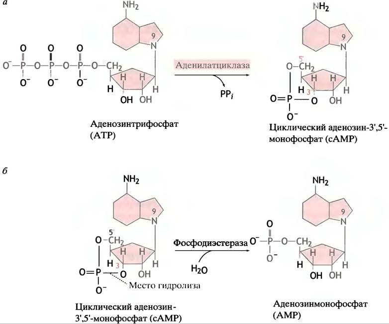
Fig. 26.10. Organization OF THE β2-adrenergic receptor within the cell membrane
Associated with the inner (cytoplasmic) surface of the receptor protein is a regulatory G protein composed of 3 subunits: α, β, and γ. Because these are three distinct Polypeptides, the protein is referred to as a heterotrimeric G protein, or simply a trimeric G protein. The α-subunit contains a site capable of binding either GTP or GDP. When this site is occupied by GDP, the protein remains inactive (Fig. 26.11, a).
Fig. 26.11. Regulation of adenylyl cyclase activity by epinephrine
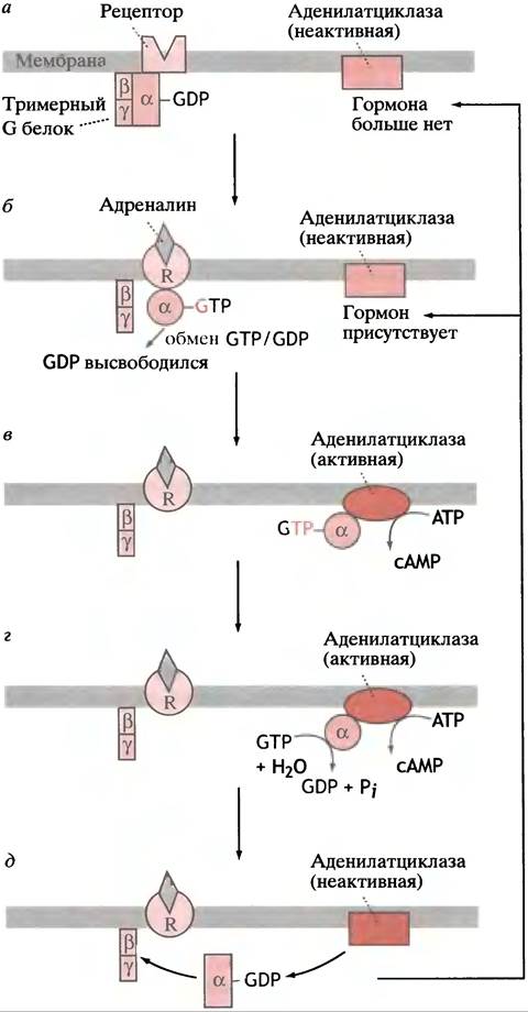
When an epinephrine molecule binds to the receptor, the cytoplasmic domain of the receptor undergoes a conformational change. This, in turn, induces a conformational shift in the associated G protein, causing the G protein (designated here as Gs; where "s" stands for stimulatory) to exchange GDP for GTP. The G protein cannot perform this exchange unless it is coupled to the hormone-bound receptor. The α-subunit–GTP complex then dissociates from the G protein, migrates to an adenylyl cyclase molecule, and activates it, leading to The production of ATP (Fig. 26.11, b, c).
Thus, the hormone stimulates cAMP synthesis by employing a G protein as an intermediary (see Fig. 26.11, c). The activation of cAMP production by the α-GTP subunit must be time-limited; otherwise, a single hormonal stimulus would act indefinitely. Once the receptor no longer binds the hormone, cAMP production must cease; the intracellular hydrolysis of cAMP by phosphodiesterase completes this process (see Fig. 26.9, b).
This situation is analogous to a staircase equipped with a timed lighting system: when you press the button, the light turns on, and then the button slowly returns to its initial position, turning the light off after a minute or two. Periodically, you must press the button again to keep the light from going out. The G protein acts precisely as the "timing device" that limits the duration of adenylyl cyclase activation.
The GTP-bound form of the G protein α-subunit (which activates adenylyl cyclase) possesses intrinsic GTPase activity. It hydrolyzes GTP to GDP and Pi (Fig. 26.11, d). This GTPase activity is low, so GTP hydrolysis does not occur instantaneously. As soon as GDP is formed via hydrolysis, the α-subunit reverts to its initial state. It dissociates from adenylyl cyclase—rendering the enzyme inactive—and, in its GDP-bound form, reassociates with the β
and γ subunits to reform the G protein–GDP complex capable of interacting with the receptor (Fig. 26.11, b). If the receptor still has a hormone molecule bound to it, the entire cycle can begin anew (see Fig. 26.11, b). GDP is replaced by GTP, and the α-subunit dissociates to activate another adenylyl cyclase molecule. If the receptor no longer binds a hormone molecule (see Fig. 26.11, a), the process halts. Therefore, to sustain cAMP synthesis, the α-subunit
must shuttle back and forth between the receptor and adenylyl cyclase. The duration of its time spent interacting with adenylyl cyclase is determined by the time required to hydrolyze the attached GTP molecule.
The system exhibits signal Amplification: a single receptor-bound hormone molecule can sequentially activate multiple G protein molecules. Consequently, a single hormone molecule binding to its receptor can lead to the activation of several adenylyl cyclase molecules and the generation of a large number of cAMP molecules. The critical role of GTP hydrolysis in this regulatory pathway can be illustrated by cholera. Intestinal mucosal cells secrete Na+ into the intestinal lumen, a process stimulated by cAMP. Cholera toxin irreversibly inhibits the GTPase activity of the G protein α-subunit. As a result, the hormonal signal activating adenylyl cyclase cannot be turned off, leaving the enzyme "locked" in the α-GTP state. Sustained cAMP production leads to massive losses of Na+ ions followed by water molecules, causing debilitating diarrhea and potentially fatal fluid and electrolyte depletion.
In the pathway shown in Fig. 26.11, the GTP–α-subunit stimulates adenylyl cyclase activity. Other types of adrenergic receptors (α2-adrenergic receptors) operate similarly, but they utilize a different α-GTP subunit (Gi) that inhibits adenylyl cyclase activity. Alpha- and beta-adrenergic receptors mediate distinct effects of catecholamines. As we already know (see p. 167), in "fight-or-flight" situations, Epinephrine stimulates the liver and skeletal muscles via β2-adrenergic receptors, which activate adenylyl cyclase. In contrast, α2-adrenergic receptors inhibit adenylyl cyclase. Another subtype—α1-adrenergic receptors—exhibits yet another mechanism: they do not use cAMP as a secondary messenger, but instead trigger Ca2+ release via the phosphoinositide pathway (see p. 352). Thus, a single hormone can elicit completely opposing responses depending on the receptor subtype.
How Does cAMP Act on Gene Transcription?
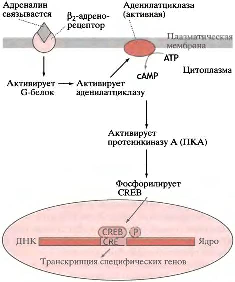
In Chapter 12, we described how cAMP activates protein kinase A (PKA). Beyond its role in controlling enzyme activity, PKA is a key component of the machinery regulating gene expression. The promoters of several cAMP-inducible genes contain cAMP response elements (CREs). When PKA phosphorylates
the CRE-binding protein (CREB), it becomes an active leucine zipper-type transcription factor (Fig. 26.12). There is a family of CREB proteins that presumably fulfill distinct functions within the cell.
Fig. 26.12. Schematic diagram illustrating the function of the β2-adrenergic receptor. Receptor-mediated stimulation of Adenylyl Cyclase and the resulting increase in cAMP levels activate cAMP-dependent protein kinase (PKA). Phosphorylation of CREB generates an active transcription factor complex for specific genes
As mentioned earlier, cAMP acts as a secondary messenger (see p. 166) in transmitting extracellular signals to functional elements in both the cytoplasm and the cell nucleus. Another cyclic nucleotide, cGMP, performs a similar function in other regulatory systems, which we will examine below.
Signal transduction using cyclic GMP as a secondary messenger
A variety of signals trigger an increase in intracellular cGMP levels, which in turn activates protein kinase G (PKG). The formation of cGMP from GTP is catalyzed by
both membrane-bound and soluble forms of the enzyme guanylyl cyclase (Fig. 26.13). In the first case, guanylyl cyclase serves as the intracellular domain of a membrane receptor protein. Hormone binding to the extracellular receptor leads to activation of the intracellular domain and enhanced cGMP synthesis. This pathway differs from the mechanism by which adrenergic receptors trigger cAMP formation: here, the intrinsic domain of the receptor itself functions as a guanylyl cyclase and is activated directly upon hormone attachment (Fig. 26.14). The resulting cGMP mediates the cellular response by activating a specific protein kinase (PKG). An example of this type of regulation is atrial natriuretic peptide, produced by endothelial cells, which stimulates renal Na+ excretion. The effects of cGMP are more specialized compared to those of cAMP; they include smooth muscle relaxation and play key roles in The Nervous system and Vision (see below).
Fig. 26.13. Formation of 3',5'-cyclic GMP catalyzed by guanylyl cyclase
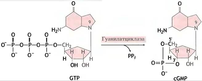
Fig. 26.14. Schematic diagram illustrating the core principles of cyclic nucleotide action as secondary messengers in cellular signaling. R1 – α2-adrenergic receptor; R2 – β2-adrenergic receptor; R3 – atrial natriuretic peptide binding site, with one domain embedded in the membrane and the catalytic domain oriented laterally. AC – plasma membrane adenylate cyclase, GC1 – plasma membrane guanylyl cyclase, GC2 – cytoplasmic nitric oxide (NO)-activated guanylyl cyclase
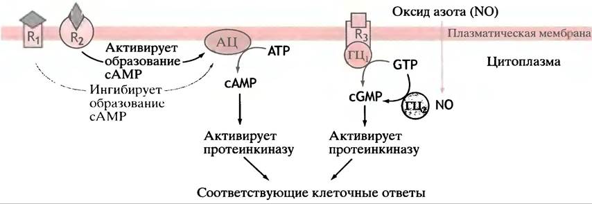
The Cytosol contains another, soluble form of guanylyl cyclase. It contains a heme group as a prosthetic group, which binds to arguably the simplest Intercellular signaling molecule: NO, or nitric oxide. The heme group acts as an exceptionally sensitive NO detector, transmitting the activation signal to the enzyme. NO is produced in the endothelial cells of The Vascular System from the guanidino group of Arginine through the action of the enzyme nitric oxide synthase. NO diffuses into the smooth muscle of blood vessels, prompting cGMP formation, which subsequently promotes muscle relaxation and vasodilation. Acetylcholine stimulates nitric oxide production, indicating that this process can be mediated by neural regulation. Nitric oxide is also synthesized in response to the mechanical stress exerted by blood flow on the endothelial lining of blood vessels, leading to vasodilation.
Trinitroglycerin, a long-established medication for angina pectoris, slowly releases nitric oxide, thereby inducing vasodilation and reducing cardiac workload. Because NO is oxidized to NO2 and NO3 within seconds, it functions as a localized signaling molecule; being highly lipid-soluble, nitric oxide readily escapes the cells that produce it and diffuses into neighboring cells. Nitric oxide is part of a complex regulatory network with diverse physiological effects. Figure 26.14 summarizes the fundamental principles of Cyclic NUCLEOTIDES acting as secondary messengers in intercellular signaling.
Hormones transmitting signals via alternative secondary messengers: the phosphoinositide cascade
Thus far, we have examined a diverse group of hormones that utilize cyclic nucleotides—cAMP and cGMP—as secondary messengers. Another group of hormones and growth factors employs different intracellular mechanisms for signal transduction. Candidate hormones bind to specific cell surface receptors that also interact with G proteins (i.e., GTP-binding proteins), yet cyclic nucleotides are not involved in this signal transmission. Examples of hormones utilizing this mechanism include thyrotropin-releasing hormone, gonadotropin-releasing hormone, and platelet-derived growth factor (PDGF).
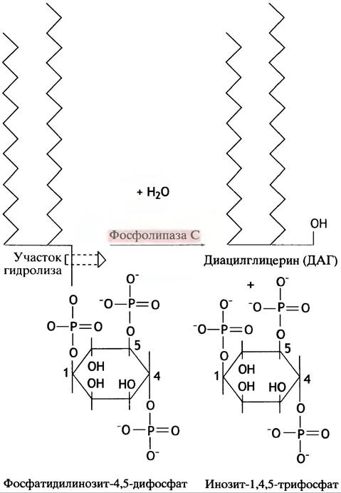
The phospholipid phosphatidylinositol 4,5-bisphosphate (PIP2) is generated in the membrane through the phosphorylation of phosphatidylinositol, The structure of which was introduced in Chapter 10. To reiterate, the G protein couples the receptor stimulus to the intracellular signaling pathway, acting as a "molecular timer." The Interaction of a hormone with its receptor induces the receptor-bound G protein to exchange GDP for GTP. The Gp-GTP complex then migrates and activates the membrane-bound enzyme phospholipase C, which cleaves PIP2 into Inositol trisphosphate (IP3) and diacylglycerol (DAG) (Fig. 26.15). The G protein hydrolyzes GTP to GDP and Pi, leading to the self-inactivation of the enzyme.
Fig. 26.15. Hydrolysis of phosphatidylinositol 4,5-bisphosphate (PIP2) into diacylglycerol (DAG) and inositol trisphosphate (IP3)
IP3 triggers the release of Ca2+ from the lumen of The Endoplasmic reticulum (ER), which maintains high concentrations of this ion. IP3 opens ligand-gated Ca2+ channels in the membrane (see below), facilitating the influx of the ion into the cytoplasm. Driven by Ca2+-ATPases, Ca2+ ions are continually pumped back into the ER lumen, while IP3 and DAG are cleared enzymatically. Consequently, the binding of a hormone or any other agonist to a receptor involved in the phosphoinositide cascade leads to an increase in intracellular DAG and Ca2+ (Fig. 26.16), with the process reversing upon dissociation of the hormone-receptor complex.
Fig. 26.16. The phosphoinositide cascade: interplay of DAG, IP3, and Ca2+ as secondary messengers. The Gp protein associates with phospholipase C exclusively in its GTP-bound complex; the process is reversed by the hydrolysis of GTP to GDP
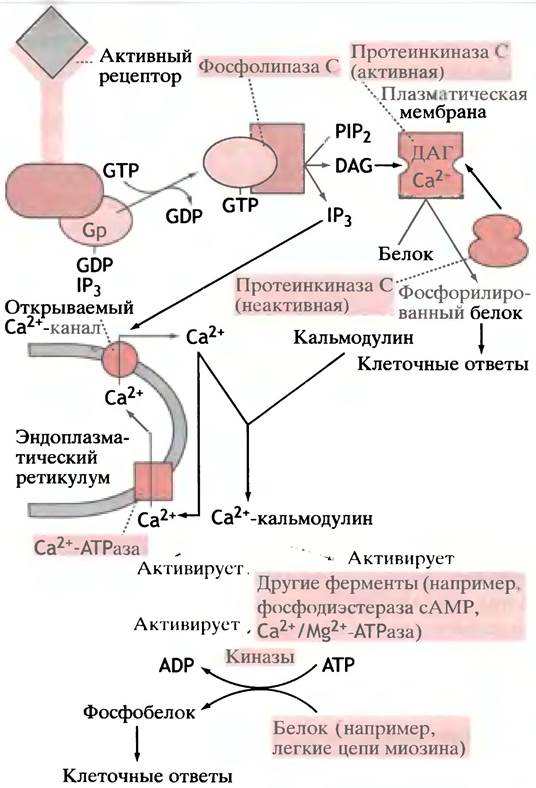
DAG serves as the physiological activator of a distinct protein kinase, differing from PKA and PKG. Maximal activation of this enzyme also requires Ca2+.
The DAG-activated protein kinase is designated as PKC; it associates with phosphatidylserine molecules located on the cytosolic leaflet of the cell membrane. PKC participates in The regulation of numerous cellular processes by phosphorylating various target proteins and certain growth factors. The critical role of PKC in cell division control is highlighted by the oncogenic effects of phorbol esters, which act as tumor promoters. Phorbol esters are structural analogs of DAG that activate PKC. The chemical structures of DAG and a phorbol ester are shown below (memorizing the STRUCTURE OF THE latter is not required).
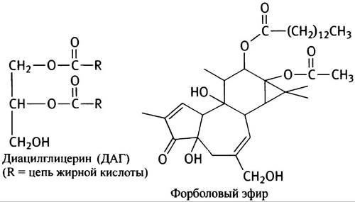
It might seem paradoxical that DAG exerts the same activating effect on PKC as a tumor-promoting agent. The difference in their action apparently lies in the fact that DAG is rapidly degraded, activating PKC only when required, whereas phorbol esters persist much longer, thereby prolonging a signal that is detrimental to the organism.
As illustrated in Fig. 26.16, stimulation of this receptor type results in the generation of two messengers—DAG and Ca2+—both of which are necessary for the maximal activation of PKC. However, Ca2+ also functions independently as an important secondary messenger.
What are the other messenger effects of calcium?
The mediation of Ca2+ effects as a secondary messenger involves calmodulin. This ubiquitous protein features four high-affinity binding sites for Ca2+ ions; their binding induces a conformational change in calmodulin. Calmodulin thus activated alters the activity of enzymes (and other proteins) with which it interacts. There is a range of Ca2+-calmodulin-activated protein kinases, implying that the execution of many Ca2+ effects within the cell also entails protein phosphorylation.
The list of target proteins for these kinases includes Glycogen phosphorylase and glycogen synthase (pp. 88-89). Since one type of adrenergic receptor (α1) stimulates the phosphoinositide pathway, thereby elevating cytoplasmic Ca2+ levels, adrenaline can influence Glycogen Metabolism both through this mechanism and via cAMP. Other target proteins of Ca2+-calmodulin-dependent kinases are Myosin light-chain kinases (see p. 386).
Tyrosine Kinase-Associated Receptors
Although we are still exploring membrane receptors in this section, we are now about to examine a critically important domain that is entirely distinct from everything discussed above.
Phosphorylation is a primary regulatory step; however, the phosphorylation target is the tyrosine amino acid residue of the receptor protein itself, meaning that self-phosphorylation (autophosphorylation) takes place.
All the protein kinases described thus far phosphorylate the hydroxyl groups of Serine or Threonine residues in their target proteins. We now turn to
receptor activation driven by the phosphorylation of the hydroxyl groups of specific tyrosine residues in proteins in response to signaling molecule binding (Fig. 26.17a). While this might seem rather trivial at first glance, tyrosine kinase-associated receptors represent a unique class of receptors that trigger specific signaling cascades. Their significance is underscored by the fact that most growth factors and certain hormones, such as insulin, utilize receptors of this type.
Fig. 26.17. Activation of various enzymatic and genetic processes upon the interaction of the EGF receptor with specific ligands. a - Protein level: tyrosine phosphorylation by tyrosine kinase; b - Cellular level: cumulative effect of EGF on the target cell
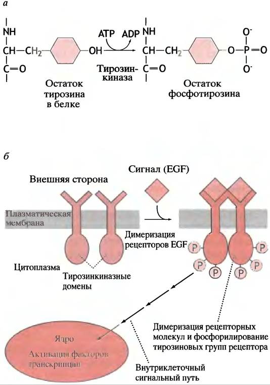
To illustrate this signaling system, we will examine epidermal growth factor (EGF).
Ligand binding induces the dimerization of EGF receptors within the cell membrane (Fig. 26.17b). This results in the multiple phosphorylation of tyrosine residues located in the cytoplasmic domains of the receptors. These cytoplasmic domains function as tyrosine kinases that likely phosphorylate one another during dimerization. Phosphorylation triggers a signaling cascade that extends from the membrane receptor to the cell nucleus, culminating in the activation of a transcription factor.
The Ras Pathway: A Major Eukaryotic Membrane-to-Gene Signaling Cascade
A protein known as the Ras protein is present in all Eukaryotic cells. It is a small, monomeric GTP-binding protein with intrinsic GTPase activity. It has been shown to constitute a core component of the primary signaling pathway linking growth factor receptor-associated tyrosine kinases to gene transcription factors. A distinctive feature of this pathway is the absence of low-molecular-weight secondary messengers: all its components are proteins.
The Ras protein and other constituents of this intracellular signaling pathway are crucial for cellular regulation; Mutations that give rise to abnormal forms of these proteins are oncogenic, driving uncontrolled cell division.
Figure 26.18 illustrates this system. The cytoplasm contains a growth factor receptor-bound protein (GRB) associated with the so-called SOS protein. Via its SH2 domain, GRB interacts with the phosphorylated receptor. This domain is also found in a family of cytoplasmic proteins thought to play a role in binding phosphorylated receptors. The designation SH2 originates from the presence of a domain homologous to domain 2 of the Src protein encoded by the Rous Sarcoma virus oncogene (see p. 314), hence the abbreviation SH2.
Fig. 26.18. Activation of the EGF Ras pathway (simplified diagram). Light shading indicates the inactive state, dark shading indicates the active state. a - Inactive state without EGF;
b - Activated state with EGF; GRB - growth factor receptor-bound protein; SOS (son of sevenless) - a protein involved in this signaling pathway, named after the corresponding "sevenless" mutation in Drosophila. Raf, Ras, Jun, and Fos derive their names from their respective oncogenes. Note that while the receptor is phosphorylated on a tyrosine residue, the subsequent kinases in the cascade belong to the serine/threonine type. The mechanism by which Ras-GTP activates Raf kinase remains to be fully elucidated

The association of the GRB-SOS complex with the activated receptor prompts the Ras protein to exchange GDP for GTP (Fig. 26.19). This is directly analogous to G-protein activation during cAMP generation (see Fig. 26.11). The Ras protein possesses a low intrinsic GTPase activity that acts as a molecular "timing device."
Fig. 26.19. Activation of the Ras protein
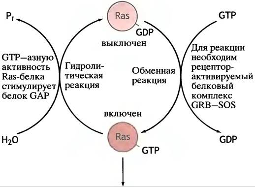
The next component in this pathway is the Raf protein, a serine/threonine-specific protein kinase whose activation requires Ras-GTP. This marks the beginning
of the serine/threonine protein kinase cascade, which concludes with the phosphorylation of transcription factor proteins; the activation of these factors promotes their binding to corresponding response elements and drives the transcription of specific genes.
The Ras Pathway and Oncogenesis
In Chapter 23, we explained that Cancer-causing genes carried by Retroviruses are mutated forms of Proto-oncogenes present in normal cells. The Ras and Raf proteins are products of proto-oncogenes; when the corresponding oncogenes are introduced into cells by retroviruses, they induce cancer. It appears that oncogenic copies of Ras and Raf proteins (as well as Other components of the Ras pathway illustrated in Fig. 26.18) are locked in a persistently "activated" state. The intracellular signaling pathway behaves as though a signaling molecule were bound to the receptor continuously. One such oncogene is v-erbB, which encodes an aberrant EGF receptor. It lacks the extracellular domain, yet its cytoplasmic domain remains constitutively phosphorylated (Fig. 26.20). Consequently, in the presence of any of these oncogenic proteins, the Ras pathway operates in an uncontrolled manner. Oncogenes can arise not only through retroviral infection; normal proto-oncogenes may also be transformed into oncogenes via mutation.
Fig. 26.20. Oncogene products as components of signal transduction pathways from the membrane to the nucleus: the EGF receptor is used as an example. a - Normal receptor - inactive; b - normal receptor - active; c - constitutively active truncated receptor; d - normal receptor or signal pathway component is constitutively active

The conversion of proto-oncogenes into oncogenes can occur not only as a result of a single base mutation. Chromosomal rearrangements promote the translocation of genes involved in the control of cell differentiation and division. This leads to the formation of oncogenes via several pathways.
1. A proto-oncogene can fall under the control of another regulatory element that causes the overexpression of a specific protein. For example, Burkitt's lymphoma occurs when the c-myc gene comes under the control of an immunoglobulin gene promoter.
2. Translocation can cause Gene Fusion and lead to the formation of oncogenic hybrid proteins.
3. Amplification can cause the overexpression of a regulatory protein.
Cancer is the result of a cell acquiring multiple genetic anomalies. In addition to the conversion of proto-oncogenes into oncogenes, cancer development often involves the loss of a functional tumor suppressor gene. About half of all human malignancies are associated with the loss of the p53 tumor suppressor gene. The p53 gene product exerts complex effects, including The stimulation of DNA Repair Mechanisms; however, most importantly, it has The ability to control the transition from the G1 phase to the S phase of the cell cycle, thereby helping to prevent uncontrolled cell division.
Terminology Note
As noted earlier, oncogenes are named after the retroviruses that carry them and are italicized. For example, the ras oncogene was discovered in the Rat Sarcoma virus. The oncogene is identified with the letter "v" (for virus), and the proto-oncogene with the letter "c" (cytoplasmic). Thus, these genes are designated as v-ras and c-ras, respectively, and so on. The protein products are written as Ras, Raf, etc. (not italicized).
The SOS protein is named after a receptor gene mutation in fruit flies (sevenless): the protein product of this gene is called son of sevenless, hence the name SOS. It was later discovered that this protein is universally present in eukaryotes. (Do not confuse this protein with the E. coli SOS Response)
Why is the Ras pathway so long?
It is tempting to suggest that the protein kinase cascade is an amplification mechanism similar to those of glycogen breakdown and blood clotting.
It is quite possible that the Ras pathway is interconnected at several points with other regulatory pathways.
There is growing Evidence for the existence of extensive crosstalk among regulatory mechanisms. For instance, the Ras protein can be phosphorylated and activated by PKC after the latter is activated in the phosphoinositide pathway. Different protein kinases in the Ras pathway may have other target proteins, thereby activating alternative pathways and regulatory cascades. Furthermore, an activated receptor tyrosine kinase can couple with SH2 proteins of other signaling pathways, allowing a single receptor type to activate multiple pathways (Fig. 26.21). Using an electrical analogy, Ras and other signaling pathways are electronic circuits on a highly complex, interconnected instrument panel, although we cannot yet see how this "instrument" functions as an integrated regulatory system. A characteristic feature of signaling pathways is the presence of Protein-Protein Interactions that apparently form networks of a complexity comparable to metabolic pathways.
Fig. 26.21. Hypothetical diagram showing how multiple proteins with SH2 domains could act as linking elements in an integrated regulatory system
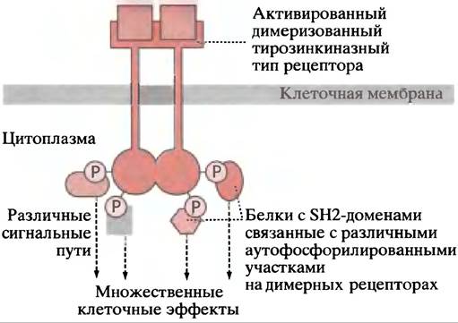
In sharp contrast to the Ras pathway is another intracellular receptor mediating a relatively simple signaling pathway that does not involve tyrosine kinase. Let us examine it in more detail.
Direct intracellular signaling pathway from the receptor to the nucleus
Interferon signaling
In 1957, it was shown that virus-infected vertebrate cells secrete proteins called interferons, which protect against the same or other Viruses (this phenomenon was termed Interference). The antiviral activity of interferons is not yet fully understood, but it is known that genes contain interferon-stimulated response elements (ISREs), so that the binding of interferon to a cell receptor triggers specific cellular gene responses.
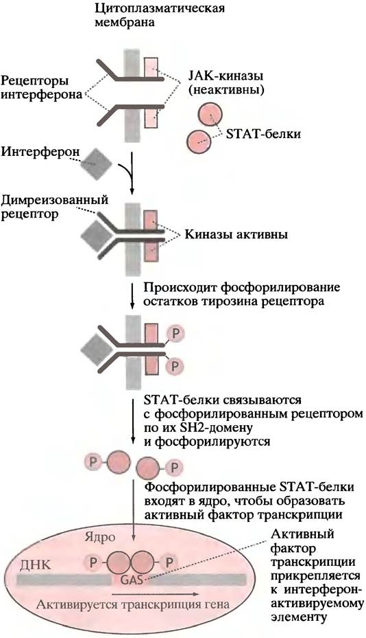
The y-interferon receptor differs from the one described for EGF. Two molecules of cytoplasmic tyrosine kinase (JAK kinases) are associated with it. Ligand binding causes receptor dimerization; this leads to tyrosine kinase activation, resulting in the phosphorylation of tyrosine residues on the receptor dimer. This, in turn, induces the binding of two cytosolic proteins to the phosphorylated receptor. These proteins contain the same SH2 domains as the GRB protein of the Ras pathway; as already mentioned, the SH2 domain is found in proteins that interact with phosphorylated receptor tyrosine kinases. Protein kinases still bound
to the receptor phosphorylate two SH2 proteins, which then migrate to the nucleus, where they assemble into an active transcription factor and promote the transcription of ISRE elements (Fig. 26.22).
Fig. 26.22. Signaling pathway by which y-interferon activates specific gene transcription. STAT - signal transducer and activator of transcription; GAS - y-interferon-activated sequence element. Note that unlike the EGF receptor, which is itself a tyrosine kinase, in this pathway the dimeric receptor activates cytoplasmic tyrosine kinases, or Janus kinases (JAKs), named after the two-faced Janus (they have two kinase domains). α-Interferon and several cytokines utilize analogous signaling pathways
How general is this direct pathway?
The main question is whether this system is generally applicable or if it is specific to only a few signaling molecules. A mechanism in which an unlimited number of signals could exert specific effects on cells would ensure the system's widespread distribution. It is known that the second interferon and several cytokines involved in regulating red blood cell production utilize this exact pathway. It can be hypothesized that various phosphorylated receptors are capable of binding different cytosolic SH2 proteins, which are subsequently converted into various specific transcription factors, thereby ensuring the transmission of a large number of signals to specific cellular "control centers."
In Conclusion, we can quote a review from the journal Nature, which states: "Over the next few years, hundreds and even thousands of papers will appear describing how various proteins transmit their signals through intracellular signaling pathways."
External Signals Regulating Ligand-Gated Ion Channels
We now turn to a completely distinct type of regulation in which the immediate result of ligand binding to a membrane receptor is the opening of channels in the membrane (Fig. 26.23). Neuronal signaling will be used to illustrate this.
Fig. 26.23. Ligand-gated channel. Channels specific for Ca2+, Na+, and K+ ions are shown.

How does the binding of acetylcholine to a membrane receptor lead to a nerve impulse?
As mentioned earlier, a motor neuron that sends a signal to contract a voluntary striated muscle receives acetylcholine from the presynaptic neuron as a signaling molecule; it is released into the synapse and ensures the conduction of a nerve impulse to another neuron, which prompts the muscle to contract (see Fig. 26.24).
Neurons, like other animal cells, maintain a high concentration of K+ inside the cell and a low concentration outside, whereas the level of Na+ is high outside and low inside the cell. This ion distribution is the result of the Na+/K+-ATPase ion pump in the membrane.
At rest (when no nerve impulses are being transmitted), the cell membrane is more permeable to K+ than to Na+. Due to the higher concentration inside the cell, K+ flows outward, and since the membrane is impermeable to anions, the latter do not leave the cell. This creates a positive charge on the outside and a negative charge on the inside, i.e., membrane polarization. The leakage of K+ is a self-limiting process. Further leakage is prevented by the resulting charge difference. An equilibrium state is reached in which the cell's Resting Potential is about -70 mV (negative charge on the inside).
The Acetylcholine Receptor is a ligand-gated Na+/K+ channel. Upon acetylcholine binding, the channel opens, allowing Na+ to move into the cell and K+ to move out (see Fig. 26.24). However, due to much higher concentration gradients, Na+ enters the cell faster than K+ leaves it, which leads to a decrease in the internal negative charge. In other words, the membrane becomes depolarized, specifically only in the immediate vicinity of the synaptic acetylcholine receptors. This is the initial stimulus.
Fig. 26.24. Chain of events resulting in motor neuron stimulation leading to acetylcholine release at the Neuromuscular Junction, accompanied by striated muscle contraction (simplified)
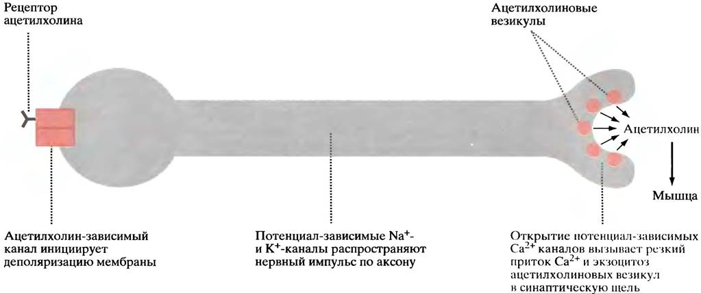
The propagation of a nerve impulse along the axon occurs due to acetylcholine-induced repeated depolarization of the membrane; however, this involves a different type of Na+ and K+ channels, termed voltage-gated channels, which open when the resting potential drops (from -70 to -40 mV). The potential change occurs in a narrowly restricted region of the membrane and is associated with the opening of only adjacent voltage-gated channels. The channels remain open for a very short time, after which they close, and a recovery period ensues. Only after recovery are the channels able to respond to the next signal. Once the wave of excitation has passed, the Na+/K+ pump restores the initial ion concentrations.
A detailed examination of the mechanism of nerve conduction is beyond The Scope of this section and the entire book. What is important is that the wave of membrane depolarization travels along the axon to the nerve terminals. The membranes of the latter contain voltage-gated Ca2+ channels that open when the depolarization wave reaches them, allowing extracellular Ca2+ to enter the cell. This triggers the fusion of acetylcholine vesicles with the Cell Membrane and their release via exocytosis (see Fig. 26.24). The Ca2+-ATPase pumps Ca2+ out, restoring the resting state. The released acetylcholine binds to muscle cell receptors. The subsequent sequence of events is described in Chapter 28.
Ca2+-induced vesicle exocytosis has a broader biological significance than merely participating in neurotransmitter release. Stimulated Ca2+ vesicle exocytosis occurs during the secretion of digestive enzymes from the pancreas; however, here Ca2+ release occurs via the phosphoinositide pathway (see p. 353).
Egg Fertilization also triggers Ca2+-induced vesicle exocytosis, which creates a barrier to prevent subsequent sperm penetration.
Another important process involving ligand-gated pore regulation is vision.
Vision: A Process Dependent on Ligand-Gated Pore Control
The basis of vision is the conversion of a light signal into chemical changes that lead to the generation of impulses in the Optic nerve, which carries signals to the Brain.
Light is absorbed by the vertebrate retina, which contains two types of cells: rods—for black-and-white and dim-light vision—and cones—for color and bright-light vision. Rods have three segments. The middle region contains the usual organelle complex. One end forms a synapse with a bipolar cell, which is connected to the optic nerve. The other end is a cylindrical (rod-shaped) segment containing a "battery" of membrane discs (about 2,000) immersed in the cytoplasm (Fig. 26.25). The discs contain the light-sensing apparatus.
Fig. 26.25. Structure of a rod cell

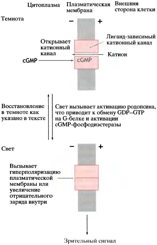
Fig. 26.26. The visual process (simplified diagram). The action of light on rhodopsin leads to the activation of cGMP-phosphodiesterase. A decrease in cGMP levels results in the closure of cation channels, hyperpolarization of the cell membrane, and transmission of the impulse along the optic nerve.
Transmission of the Light Signal
The details of light perception are complex; we will examine only the basic principles (Fig. 26.26).
In the dark, guanylyl cyclase maintains a fairly high level of cGMP in cone cells. The cell membrane contains ligand-gated cation channels that are kept open by cGMP.
The constant influx of Na+ in the dark is determined by a lower potential across the cell membrane; the equilibrium between Na+ entering and leaving the cell
sets the potential at about -30 mV (which differs from the situation in a neuron, where resting Na+ channels are closed and the Membrane Potential is -70 mV).
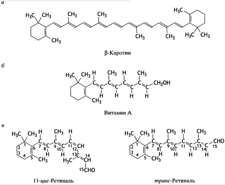
The receptor that captures light photons in rod discs is rhodopsin, a complex of the protein opsin and 11-cis-retinal. Light triggers the activation of rhodopsin through a conformational change in the visual pigment, converting it entirely to trans-retinal. Look at the structure shown in Fig. 26.27 and note the similarity of the retinal molecule to vitamin A and β-carotene.
Fig. 26.27. Structures of β-carotene, vitamin A, and retinal. a - Structure of β-carotene, the primary photosynthetic pigment in plants and a precursor of vitamin A in animals; b - structure of vitamin A; c - structure of 11-cis-retinal and trans-retinal
Rhodopsin activation triggers a cascade of reactions involving a trimeric G protein (and the hydrolysis of GTP, which plays its "usual" role as a molecular clock). All of this ultimately leads to the activation of the GTP-hydrolyzing enzyme, cGMP phosphodiesterase. Its action is similar to that of cAMP phosphodiesterase (see Fig. 26.9, b).
The decrease in cGMP levels leads to the closure of Na+ channels and hyperpolarization of the membrane. Thus, the visual signal is transmitted to the optic nerve.
Cell recovery following the absorption of a light photon is a complex process. It includes: 1) inactivation of rhodopsin; 2) a decrease in intracellular Ca2+ levels and stimulation of cGMP synthesis; and 3) a complex cycle of transformations of the rhodopsin molecule. Ultimately, the level of cGMP drops in the light, Na+ channels close, and hyperpolarization of the cell membrane leads to the transmission of the visual signal to the optic nerve. Upon illumination, cGMP levels are restored, Na+ channels open, and we are ready to perceive the next photon.
Questions for Chapter 26
1. Which classes of signaling molecules mediate communication between cells?
2. Using a simple diagram, compare the cellular effects of lipid-soluble and lipid-insoluble extracellular signaling molecules.
3. How is cAMP formation regulated? Describe The Role of GTP in this process. What is The connection between GTP and cholera?
4. How does cAMP function as a secondary messenger?
5. In what way does nitric oxide exert its regulatory effect?
6. cAMP and cGMP are not the only secondary messengers. Describe another similar system.
7. There are important gene-activating regulatory pathways that do not involve low-molecular-weight secondary messengers. Describe such a pathway.
8. If a protein has an SH2 domain, what is its likely function?
9. Explain how abnormal components of cellular signaling pathways can contribute to cancer development.
10. Describe the interferon-mediated pathway of gene activity regulation.
11. Provide a general comparison of the activation mechanisms of three receptor types: A - an adenylyl cyclase-activating adrenergic receptor; B - an EGF receptor of the Ras pathway; C - an interferon receptor.
12. WHAT IS A voltage-gated Ca2+ channel? Illustrate with an example.
13. What is a ligand-gated channel? Give an example of such a channel involved in the visual process.
Last update: 06/08/2026
Editorial and Educational Adaptation: This material has been compiled based on the primary/original source text. The project team performed an editorial review, corrected technical inaccuracies, structured sections, and adapted the content for an educational format.
What was processed:
- elimination of formatting defects (OCR errors, structural breaks, corrupted characters);
- editorial organization of content;
- standardization of terminology in accordance with academic sources;
- verification of factual statements against the original source text.
All mentions of the author, publication year, and origin of the primary text have been preserved in accordance with the source.