Molecular Biology of the Cell - Volume 1 - Alberts B., Bray D., Lewis J., Raff M., Roberts K., Watson J. 1994
Molecular Organization of Cells
Basic Genetic Mechanisms
DNA Repair Mechanisms
While the long-term survival of a species may be enhanced by changes in its genetic makeup, survival at any given moment demands the absolute preservation of Genetic information. Maintaining this constancy of genetic material requires not only an extremely precise mechanism for copying DNA nucleotide sequences in each new Cell generation, but also a mechanism for repairing spontaneously occurring DNA damage. Most of this damage is temporary because it is efficiently removed by a specialized mechanism known as DNA repair. If this mechanism, which ensures cellular constancy, fails, the change becomes permanent. Such a change is called a mutation. It can be fatal to the Organism if it affects a vital DNA sequence.
Before discussing DNA repair mechanisms, we will briefly address how DNA nucleotide sequences are replicated from generation to generation.
5.2.1. High Fidelity of DNA Nucleotide Sequence Maintenance [17]
The rate of change in DNA nucleotide sequences (the mutation rate) can only be determined indirectly. One method involves comparing the Amino acid sequences of the same protein across several biological species. The proportion of Amino Acids that differ is then compared with the number of years that have passed since the two species diverged from a common ancestor in the course of evolution (this time span is estimated from fossil record data). Based on this, one can calculate the average number of years required for a stable change to affect a single amino acid in the protein. Since each such change typically reflects a single nucleotide change in The sequence of the Gene encoding the protein, we can also determine the average number of years required for a single stable mutation to occur in that gene.
Such determinations always yield heavily underestimated mutation rates, as most Mutations disrupt the protein's function and are eliminated from the population by natural Selection. However, among the Proteins studied, there is one family for which this objection is virtually invalid. These are the fibrinopeptides—fragments of 20 amino acid residues cleaved from the fibrinogen protein when it is activated and converted into fibrin during Blood clotting. Fibrinopeptides perform no direct function and are therefore tolerant to almost all possible Amino Acid Substitutions. Analysis of fibrinopeptides shows that an average-sized protein of 400 amino acids changes randomly by a single amino acid substitution approximately once every 200,000 years. Later, with The Development of DNA Sequencing Methods (see Section 4.6.6), it became possible to determine the degree of similarity between DNA nucleotide sequences in homologous non-coding Regions of the genome in different mammalian species. The mutation rate estimates obtained in this way are in excellent agreement with those derived from fibrinopeptide analysis.
5.2.2. The Mutation Rate in Growing Cells Agrees with Estimates Derived from Evolutionary Studies [18]
The mutation rate can be estimated directly by counting the number of genetic changes that occur spontaneously in a large population of cells over a relatively short period of time. This is done by determining the frequency of new mutants in very large animal populations (for example, in fruit flies or mouse colonies) or by screening for changes in The activity of specific Enzymes in cells growing in culture. Although these estimates are only approximate, in both cases they agree with the idea that Replication produces, on average, one error (i.e., one base-pair substitution) per 109 Base Pairs. Consequently, for a mutation to occur in a gene encoding an average-sized protein and containing approximately 103 coding base pairs, about 106 cell generations are required. This value is in reasonable agreement with the estimate obtained from evolutionary studies, according to which a mutation in an average germ-line gene occurs once every 200,000 years.
5-16
5.2.3. Most Mutations That Alter Proteins Are Harmful and Are Eliminated by Natural Selection [17]
If the number of amino acid differences in the same protein between two different biological species is plotted as a function of the time elapsed since their divergence, we obtain a straight line. In other words, the longer the period since divergence, the greater the number of such differences. For convenience, the slope of the line can be characterized by the unit of evolutionary time for a given protein (the average time required for one amino acid substitution to appear in a sequence of 100 amino acids). By doing this for different proteins, we can see that each is characterized by its own specific rate of evolution (Fig. 5-30).
Class="center">
Fig. 5-30. Comparison of the rates of amino acid substitution in Hemoglobin and cytochrome c with the corresponding rate for fibrinopeptides. Hemoglobin and cytochrome c evolve much more slowly than fibrinopeptides. When determining substitution rates (Table 5-2) per year, It is important to remember that two organisms that diverged from a common ancestor 100 million years ago are separated from each other by 200 million years of evolutionary time.
5.2.5. Low Mutation Rates Mean That Related Organisms Are Built of Virtually the Same Proteins [17]
Humans, as a genus distinct from the great apes, emerged only a few million years ago. Consequently, only a relatively small number of nucleotide substitutions could have occurred in each gene during this time, and natural selection must have eliminated most of them. A comparison of humans and apes shows, for example, that their Cytochromes c differ by only 1% of amino acid residues, and their Hemoglobins by approximately 4%. A significant portion of our genetic heritage was undoubtedly shaped long before the appearance of Homo sapiens, during mammalian evolution (which began approximately 3 ∙ 108 years ago) and even earlier.
|
Protein |
Unit of evolutionary time 1), |
|
million years |
|
|
Fibrinopeptide |
0.7 |
|
Hemoglobin |
5 |
|
Cytochrome c |
21 |
|
Histone H4 |
500 |
1) Unit of evolutionary time is the average time required for one acceptable amino acid substitution to appear per 100 amino acids in a given protein.
It is not surprising, therefore, that in such phylogenetically distant mammals as the whale and the human, Proteins are very similar. The evolutionary process that produced the dramatic morphological differences among mammals achieved these differences with remarkably small Changes in the material from which we are all built. It is assumed that these morphological differences are largely determined by differences in the temporal and spatial patterns of Gene Expression during embryonic development. In Chapter 10, we will discuss how such changes in gene expression might have arisen (see Section 10.5.8).
5-13
5-17
5.2.6. Without Repair, Spontaneous DNA Damage Would Rapidly Alter Its Nucleotide Sequences [20]
In 1945, the physicist Erwin Schrödinger suggested that a gene, regardless of its chemical nature (which was not yet known at the time), must be extremely small—consisting of no more than a few atoms. Otherwise, Schrödinger reasoned, the vast number of genes thought to be required by every organism could not fit inside the Cell Nucleus. It was clear, however, that at such a small size, a gene would be subject to significant changes due to spontaneous reactions caused by random thermal collisions with surrounding molecules. This created a serious dilemma, as genetic evidence indicated that gene material is highly stable and spontaneous changes (mutations) occur extremely rarely.
This Schrödinger dilemma is very real. DNA does indeed undergo significant changes due to thermal fluctuations. We know, for example, that the DNA in every human cell loses about 5000 purine bases (adenine and guanine residues) per day due to the thermal Cleavage of N-glycosidic bonds between purine and deoxyribose (a process called depurination). A similar example is the Spontaneous deamination of cytosine to uracil in DNA, which is estimated to occur at a rate of 100 times per genome per day (Fig. 5-31). The bases in DNA are also altered by reactive metabolites that disrupt their normal pairing, as well as by ultraviolet radiation from the sun, which can cause The formation of a covalent bond between two adjacent pyrimidine base residues in DNA (thymine dimer formation; Fig. 5-32). All of the above is only a small fraction of the many changes that occur spontaneously in our DNA. Most of these would lead either to the loss of one or more base pairs in the daughter DNA strand after a replication cycle, or to a base-pair substitution (for example, each C → U deamination would eventually cause a C-G to T-A transition, since U behaves similarly to T and forms a complementary pair with A). As we already know, if such changes occurred frequently enough, they would have fatal consequences for living organisms.

Fig. 5-31. Deamination and depurination are two common spontaneous Chemical Reactions that cause serious DNA damage in cells. Only one example of each of these Two Types of reactions is shown here.
5-14
5.2.7. Gene stability is preserved by DNA repair [21]
Although thermal energy causes thousands of random changes in the DNA of any human cell every day, only a very small number of stable changes in the DNA nucleotide sequence accumulate in each cell over a year (if indeed any accumulate at all). We now know that among the many random base substitutions in DNA, only about one in a thousand leads to a mutation; all other damage is highly efficiently eliminated by DNA repair processes. All repair mechanisms rely on The Cell having two copies of the genetic information—one in each of the two strands of the DNA molecule. If The nucleotide sequence of one strand is accidentally altered, the information is not lost because its backup copy is preserved in the nucleotide sequence of the complementary DNA strand. As shown in the diagram in Fig. 5-33, the main pathway of DNA repair involves three steps.
1. The altered region of the damaged DNA strand is recognized and removed by specific enzymes called DNA repair Nucleases; they hydrolyze the phosphodiester bonds between the damaged NUCLEOTIDES and the rest of the DNA molecule, leaving a gap in the DNA helix at this site.
2. Another enzyme, DNA polymerase, binds to the 3'-end of the damaged DNA strand and fills this gap by adding nucleotides one by one, copying the information contained in the "good" (template) strand.
3. Finally, an enzyme called DNA ligase "seals" the DNA, thereby completing the restoration of an intact molecule.

Fig. 5-32. Thymine dimer formation is a common type of DNA Damage caused by ultraviolet radiation (specifically, by sunlight). Any two adjacent pyrimidine bases (C or T) can form such a dimer.
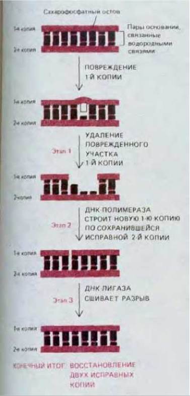
Fig. 5-33. The three steps of DNA repair. In the first step, the damaged region is excised; In the second and third steps, the original DNA nucleotide sequence is restored. DNA polymerase fills the gap left by the removal of the damaged part of the strand (step 2), and DNA ligase seals the remaining nick in the repaired strand (step 3). Sealing is achieved by restoring the broken phosphodiester bond (see Fig. 5-35).

Fig. 5-34. The enzyme DNA polymerase. A. The reaction catalyzed by DNA polymerase. This enzyme catalyzes the stepwise addition of deoxyribonucleotides to the 3'-end of a polynucleotide (primer) chain paired with another (template) polynucleotide chain. Thus, the new DNA strand grows in the 5' → 3' direction. Because each incoming deoxyribonucleoside triphosphate must pair with the template strand to be recognized by DNA polymerase, it is the template strand that determines which of the four possible deoxyribonucleotides (A, C, G, or T) will be added to the 3'-end of the synthesized chain. As with RNA polymerase, the reaction is driven by a large favorable change in Free energy (see Fig. 5-2). B. The Structure of E. coli DNA polymerase, determined by X-ray crystallography. DNA polymerase is shown here engaged in DNA Synthesis. (Courtesy of Tom Steitz.)

Fig. 5-35. The enzyme DNA ligase seals a broken phosphodiester bond. The diagram shows that DNA ligase first uses an ATP molecule to activate the 5'-end of the damaged strand at the site of the nick (step 1), and only then forms a new bond (step 2). The energetically unfavorable sealing reaction is thus driven by coupling to the energetically favorable Hydrolysis of ATP. Patients with Bloom's syndrome (an inherited disorder) have been found to have a partial deficiency of DNA ligase. Consequently, their DNA repair is impaired, resulting in an increased incidence of Cancer.
The enzymes DNA polymerase and DNA ligase play crucial roles in DNA METABOLISM: both enzymes are involved not only in DNA repair but also in DNA replication. The reactions they catalyze are illustrated in Figs. 5-34 and 5-35, respectively.
5-18
5-19
5.2.8. Different types of DNA damage are recognized by different enzymes [22]
The way a damaged region is removed during repair depends on the type of damage. For example, in depurination (the most common type of DNA damage), a deoxyribose residue loses its associated base (see Fig. 5-31). An AP endonuclease rapidly recognizes this deoxyribose residue and cleaves the phosphodiester bond at this altered site. The damaged nucleotide is then removed, and the correct nucleotide sequence is restored by the mechanism shown in Fig. 5-33.
Another closely related repair pathway involves a specific set of enzymes called DNA glycosylases. Each of these enzymes recognizes a single specific type of altered base in DNA and catalyzes its hydrolytic removal. There are believed to be at least six types of enzymes in this group. These include enzymes that remove deaminated cytosine, deaminated adenine, various types of alkylated bases, open-ring bases, and bases in which a carbon-carbon double bond has been converted to a single bond. The general mechanism is illustrated with a specific example in Fig. 5-36, which shows the removal of deaminated cytosine. First, the enzyme uracil-DNA glycosylase removes the altered base (uracil). The deoxyribose that has lost its base is then recognized by another enzyme, AP endonuclease. Since this is the same enzyme that recognizes apurinic sites in DNA, the restoration of the correct sequence proceeds via the same pathway described for depurination. As a result, the U generated by accidental deamination is replaced back with C. The Importance of removing accidentally deaminated bases from DNA has been demonstrated directly. For instance, in bacterial strains lacking the enzyme uracil-DNA glycosylase due to a mutation, the frequency of spontaneous C-G to T-A transitions (normally very low) increases approximately 20-fold.
Cells possess a special pathway for removing almost any type of DNA damage that affects a large region. Such bulky lesions occur, for example, from covalent interactions between DNA bases and large Hydrocarbons, such as the carcinogen benzopyrene. They also include various pyrimidine dimers (T-T, T-C, and C-C) caused by sunlight (see Fig. 5-32). In such cases, a large multienzyme complex recognizes a major distortion in the DNA double helix rather than a single specific base alteration. The phosphodiester bonds of the damaged strand are cleaved on both sides of the lesion, and the entire altered segment is excised. After this, the restoration of the normal sequence proceeds as usual.
The importance of repair processes is highlighted by the fact that cells devote a significant portion of their resources to producing repair enzymes. Extensive studies in Yeast have identified over 50 different genes encoding such enzymes. DNA repair pathways in humans are no less complex. For example, patients with xeroderma pigmentosum have a defect in the repair of bulky lesions, a process that genetic analysis shows involves at least 7 different gene products. In these patients, pyrimidine dimers accumulate in cells, leading to severe Skin lesions, including cancer.
5-15
5.2.9. Cells synthesize repair enzymes in response to DNA damage [23]
Throughout evolution, cells have developed many different mechanisms to ensure their survival in a world full of hazards. Often, a sudden environmental stress activates a specific set of genes whose products can protect the cells from that stress. For example, All cells share a mechanism known as the heat-Shock response, which can be observed in cells exposed to excessively high temperatures. This response induces the Synthesis of specific "heat-shock" proteins, some of which apparently help stabilize and repair other cellular proteins that have been partially denatured by the heat shock.
Many cells also possess mechanisms that allow them to synthesize DNA repair enzymes under emergency conditions, so to speak, in response to severe DNA damage. Among such Examples, the SOS Response (SOS repair) in E. coli is the best studied. In this bacterium, any disruption of DNA replication caused by damage leads to a signal (apparently an excess of single-stranded DNA) that enhances the Transcription of more than 15 different genes, many of which encode proteins involved in DNA repair. The signal activates a protein in E. coli (see Section 5.4.4) that then destroys another protein—a negative regulator of gene activity (repressor). The function of this repressor is to suppress the Transcription of the entire set of genes involved in the SOS response in E. coli. Studies of bacterial mutants with various defects in SOS repair have shown that the Newly synthesized proteins produce two effects. First, their induction increases cell survival: if Mutants with Impaired synthesis of these enzymes are exposed to DNA-damaging agents (such as ultraviolet light), the percentage of dead cells is unusually high. Second, some of the induced proteins cause a temporary increase in mutation rate, thereby increasing the genetic Water/126.html">Diversity of the bacterial population. The advantage here is presumably that it increases the chances of producing a mutant cell with enhanced fitness.
Other inducible DNA repair systems also exist. For example, one such system in Bacteria is known to be activated by the presence of methylated nucleotides in DNA. A similar system Functions in yeast cells. There is evidence that some higher Eukaryotic cells also adapt to DNA damage in a similar manner.
5.2.10. Structural features and Chemical properties of the DNA double helix facilitate its repair
The DNA molecule has a structure that seems uniquely suited for repair. If the hypothesis that RNA evolved before DNA is correct (see Section 1.1.7), the question arises as to why uracil (U), present in RNA, was replaced by thymine (T) in DNA. This can clearly be explained by the fact that the mechanism responsible for removing deaminated cytosine residues (Fig. 5-36) could not function if the fourth nucleotide in DNA were uracil rather than thymine (i.e., not 5-methyluracil). Spontaneous deamination of C yields U, and therefore the enzyme that recognizes and removes such accidentally generated U residues would also remove those U residues that were normal components of this DNA.
A similar situation arises in another case, namely the choice of guanine over hypoxanthine. The simplest purine that specifically pairs with C is hypoxanthine. However, hypoxanthine is the direct product of the deamination of A (Fig. 5-37). By adding a second amino group to hypoxanthine, evolution created guanine, which cannot be formed from A by spontaneous deamination. Thus, any potential deamination in DNA leads to an unnatural base, which, by virtue of its very abnormality, can be immediately recognized and removed by a specific DNA glycosylase (Fig. 5-37).
Thus, the Chemical Nature of the bases themselves ensures that deamination does not go unnoticed. However, accurate repair (and with it, a radical solution to Schrödinger's dilemma) is possible because of the existence of two copies of genetic information, each represented by one of the two strands of the DNA double helix. Only in the highly unlikely event of simultaneous damage to both members of the same base pair will the cell lack a correct copy to serve as a template for DNA repair.
Genetic information can also be stored in single-stranded DNA or RNA, and some very small Viruses have single-stranded genomes containing only a few thousand nucleotides. The mechanisms described above are unable to repair such Nucleic Acids, and consequently, the mutation rate in these viruses is very high. Only organisms with extremely tiny genomes can afford to store their genetic information in structures other than a DNA double helix.

Fig. 5-36. The DNA repair pathway involving uracil-DNA glycosylase, which restores cytosine in the DNA strand after its accidental deamination. Following the action of DNA glycosylase, the sugar-phosphate lacking its base is removed from the strand by an AP endonuclease, the same enzyme involved in the repair of apurinic sites. This is followed by the steps shown in Fig. 5-33. The name "AP endonuclease" reflects the fact that this enzyme recognizes any site in the DNA helix containing a deoxyribose residue that has lost its base. The lost base can be either a purine (apurinic sites) or a pyrimidine (apyrimidinic sites).
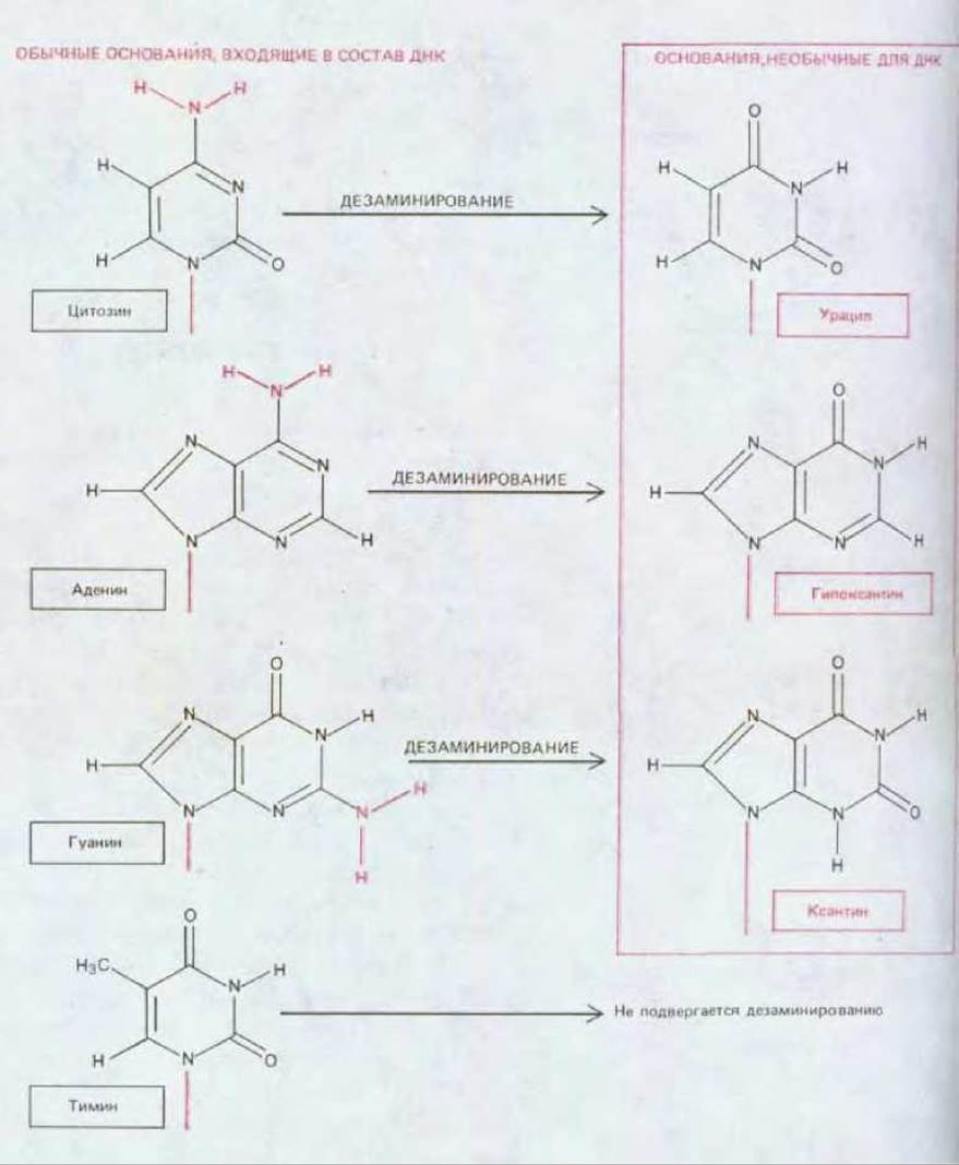
Fig. 5-37. Products of the spontaneous deamination of various DNA bases. All of these deamination products are unnatural in DNA and are recognized for this very reason.
Summary
The fidelity with which DNA nucleotide sequences are maintained in higher eukaryotes can be judged from the rate at which The amino acid sequences of nonessential proteins and DNA nucleotide sequences change over evolutionary time. This fidelity is so high that, on average, only 10-20 base changes affecting germ-line cells occur per year in a mammalian genome of 3 ∙ 109 base pairs. At the same time, in a genome of this size, thousands of DNA nucleotides are damaged daily due to unavoidable chemical decay processes. Genetic information can be reliably stored in DNA nucleotide sequences only because A wide variety of different repair enzymes continuously "scan" the DNA and remove damaged nucleotides from it.
The process of DNA repair relies on the fact that genetic information is stored in two copies within the molecule—one in each of the two strands of the DNA double helix. Consequently, accidental damage in one of the strands can be removed by a repair enzyme, and this section of the strand can be resynthesized in its normal form using the information contained in the undamaged strand.
Last update: 12/08/2026
Editorial and Educational Adaptation: This material has been compiled based on the primary/original source text. The project team performed an editorial review, corrected technical inaccuracies, structured sections, and adapted the content for an educational format.
What was processed:
- elimination of formatting defects (OCR errors, structural breaks, corrupted characters);
- editorial organization of content;
- standardization of terminology in accordance with academic sources;
- verification of factual statements against the original source text.
All mentions of the author, publication year, and origin of the primary text have been preserved in accordance with the source.