BIOCHEMISTRY AND MOLECULAR BIOLOGY - W. H. ELLIOTT - 2002
CHAPTER 25. THE IMMUNE SYSTEM
The body is constantly under the threat of infection by various pathogenic agents, such as Bacteria and Viruses, which are countered by The Immune System. The mortality of patients with genetic (e.g., adenosine deaminase deficiency, see Fig. 18.6) or acquired (AIDS) immune system disorders vividly demonstrates the vital importance of Immunity.
The immune system protects the body against several types of macromolecules, of which Proteins are the most important. Protection against foreign proteins is especially crucial. At the same time, the immune system does not react to small foreign molecules, such as drugs, which upon entering the body become the "prey" of other chemical defense systems (see Chapter 17). The entry of a foreign macromolecule into the body serves as a warning signal of the need to defend against the "invader."
However, patients may experience allergic reactions even to small molecules, such as penicillin. In this case, however, the body reacts not to penicillin itself, but to its combination with another macromolecule, such as a protein. As a result of this combination, the body's own protein begins to "look" foreign to the immune system.
As you may recall from Chapter 4, one of the primary Functions of the Digestive System is to prevent autodigestion, i.e., the destruction of the Organism by itself. A similar challenge faces the immune defense.
Unlike the recognition of lipopolysaccharide components of a bacterial Cell—which differ from the corresponding components of any bacteria normally present in our body and can therefore be easily classified as foreign—Structure/149.html">The problem of recognizing foreign proteins is much more complex. The body itself contains thousands of proteins that differ from foreign ones only by individual Amino acid sequences, so sorting "self" from "non-self" proteins must be carried out with great care.
When the immune system makes a mistake and attacks one of its own proteins, an autoimmune disease may occur. For example, myasthenia gravis is an autoimmune reaction that destroys Muscle acetylcholine receptors (see p. 384), making nerve-stimulated contraction impossible. In rheumatic fever, the Immune Response against a protein produced by certain Streptococcus strains is accompanied by The production of Antibodies that cross-react with Heart proteins, causing heart valve damage. Insulin-dependent diabetes arises from an autoimmune attack on pancreatic Cells.
The immune system employs two major defense mechanisms
The first is the production of antibodies. Antibodies are soluble proteins secreted by Cells of the immune system that specifically bind to foreign substances called Antigens (from antibody generation). An antigen is defined as any molecule capable of triggering a specific immune response, in this case, the production of antibodies. The formation of the antigen-antibody complex leads to defense reactions that will be discussed later. This type of immunity is called humoral immunity (from the Latin humor, meaning fluid); this defense mechanism relies on the presence of soluble antibodies in the Body Fluids.
The second type of immunity, in which specialized cytotoxic or killer cells recognize abnormal cells within the body, is known as cell-mediated immunity. It operates at THE CELLULAR LEVEL, for instance, during a viral infection. A killer cell attaches to the virus-infected cell and destroys it. This is an effective way to prevent the further spread of a viral infection, as cell destruction halts viral Replication. The key feature of this mechanism is direct contact between the killer cell and the target cell—hence the term cell-mediated immunity. Humoral and cell-mediated immunities Complement each other. In the viral infection example discussed above, the humoral system provides protection before The Cell is infected by the virus, whereas the cellular system destroys the host cell after infection, thereby halting viral replication within that cell.
Antibodies are produced by a specific group of lymphocytes called plasma cells (a type of WHITE Blood CELL), which are generated upon
appropriate stimulation of B cells (B lymphocytes). The cytotoxic (killer) cells are T lymphocytes (the letter "T" indicates their site of formation—the Thymus, a gland located behind the Sternum in humans). All lymphocytes are continuously produced in the Bone Marrow (in the Liver during the fetal stage). B cells mature in the bone marrow.
There is a very important condition regarding the "division of labor" between B AND T lymphocytes. For a B cell to produce antibodies, it must, firstly, encounter an antigen, and secondly, come into contact with a special type of T cell that has already "met" the same antigen. Because this T cell "helps" the B cell perform its function, it is called a helper T cell (derived from the English word help). Helper T cells constitute a distinct group of cells, separate from cytotoxic T cells.
Thus, the body contains mature B cells that produce antibodies, helper T cells that assist B cells in producing antibodies, and cytotoxic (killer) T cells responsible for cell-mediated immunity.
Where in the body is the immune system located?
The cells of the immune system are not grouped into a single organ, but are distributed throughout the body: 30% are in the Spleen, 20% in the Lymph Nodes, 40% in the intestinal and mucosal lymphoid tissue, and 10% in the Blood and Lymphatic vessels. For both humoral and cell-mediated immunity, the primary cells are lymphocytes, but other white Blood Cells, such as macrophages, also participate in the immune response. As noted above, both B and T cells are lymphocytes. They circulate in lymph (the body has two circulating fluids: BLOOD AND LYMPH). In Tissues, Blood Plasma passes from capillaries into the tissue (interstitial) fluid, which bathes all cells. This fluid supplies them with nutrients brought by the blood and collects Metabolic waste products. Subsequently, this fluid enters venous and lymphatic capillaries, and from there into larger Lymphatic vessels (which ultimately drain into the two central neck Veins). At certain points, lymphatic vessels expand to form lymph nodes, where the concentration of B and T cells is particularly high. Nodes are located in the armpits, groin, Tonsils, adenoids, and intestines; the lymphocytes residing within them can encounter any foreign antigen entering the tissues drained by the lymph. Therefore, lymph nodes constantly "monitor" tissue infection by acting as a filter, while also providing a suitable microenvironment for the proliferation and differentiation of lymphocytes upon contact with antigens. Lymphocytes are formed in the bone marrow. All blood cells originate from stem cells; as they differentiate, some become B cells, others T cells, red blood cells, and so on (Fig. 25.1). As previously mentioned, B cells develop in the bone marrow, whereas T cells migrate to the thymus, where they proliferate and undergo initial maturation. The migration of T cells from the bone marrow and the release of mature T cells from the thymus occur mainly during the early Selection/3.html">Stages of development, meaning that removal of the thymus in adults has little effect on immune responses. The production of B cells and their release from the bone marrow continue throughout adult life.
Class="center">Fig. 25.1. Stages of hematopoiesis (simplified). Each arrow indicates potential cell proliferation, which is specifically regulated by various proteins known as cytokines. These include colony-stimulating factors, interleukins, and erythropoietins, the latter of which stimulate red blood cell production.

The bone marrow and thymus are referred to as Primary Lymphoid Organs, whereas the lymph nodes are known as Secondary Lymphoid Organs. Lymphocyte maturation in the primary lymphoid organs is antigen-independent, whereas all subsequent differentiation of released mature lymphocytes within the secondary lymphoid tissue is antigen-dependent.
In this chapter, we explore the immune process, beginning with a Brief Overview of the body's immune defense strategy.
Basic strategy of immune defense
The immune system must solve two main tasks: 1) orchestrating the production of antibodies by B cells in response to foreign antigens, but not to the body's own components; 2) ensuring that cytotoxic T cells selectively attack only abnormal body cells. The fundamental principle of immune defense is that a given B cell can produce only one specific antibody (to bind its specific antigen), and a given T cell responds to only one antigen. A vast array of different antibodies is produced in the body. They must correspond to an equivalent number of B cells, each containing the Gene encoding the antibody produced by that cell. This raises a natural question: how can such A large number of genes exist? We will discuss this later.
The body also contains a multitude of T cells with diverse receptors, each specific to its own antigen.
Immature B cells are capable of generating antibodies that attack virtually any macromolecule in the body. To prevent autoimmune reactions, any cell producing an antibody against the body's own components must be eliminated in the first place. This initially occurs during the primary maturation of B cells in the bone marrow; as they mature, B lymphocytes presumably adapt to most autoantigens they might ever encounter. Other mechanisms for suppressing autoimmune reactions are hypothesized to exist, but they remain unknown.
In the body, an immature B cell produces about 105 molecules of its own antibody and displays them on its surface (anchored in the membrane); at this stage, the B lymphocyte does not secrete the antibody, which instead acts as a receptor. During primary B-cell maturation in the bone marrow, only antigens that are part of the organism's own body can interact with the surface-bound antibody. The immune system must not react to self-antigens; otherwise, B cells at this stage of development are destroyed (or functionally eliminated in another way). Following this selective "culling," the surviving cells must respond exclusively to foreign antigens. They are then released into the bloodstream.
The newly released population of naive B cells—as they are now called—is activated upon encountering matching antigens, which is entirely appropriate since the encountered antigen is foreign.
An activated B cell does not mature into an antibody-secreting plasma cell until it encounters a mature T cell that has already been activated by the same antigen. T cells recognize specific antigens via their receptors; during maturation in the thymus, they produce an antigen-specific receptor whose reaction to self-components leads to the elimination of immature cells. T-cell selection takes place early in development during their maturation in the thymus, where, much like B cells, they encounter most autoantigens. T cells that survive this selection are released as inactive helper T cells (or cytotoxic T-cell precursors, see below), which become activated upon encountering specific antigens. The Role of activated helper T cells is to deliver a signal to specifically activated B cells, prompting them to proliferate and mature into antibody-secreting plasma cells (Fig. 25.2). All of the above outlines the fundamental principles of clonal selection theory. The antigen selects the appropriate cells for proliferation. There is an exception to the mechanism described above: certain Components of the Cell wall of Gram-negative bacteria—lipopolysaccharides—can stimulate antibody production by corresponding naive B cells even without a helper T-cell signal. Contact between this type of antigen and a B cell is fully sufficient for the latter to mature into an antibody-secreting plasma cell.
Fig. 25.2. B-cell activation for antibody secretion (main stages). The Mechanism of helper T-cell activation by an antigen differs somewhat from the activation of B cells by the same antigen
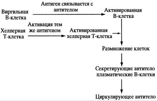
The plasma cell represents the final product of B-Cell Differentiation. It specializes in the Synthesis and Secretion of antibodies. Given the role of The Endoplasmic reticulum in protein secretion, it is hardly surprising that these Organelles are exceptionally well developed in plasma cells. (The regulation of B-cell and helper T-Cell Division will be discussed later.)
In the thymus, some immature cells develop into precursors of cytotoxic, or killer, T cells, which play a vital role in destroying virus-infected cells. A similar selection process occurs here: any potentially cytotoxic cell whose receptor reacts with a self-component is inactivated or eliminated. The selected cells are released from the thymus into the bloodstream. Upon recognizing a foreign antigen On the surface of an infected host cell, they transform into functionally active cytotoxic cells. When an activated cytotoxic T cell encounters a cell bearing a foreign antigen, its receptor interacts with it. As a result, the T cell releases perforin—a protein that kills the target cell by increasing its membrane permeability or by inducing apoptosis, which is programmed cell self-destruction.
Immune Mechanisms: B Cells, Helper T Cells, and Antibody Production
Antibody Structure
Antibodies are IMMUNOGLOBULINS. The term globulin stems from an early Classification system used to designate proteins soluble in salt solutions. Immunoglobulin is commonly abbreviated as Ig. There are several classes of immunoglobulins, one of which is IgG, produced in large quantities by cells during prolonged contact with an antigen. Therefore, its structure will be examined first. The IgG molecule has a Y-shape. It consists of 4 polypeptide chains: 2 identical light (L) chains and 2 identical heavy (H) chains, joined by Disulfide Bonds (Fig. 25.3). The tips of the "Y" arms feature variable regions on both heavy and light chains. It is these fragments that form the antigen-binding site on each arm. Binding to the antigen occurs via non-covalent bonds. Thus, an antibody has two identical antigen-binding sites and is consequently able to cross-link antigen molecules. Flexible "hinge" regions are located at the fork of the "Y," facilitating the formation of cross-linked molecular structures. A similar (though not identical) structure is characteristic of other immunoglobulins as well. Some of them (such as IgM) are polymers consisting of 5 subunits, each closely resembling an IgG molecule.
Fig. 25.3. STRUCTURE OF THE IgG molecule. a — IgG molecule. IgM and IgA molecules differ in their Fc fragments but share a similar variable region. H — heavy chain; L — light chain. N and C denote the amino and carboxy terminals of the polypeptide, respectively. The Fc fragment is the C-terminal portion of the H chains, covalently joined into a single domain by S-S bonds; b — cross-linking of an antigen with distinct specific epitopes; c — antigens with multiple antigenic determinants form a cross-linked insoluble cluster with the antibody
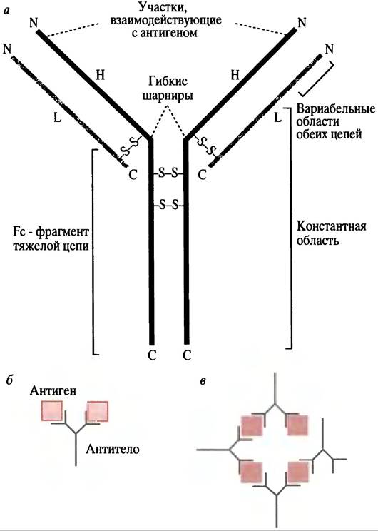
Despite the large size of the antigen triggering the immune response, a given antibody binds to only a small portion of it (in the case of a protein antigen, just a few Amino Acids). The specific part of the antigen recognized by the antibody is called an epitope. A large protein antigen will stimulate the production of a series of different antibodies, each binding to a specific epitope and produced by a separate clone of B cells.
The body can encounter a vast number of various antigens and is potentially capable of producing diverse antibodies that interact with them. Specific antibodies differ in the Amino Acid Sequence of their antigen-binding regions. In antibodies produced by different cells, both L and H chains contain variable amino acid sequences (V domains) that determine the Structural Features of the antigen-binding sites and thereby account for the immense diversity of antibodies. As already noted, the organizational principle of the immune system involves the random GENERATION OF B cells differing in the genes encoding antibodies. Each newly developed B cell can produce antibodies of only a single antigenic Specificity, the molecules of which are anchored in The Plasma Membrane at this stage, with their antigen-interacting regions exposed on the outside. Metaphorically speaking, the cell displays antibodies on its surface like goods in a shop window. When an antigen approaches, one or more membrane-bound antibodies will bind to it, initiating B-cell activation.
What are the Functions of Antibodies?
The IgG antibody molecule is bivalent: it contains 2 identical binding sites, each capable of binding to the same epitope of an antigen molecule. If an antigen has more than two identical epitopes, a single antibody will form an extensive network within the antigen-antibody complex. Most antibodies produced in animals contain multiple recognition sites for various epitopes of a given antigen, so that the interaction of antibodies with antigens leads to the formation of a sprawling molecular network that activates the complement system. The name of the latter indicates that it "complements" the action of antibodies in the lysis of bacterial or other invasive cells. Complement is the term for a complex of approximately 20 blood proteins. Following the interaction of an antigen with an antibody on a bacterial cell, fixation of some of these components occurs. Complement proteins surround the antigen-antibody complex, causing the destruction of the bacterial or other cell through perforation of its cell membrane. In addition, the complex of an antibody-coated bacterium can be engulfed by phagocytic leukocytes (such as macrophages and neutrophils), which kill and digest the microorganisms. Phagocytic cells possess receptors on their surface (Fc receptors). Some of these bind to the constant Fc region of antibodies (the stem of the "Y," see Fig. 25.3, a), which projects from the antigen-antibody complex, while others (complement receptors) bind the attached complement components. These events trigger the adhesion of the phagocyte to its target and initiate the particle uptake mechanism. The process of complement activation is accompanied by the release of BIOLOGICALLY ACTIVE SUBSTANCES that dilate Blood Vessels, thereby facilitating the recruitment of phagocytes to the infected site.
Antibody Classes
There are 5 main classes of immunoglobulins, which differ from one another in the constant regions of their H chains. These differences have nothing to do with the antigen-binding sites, but are responsible for the physiological function of the antibody.
When the body responds to an antigen, IgM is the first antibody produced. It is a multimeric antibody form with 10 antigen-binding sites, which makes it particularly effective in neutralizing viruses and bacteria due to these Multiple binding sites. This immunoglobulin participates in complement activation and the promotion of phagocytosis.
Repeated exposures to the same antigen lead to the massive production of IgG. It also activates The Complement System and is efficiently transferred across the Placenta to the fetus.
Immunoglobulin A (IgA) is virtually indispensable on the front line of defense. It is transported across the epithelial cell membrane (in combination with a special secretory peptide) into the mucous layer of the gut, Respiratory system, etc., and is secreted into milk. IgA plays a crucial role, for instance, in establishing immunity against the cholera vibrio, which during the infectious process must attach to the epithelial lining of the intestine at Peyer's patches and other sites. Bacteria coated with specific IgA are unable to interact with intestinal epithelial cells and thus cannot take part in the infection.
IgA possesses properties that determine its role in mucosal membranes. It is not readily degraded by intestinal Proteolytic Enzymes and cannot efficiently activate complement.
Immunoglobulin E (IgE) is involved in histamine release and contributes to The Development of allergy symptoms in the presence of a specific antigen or allergen. In hypersensitive individuals, it can trigger the synthesis of platelet-activating factor (PAF). PAF resembles lecithin, except that its central fatty acyl group is replaced by an acetyl group. It promotes airway inflammation and causes platelet aggregation, a drop in blood pressure, and other effects. The functions of IgD remain unknown.
Although each B cell can produce antibodies of only a single antigenic specificity, it is capable of switching from the production of one antibody class to another while retaining its antigenic specificity. This switching enables a B-cell clone to expand the range of physiological functions performed by its antibodies. The class switch is accomplished through DNA recombination.
How Is Antibody Diversity Achieved?
A human cell contains approximately 50,000 to 100,000 genes encoding all the proteins in the organism. To encode, say, a million antibodies, a million different genes would seem to be required. Obviously, to synthesize such a vast diversity of antibodies, a special mechanism is necessary.
To understand how this diversity is achieved, let us examine the light (L) chain in detail. It has two regions: an amino-terminal variable region for antigen binding and a constant region identical for all Ig molecules. The assembly of a functionally active gene encoding an immunoglobulin L chain involves joining the segments that encode the constant region to one of numerous segments encoding the variable regions (Fig. 25.4).
Fig. 25.4. The process of DNA recombination leading to the formation of a functional gene encoding an immunoglobulin L chain. a - Arrangement of gene segments in a stem cell where immunoglobulin genes are not expressed; b - a randomly selected V gene (V24 in this example) moves toward one of the J genes (here J3), and the DNA segment between them is excised; c - METABOLISM/31.html">Transcription begins at the V24 gene segment, and the J genes (J3 and J4) are also transcribed; d - following RNA splicing, which removes the J4 and intron transcripts, the transcripts corresponding to V24, J3, and C form mRNA. Due to allelic exclusion, only one of the pair of allele genes becomes a functional immunoglobulin gene (see text), so that a given cell produces only one immunoglobulin rather than two

Consider a stem cell in the bone marrow before it has differentiated into a B cell. The DNA encoding the L chain consists of several regions. There is a segment encoding the constant (C) region, and there are 4 separate segments, each encoding different short peptide sequences that can be used to link the constant and variable regions. These sequences are called J segments (from 'joining').
Finally, there are about 300 segments (V), any of which can encode alternative variable ends of the L chain. All 300 V segments differ from one another, just as the 4 J segments do. In each specific case, the V segments will encode different peptide sequences. When a bone marrow stem cell differentiates into a B lymphocyte, a reorganization occurs, driven by DNA recombination and excision, resulting in the joining of one of the 300 V segments to one of the J segment regions. This leads to the formation of a novel complex gene in each B lymphocyte. An immunoglobulin gene may contain more than one J segment region, but during splicing of the primary RNA transcript into mRNA, only a single J segment is retained (see Fig. 25.4). DNA recombination is an entirely random process. Thus, in the final mRNA encoding the L chain of an IgG molecule, any of the 300 V segments joins with any of the 4 J segments, yielding 1,200 different combinations. Recombination is imprecise because the sites of Cleavage and rejoining vary slightly, which increases the number of L chain variants to approximately 3,000.
The heavy (H) chain gene is organized similarly, but its formation involves a much larger number of variable segments, generating even more H chain gene variants. The Formation of the antigen-binding site through the combination of H and L chain variable parts, the existence of a vast number of genes encoding these chains, and The Diversity of various binding sites explain the immune system's ability to produce an enormous array of different antibodies. Thus, each developing B cell randomly assembles genes encoding a single antibody with its own specific antigen-binding site.
The formation of immunoglobulin-encoding genes in diploid cells would normally result in each B cell producing not one, but two specific antigen-binding sites. However, the process of allelic exclusion ensures that the genes for the light and heavy chains of immunoglobulins are expressed from only one of the homologous Chromosomes.
The ingenuity of the immune system also lies in the fact that, following B cell activation, rapid Mutations occur within the V region, further increasing antibody diversity. Many of these mutations may reduce the affinity of the antibody for the antigen, but some will increase it. The antigen preferentially binds to cells bearing membrane-bound antibodies with a higher affinity for it. Because antigen binding triggers cell proliferation, the organism can select the cells that most effectively produce antibodies against a specific antigen. Upon contact with the antigen, a rapid evolutionary process begins—affinity maturation—which refines antibody efficacy by increasing their antigen affinity.
What is the role of helper T cells in activating B cells for antibody secretion?
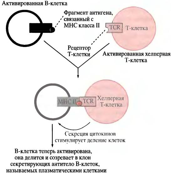
A B cell activated by the binding of an antigen to its surface-bound antibody engulfs the antigen and cleaves it into small fragments, which interact with peptide receptors called MHC proteins. The antigen fragments are then transported to the cell surface and displayed within the binding clefts of the MHC proteins (Fig. 25.5). MHC molecules are Glycoproteins of the Major Histocompatibility Complex (MHC). In this case, the MHC molecules belong to class II (see below) and are expressed by cells in the immune system that function as specialized antigen-presenting cells, including B cells. The processed antigens presented in a complex with MHC molecules on the B cell surface can be recognized by helper T cells bearing receptors specific for the MHC-antigen complex. Helper T cells do not produce antibodies; however, they express antigen-specific surface receptors known as TCR (T CELL RECEPTORS). The TCR recognizes not the free antigen fragment on the APC surface, nor the MHC molecule alone, but specifically the complex of a class II MHC molecule with the antigen fragment. The interaction between the APC and the helper T cell is facilitated by its CD4 protein, which binds to a constant region of the APC cell protein, specifically a class II MHC molecule (Fig. 25.6). The term CD4 stands for cluster of differentiation; it originates from the methodology used to identify specific cell differentiation markers. The number '4' denotes a specific region identified experimentally. The CD4 protein serves as the primary receptor to which the AIDS virus attaches. When a helper T cell, having entered the bloodstream after maturation, encounters and binds to the corresponding MHC-antigen complex displayed on a circulating APC, it becomes activated (see Fig. 25.6). If the activated helper T cell subsequently encounters a B cell displaying the same MHC-antigen complex (originally presented to the T cell by an APC), the T cell binds to this B cell and releases cytokines. These cytokines stimulate B cell proliferation (Fig. 25.7), and the resulting plasma cells (antibody-secreting B lymphocytes) 'pump out' protective antibodies. Plasma cells feature a highly developed endoplasmic reticulum, which is essential for protein secretion (see p. 300).
Fig. 25.5. The process of Activation of a naive B cell into an antibody-producing plasma cell
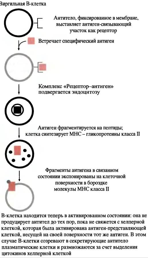
Fig. 25.6. Activation of a helper T cell by an APC (antigen-presenting cell). Autocrine stimulation of the helper T cell enables cells to proliferate after they have separated. CD4 is a glycoprotein located on the T cell that interacts with the MHC class II protein of the APC. This interaction is essential for the binding of the T cell receptor (TCR) to the APC-MHC-antigen complex. The CD4 protein also serves as the receptor utilized by the AIDS virus to infect the helper T cell

Why, in this case, is the antibody secreted rather than retained in the membrane? The answer lies in Alternative Splicing of mRNA introns in immunoglobulin genes (see p. 271). At the 3' end of the immunoglobulin gene, there is a segment encoding a polypeptide sequence that anchors the antibody in the membrane. The onset of secretion is triggered by a switch in the splicing mechanism whereby this segment is excised from the mRNA.
Fig. 25.7. Helper T cell-mediated conversion of a B cell into a clone of antibody-secreting plasma cells. Cytokines are growth factors that stimulate cells to divide. In response to B cell activation—which consequently transforms it into a plasma cell—differential Processing of RNA transcripts leads to the elimination of the antibody segment responsible for membrane anchoring. Cytokines, including those secreted by the helper T cell, are also believed to activate cytotoxic cells
Memory cells
During B cell activation and proliferation, not all cells of the clone differentiate into antibody-secreting plasma cells. Some become long-lived memory cells that can circulate in the body for years, forming The basis of long-term immunity against reinfection. If subsequent contact with the corresponding antigen occurs, the immune system responds very rapidly. T cells also play a key role in establishing immunological memory.
T cells and cell-mediated immunity
As previously mentioned, TCR receptors on helper T cells recognize antigens associated with class II MHC molecules present on APCs and B cells. Another group of T cells—cytotoxic, or killer, T cells—possess receptors that recognize antigens presented on class I MHC molecules. The latter are found on The surface of most somatic cells. Killer T cells interact exclusively with host cells displaying a foreign antigen on their class I MHC molecules. While this rule generally holds true, recent findings indicate that under certain conditions, cytotoxic cells can also recognize class II MHC proteins. Without wishing to confuse the reader, It is important to emphasize the incredible complexity of the immune system, much of which remains poorly understood.
The surface of a killer T cell bears the CD8 glycoprotein, which interacts with the constant region of the class I MHC protein. Because cell binding is mediated by this CD8-MHC interaction, cytotoxic cells exclusively attack cells bearing class I MHC. Cytotoxic cells identify and destroy body cells that have become abnormal, for example, due to a viral infection. Inside a virus-infected cell, viral proteins (synthesized within the cell) are cleaved into Peptides that are displayed on the cell surface in a complex with class I MHC molecules (Fig. 25.8). An antigen-specific killer T cell binds to the antigen-MHC complex of the infected cell and destroys it, either by perforating the membrane (see Fig. 25.8) or by initiating apoptosis.
Fig. 25.8. Sequence of cellular events in response to the appearance of a foreign antigen. The diagram omits the fact that cytotoxic cell division is stimulated by cytokines released during the interaction between a helper T cell and a B cell. This leads to the generation of a clone of cytotoxic cells specific to a given antigen, with a subset of these cells developing into memory cells. The infected cell can be destroyed either via membrane perforation through the release of the protein perforin or as a result of apoptosis

The Role of Cytokines in the Immune System
In Chapter 26, we will discuss how hormone-like signaling molecules facilitate communication between cells. An important class of such molecules is growth factors, which include proteins that signal cells to divide. When a helper T cell is activated by an antigen-presenting cell, it secretes cytokines, including interleukins, which stimulate the proliferation and maturation of both cell types. Cytokines are secreted by helper T cells upon contact with B cells, and some of them participate in the activation of killer T cells. Certain cytokines stimulate phagocytes, others induce antibody class switching in B cells, and some promote cell division and the maturation of B cells into plasma cells. The immunosuppressant cyclosporine, used in organ transplantation, inhibits the production of interleukins by helper T cells.
Why Does the Human Immune System So Fiercely Reject Foreign Human Cells?
At first glance, there is something puzzling about the very existence of an immune attack mechanism against cells transferred from one individual to another. Why does tissue graft rejection occur? After all, evolution did not anticipate such transfers. The primary reason for the rejection of a foreign tissue graft is the presence of MHC molecules on the cell. These molecules form a family of glycoproteins, in which class I and class II are structurally distinct subfamilies. The genes encoding them are clustered within the highly polymorphic major histocompatibility complex. For each gene encoding an MHC protein, there are numerous alleles (variants) within a population. Therefore, it is highly unlikely that the MHC molecules of different individuals will be identical. MHC antigens are the same molecules as the class I and class II antigen-peptide receptors. A foreign MHC molecule is recognized by a killer T cell as a self-MHC molecule complexed with a foreign antigen; consequently, the cell bearing it will be attacked by cytotoxic T cells. As a reminder, the killer T cell receptor recognizes the complex of the MHC molecule and the antigen, rather than each component separately. The evolution of such Variability in MHC molecules within a population could act as a defense for species as a whole against death from infection. Suppose that a virus happens to mutate in such a way that its processed antigen is presented on a host cell's MHC molecule: in this case, it may be recognized by the cellular immune system as normal—or not recognized at all. The virus can then multiply and kill its host. However, the next infected individual is unlikely to possess the same MHC alleles, thereby reducing the likelihood that the events described above will recur. Consequently, despite the death of individual hosts, the disease is unlikely to sweep through the entire population.
Questions for Chapter 25
1. Describe The structure of an IgG molecule.
2. Explain how genes encode a diverse array of different antibodies.
3. What types of lymphocytes do you know, and what are their functions?
4. What is an antigen-presenting cell?
5. When a host cell displays, for example, a viral antigen on its surface, on which class of MHC molecules is it presented? Which class of MHC molecules is recognized by a cytotoxic T cell?
6. What is the "clonal selection theory"?
7. What is the principle behind self-tolerance, or how does the immune system become unresponsive to autoantigens?
8. The production of antibodies following The stimulation of a B cell (to transition into a plasma cell) by a helper T cell does not alter the antigen-binding site of the antibody. However, the antibody must be secreted rather than remain membrane-bound. How is this achieved?
9. When a B lymphocyte (plasma cell) begins secreting an antibody, the latter may initially exhibit a relatively low affinity for its antigen, but this shortcoming is rapidly overcome. How does this happen?
10. Which immune cells incorporate the CD4 glycoprotein, and which incorporate CD8? What is their role in the immune response? What is the relationship between CD4 and the AIDS virus?
Last update: 06/08/2026
Editorial and Educational Adaptation: This material has been compiled based on the primary/original source text. The project team performed an editorial review, corrected technical inaccuracies, structured sections, and adapted the content for an educational format.
What was processed:
- elimination of formatting defects (OCR errors, structural breaks, corrupted characters);
- editorial organization of content;
- standardization of terminology in accordance with academic sources;
- verification of factual statements against the original source text.
All mentions of the author, publication year, and origin of the primary text have been preserved in accordance with the source.