Human Biochemistry, Volume 2 - Murray R. 1993
Structure, Function, and Replication of Information Macromolecules
RNA Synthesis and Processing
Processing of RNA Molecules
In prokaryotes, primary transcripts of mRNA-coding genes begin to serve as templates for Protein Synthesis even before METABOLISM/31.html">Transcription is fully completed. Thus, transcription and Translation are tightly coupled processes in prokaryotes. Prokaryotic rRNA and tRNA transcripts are significantly longer than the corresponding mature RNA molecules; for instance, many tRNA transcripts contain more than one tRNA molecule. Consequently, Processing of primary rRNA and tRNA transcripts is an essential step in The formation of mature RNA molecules in prokaryotic organisms.
Virtually all primary RNA transcripts in eukaryotes undergo complex processing between their synthesis and the onset of their functional activity—whether as mRNA or as independent structural factors such as rRNA, 5SPHK, or tRNA. This processing takes place primarily within The Nucleus itself and involves capping, Cleavage and ligation reactions, The addition of extra terminal NUCLEOTIDES, and nucleoside modification. Between 50% and 75% of mammalian nuclear RNA, including 5'-capped chains, is never incorporated into cytoplasmic mRNA. This volume of intranuclear RNA loss is significantly higher than can be accounted for solely by the removal of non-coding transcript regions (see below). The exact function of this "excess" RNA in mammalian Cell nuclei remains unknown.
With the advancement of restriction mapping and DNA Sequencing techniques, it has been established that in Many eukaryotic genes, extensive DNA stretches devoid of Genetic information directly translatable into protein Amino acid sequences (see Chapter 38) are located between exons (i.e., the coding sequence fragments). Such intervening sequences, or introns, are found in most, though not all, higher eukaryotic genes. The primary transcripts of structural genes include regions corresponding to these introns. During a process known as "splicing", these primary transcript segments are precisely excised, and the corresponding exons are joined together. This process occurs in the nucleus, after which the assembled mRNA molecules are exported to the Cytoplasm, where translation takes place (Fig. 39.9).
The exact mechanisms ensuring the error-free excision of introns, the ligation of exons, and The transport of RNA to the cytoplasm remain elusive. However, recent studies have provided a wealth of new insights into these processes. Although nucleotide sequences within introns are highly heterogeneous even within a single transcript, a consensus sequence can be identified for each of the two intron-exon junction sites (splicing sites) (Fig. 39.10). The consensus sequence at the intron-exon boundary is not unique enough to guarantee strictly specific cleavage mediated solely by any specialized endonuclease. Abundantly present small nuclear RNA (U1 RNA) contains a sequence complementary to the consensus sequence of the splice site (Fig. 39.11). Furthermore, it has been established that U1 RNA molecules in The eukaryotic nucleus specifically bind to certain protein factors. Such RNA-Structure/178.html">Protein Complexes selectively associate with the 5'- and 3'-splice site sequences in the RNA. Antibodies directed against the U1 protein complex inhibit intron excision in vitro.
Class="center">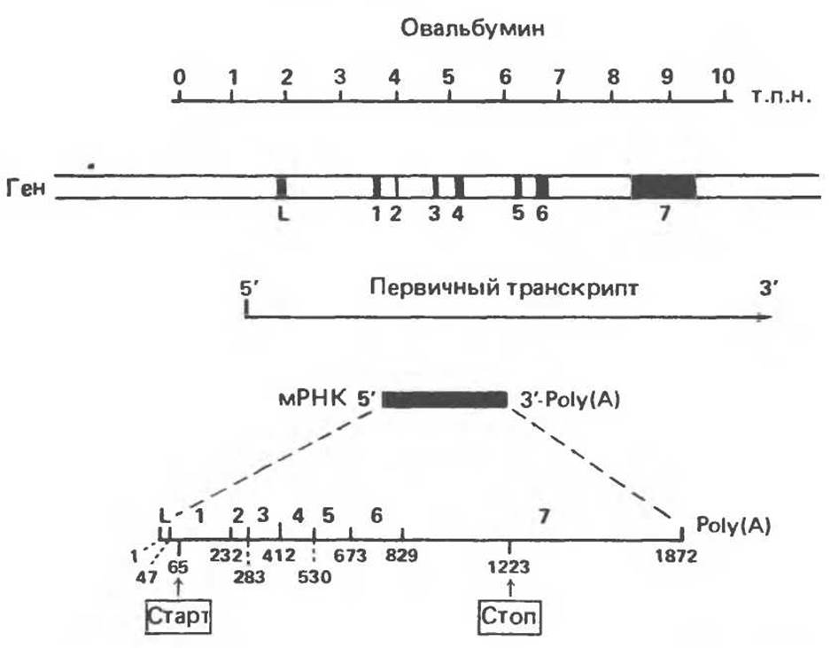
Fig. 39.9. Arrangement of coding and non-coding sequences (introns) in the chicken Ovalbumin Gene structure. Informational segments that become part of the mature mRNA are numbered and highlighted in black. The primary transcript initiates to the left of the L-exon and terminates in the 3'-untranslated region beyond exon 7. The structure of the mature mRNA is shown at the bottom of the figure; exon numbers are indicated above it, while nucleotide positions, the Translation initiation site, and the stop codon position are shown below it.
Interestingly, patients with the autoimmune disease systemic lupus erythematosus possess antibodies directed against certain specific U1 protein complexes. The direct link between this phenomenon and the Pathogenesis of the disease remains unclear.
It has recently been discovered that during the removal of introns from mRNA precursor molecules, an unusual lariat-like structure is formed. Specifically, the 5'-end of the intron sequence is joined by a 2'–5' phosphodiester bond to an adenylate residue located 28–37 nucleotides upstream from its 3'-end. This process and the corresponding structures are schematically illustrated in Fig. 39.12.
As for the enigma of the relationship between hnRNA and the corresponding mature mRNAs, it can now be considered solved. Heterogeneous nuclear RNA represents primary transcripts plus molecules undergoing Cytology/cytology/16.html">Early stages of processing, which—following capping, poly(A) tail addition, and intron removal—are transported to the cytoplasm as mature mRNAs.
hnRNA processing represents yet another potential regulatory checkpoint in Gene Expression. For instance, Alternative Splicing has been demonstrated for a single primary transcript. To give an example, rat salivary gland and Liver α-amylase mRNAs differ in the structure of their 5'-terminal sequence regions, whereas the remaining mRNA segments, including the coding region and the polyadenylation site, are identical. Further analysis revealed that different splice sites are utilized to attach two distinct capped leader sequences to the same mRNA "body". Another example of alternative splicing is the generation of mRNA molecules encoding two immunoglobulin heavy chains: one mRNA molecule encodes a membrane-bound heavy chain, whereas the other encodes a secreted heavy chain (see Chapter 41). Thus, splicing is essential for the maturation of mRNA molecules and can additionally serve as one of the mechanisms driving differential gene expression.
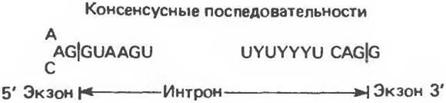
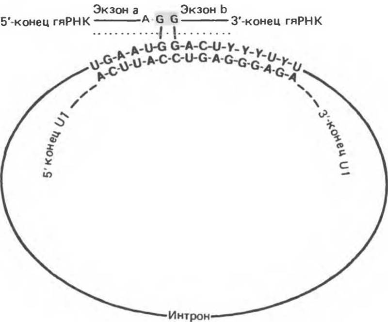
Fig. 39.11. Proposed mechanism of splice site identification during intron removal from hnRNA. The 5'-end of U1 snRNA forms a complementary complex with the distal end of the splice site consensus sequence at the 3'-end of exon a. The other end of the U1 RNA interacts with the consensus splice site of exon b. The structure indicated by the dotted line is excised, and the molecule is spliced across the G residues (shaded box).
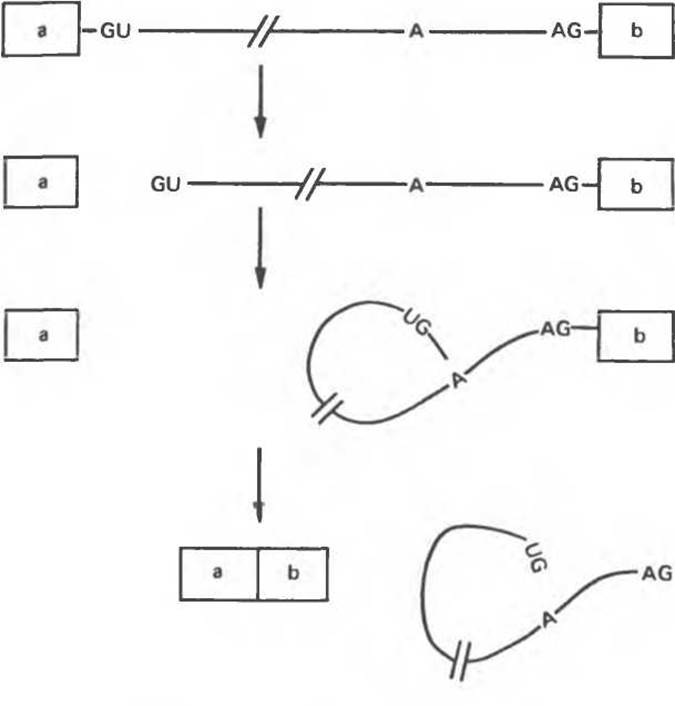
Fig. 39.12. Proposed pathway of pre-mRNA splicing. Cleavage at the 5' site is accompanied by lariat formation and its subsequent release via cleavage from exon b. The intron is depicted as a line, and exons a and b as squares. These reactions occur with the participation of snRNAs and pre-mRNA assembled into a stable complex that forms part of a ribonucleoprotein structure termed the "spliceosome."
It turns out that at least one form of ß-thalassemia—a disease characterized by a markedly reduced expression level of one of the Hemoglobin chains—results from a nucleotide Substitution at the intron-exon boundary, which prevents intron removal and leads to a decrease or complete suppression of ß-chain synthesis.
Messenger RNAs (mRNAs)
As mentioned above, mammalian mRNA molecules contain a "capping" structure at the 3'-end and, in most cases, a polyadenylate "tail" at the 3'-end. The capping structure is added to mRNA in the nucleus prior to mRNA export to the cytoplasm. The polyA structure is attached to the 3'-end of the transcript either in the nucleus or in the cytoplasm. Secondary methylation of the mRNA molecule, including 2'-hydroxyl groups and N6 atoms of adenylate residues, occurs after the RNA molecule enters the cytoplasm. This process can also take place in the nucleus and may play a role in splicing. The 5'-capping structure appears to be required for the formation of a nucleoprotein complex, which in turn is necessary for splicing to occur. In addition, it may be involved in mRNA transport and translation initiation.
The function of the mRNA polyadenylate "tail" remains unknown. In many cases, the presence or absence of polyA is not directly linked to cytoplasmic transport, since not all polyadenylated Heterogeneous nuclear RNAs are exported to the cytoplasm, and not all cytoplasmic mRNAs are polyadenylated. In mammalian Cells, mRNA polyadenylate "tails" can undergo both lengthening and shortening during cytoplasmic processes.
The turnover of polyA-containing mRNAs in cultured mammalian cells follows a first-order process with a t1/2 value close to The Cell doubling time in culture. The degradation kinetics of histone mRNAs, which lack polyA structures, follows a zero-order process characterized by age-dependent decay with a half-life of about 6 hours. It remains unclear whether these differences are related to the presence or absence of terminal polyA sequences or to other Structural Features of this class of mRNA molecules.
Even after the removal of the polyA chain, the size of cytoplasmic mRNA molecules remains significantly larger (often 2- to 3-fold) than required to encode the corresponding polypeptide. Excess untranslated regions are present at both the 5'- and 3'-ends of the translated segment, with the 3'-untranslated region typically being much longer. The exact function of these sequences is unknown, though there is evidence suggesting they participate in RNA Processing, transport, degradation, and translation.
Transfer RNAs (tRNAs)
As described in Chapters 37 and 40, tRNA molecules function as adaptor molecules during the translation of mRNA into protein sequences. tRNA molecules contain numerous unusual ("minor") nucleic acid bases. Some of these are methylated derivatives of common bases, while others contain non-traditional glycosidic bonds. Both prokaryotic and eukaryotic tRNA molecules are initially transcribed as large precursors that frequently contain more than one tRNA molecule, which undergo nucleolytic processing by a specialized class of ribonucleases. Furthermore, the genes for certain tRNAs contain a single intron, 10–40 nucleotides in length, located immediately upstream of the region corresponding to the anticodon arm. Consequently, the processing of primary transcripts for many tRNA molecules must include an intron removal step followed by precise splicing in the codon-recognition region. This step is critical for tRNAs to function as adaptors in protein synthesis. The nucleolytic processing of tRNA precursors appears to be guided not by The nucleotide sequence itself, but by the specific three-dimensional structure that tRNA molecules can adopt, and thus it occurs exclusively on molecules capable of folding into proper functional structures.
Further modifications of tRNA molecules include nucleotide alkylation and the addition of a characteristic CCA triplet to the 3'-end of the molecule. This triplet serves as the attachment site for the specific amino acid directed by the given tRNA molecule into the polypeptide synthesis reaction. Methylation of mammalian tRNA precursors likely takes place in the nucleus, whereas cleavage and addition of the CCA triplet occur in the cytoplasm, as the turnover rate for the terminal portion of tRNA is higher than that for the molecule as a whole. The attachment of an amino acid to the CCA terminus requires specific Enzymes found in the cytoplasm of mammalian cells.
Ribosomal RNAs (rRNAs)
In mammalian cells, ribosomal RNA molecules (two major and one minor) are transcribed as part of a large common primary transcript (Fig. 39.13). The processing of this transcript to yield mature rRNAs, which are then transported to the cytoplasm, takes place in the nucleolus, where the ribosomal RNA genes themselves are localized. Hundreds of copies of these genes are present in each cell. The transcription units contain the 18S, 5.8S, and 28S rRNA sequences arranged consecutively in the 5'→3' direction. The 45S primary transcript undergoes extensive methylation directly within the nucleolus. In this 45S precursor, the region corresponding to the 28S rRNA contains 65 methylated ribose residues and 5 methylated nucleic acid bases. Methylation occurs exclusively in the regions that subsequently form mature rRNA molecules. Although the 45S precursor undergoes nucleolytic processing, the processing signals differ markedly from those found in hnRNA. The Mechanism of processing presumably also differs from the nucleolytic processing mechanism involved in hnRNA maturation.
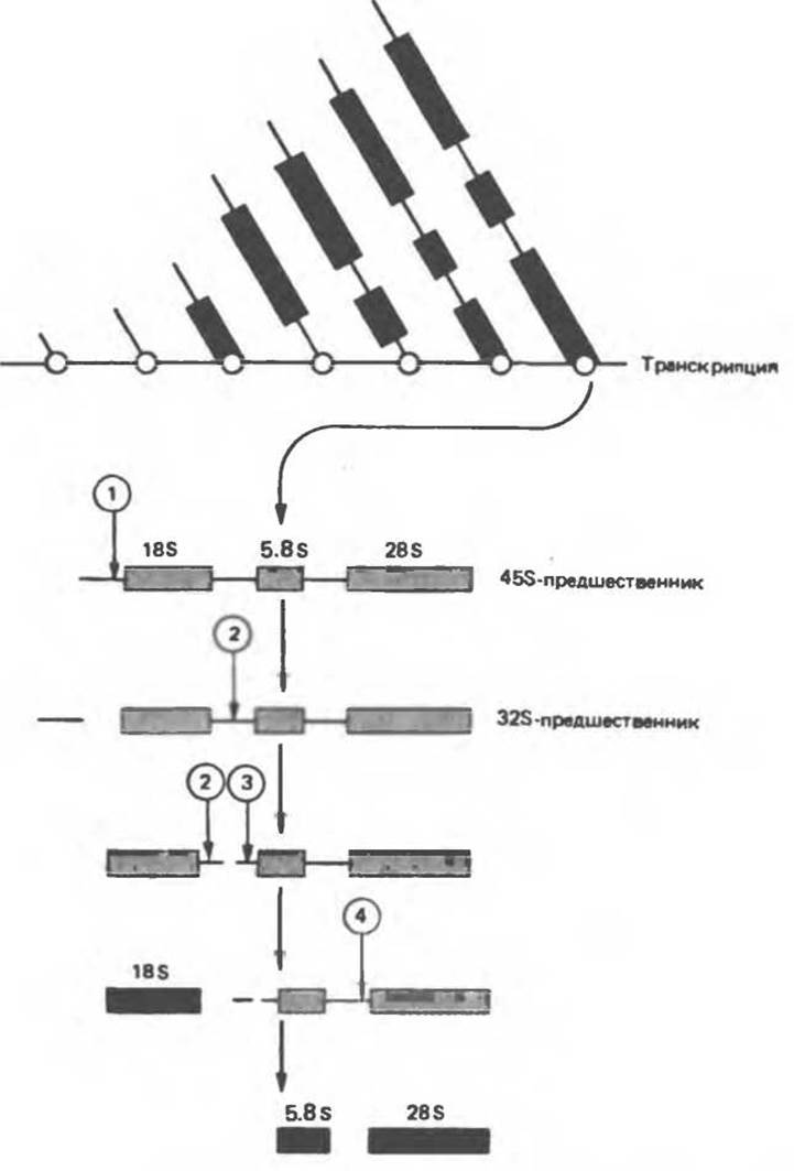
Fig. 39.13. Diagram of mature ribosomal RNA formation during the processing of precursor RNA molecules. The final product is indicated by solid rectangles. (Reproduced, with permission from Perry R. P.: Processing of RNA Annu. Rev. Biochem. 1976, 45:605.)
Nearly half of the original primary transcript (Fig. 39.13) undergoes degradation. During rRNA Processing in the nucleoli, further methylation takes place, and there the 28S rRNA associates with ribosomal Proteins to assemble the large 60S ribosomal subunit. The 5.8S rRNA molecule is also generated in the nucleolus and becomes an integral component of the large ribosomal subunit. The 18S rRNA molecule, complexed with a set of specific Polypeptides, forms the small 40S ribosomal subunit.
Last update: 06/08/2026
Editorial and Educational Adaptation: This material has been compiled based on the primary/original source text. The project team performed an editorial review, corrected technical inaccuracies, structured sections, and adapted the content for an educational format.
What was processed:
- elimination of formatting defects (OCR errors, structural breaks, corrupted characters);
- editorial organization of content;
- standardization of terminology in accordance with academic sources;
- verification of factual statements against the original source text.
All mentions of the author, publication year, and origin of the primary text have been preserved in accordance with the source.