Glycoproteins - Hughes R. 1985
Structure
Structure of N-glycans
Ovalbumin
For a long time, this major chicken egg glycoprotein has been a favored model for structural studies. It can be isolated in a pure crystalline form, i.e., as a preparation consisting of a single specific polypeptide with a single asparagine residue located at a defined position in the chain. It is precisely to this residue that the carbohydrate component is attached. The relatively recent Determination of the complete carbohydrate chain sequence of ovalbumin has significantly influenced MODERN CONCEPTS OF the biosynthetic Assembly of N-glycans in general.
The Structure of the carbohydrate component of ovalbumin was analyzed According to the scheme described above (Table 2.2). The glycopeptide fraction can be isolated by Gel filtration from the hydrolyzate obtained after exhaustive proteolysis of ovalbumin with a nonspecific protease, pronase. The Amino Acids and small Peptides resulting from pronase proteolysis are effectively separated from the asparagine-carbohydrate complex, which has a higher molecular weight. One might expect that the carbon-containing fragment obtained using pronase would be a homogeneous substance or at least a relatively simple mixture, since ovalbumin contains a unique polypeptide to which only a single carbohydrate chain is attached. However, this turned out not to be the case: the glycopeptide fraction can be separated into at least six components (I–VI) by Ion-exchange Chromatography [9]. This important observation suggested that the carbohydrate component attached to a specific amino acid residue may exhibit structural heterogeneity. Initially, this seemed surprising, and considerable effort was made to rule out several potential conventional causes for such heterogeneity: 1) similar heterogeneity was found in ovalbumin isolated from eggs of the same hen; 2) all genetic variants of chickens possessed the same set of glycopeptides; 3) the relative composition of glycopeptides was independent of egg age; and 4) since chicken egg white exhibits glycosidase activity due to the presence of a-mannosidases and ß-N-acetylglucosaminidases, the albumins of eggs from birds (such as turkeys) in which egg white glycosidase activity is either entirely absent or negligible were examined. It was found that multiple glycopeptide fractions are still detected in this case. Consequently, their presence cannot be an artifact arising from the Cleavage of a single initial carbohydrate chain by glycosidases.
Class="center">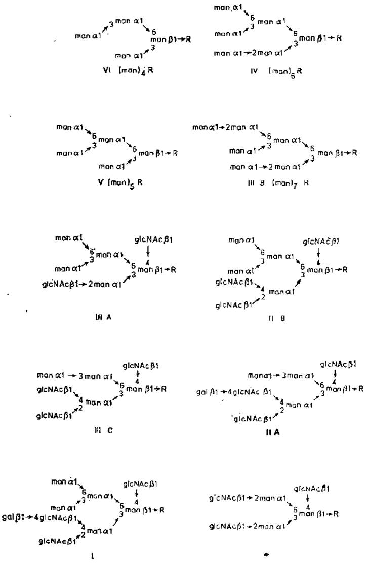
Fig. 2.6. Structure of ovalbumin glycopeptides. R — glcNAcß1→4glcNAc.Asn. The asterisk denotes the minor component of fraction V isolated by Electrophoresis [10].
All six isolated glycopeptide fractions contain only a single amino acid, asparagine. Their monosaccharide composition is also very similar: mannose and N-acetylglucosamine are always found as the major (fractions I and II) or sole (fractions III, IV, V, and VI) components. Fractions II–IV can be subdivided into two or three subfractions [10]. The complete structure of each major ovalbumin glycopeptide was elucidated by a combination of physical, chemical, and enzymatic Methods (Fig. 2.6). Strikingly, all glycopeptides share a common region (the "core") linked to asparagine. Glycopeptides VI, V, IV, and IIIB form a group of very similar oligomannose, or mannose-rich structures containing 1 to 7 mannose residues, as well as chitobiose (N-acetylglucosaminyl ß1→N-acetylglucosamine) attached to asparagine. In each case, two mannosyl residues are attached via â-glycosidic bonds to the hydroxyl groups at C-3 and C-5 of the mannose residue that is linked by a ß-glycosidic bond to chitobiose. Further elongation of the carbohydrate chain occurs via The addition of other mannose residues to these â-mannosyl units. The presence of terminal â-mannosyl residues can be readily detected by treating the glycopeptides with â-mannosidases and subsequently determining The amount of liberated mannose. Following such Treatment of glycopeptides V and IV, exactly 4 and 5 residues per mole are released, respectively, as predicted from the structure shown in Fig. 2.6. In each case, Hydrolysis halts upon reaching the mannose residue linked by a ß-glycosidic bond to N-acetylglucosamine. The exact same trisaccharide linked to asparagine remains in all cases, having the following structure: manß1→4glcNacâl→4glcNAc. In contrast, treatment with exo-ß-N-acetylglucosaminidase fails to reveal any free N-acetylglucosamine residues. As mentioned previously, exoglycosidases can act only on terminal non-reducing residues; therefore, the N-acetylglucosamine residues constituting the chitobiose core region are resistant to these Enzymes. Data obtained from exoglycosidase experiments are in full agreement with the structures of other ovalbumin glycopeptides. Following treatment of fractions I, II, and III with exo-ß-N-acetylglucosaminidase, several N-acetylglucosamine residues can be detected as free sugars, whereas a minor fraction of the mannose residues remains sensitive to â-mannosidase. In such hybrid chains, terminal ß-N-acetylglucosamine residues occupy two main positions: they are attached either 1) via a (ß1→4) bond to a mannose residue of the core region, or 2) via (ß1→2) or both (ß1→2)- and (ß1→4)-bonds to the â-mannosyl residues adjacent to the core region. In the latter case, some of the N-acetylglucosamine units are in turn substituted with galactose to form an N-acetyllactosamine sequence. The disaccharide galß1→4gIcNAc was first discovered in the molecule of Orosomucoid (also known as a1-acid glycoprotein).
Last update: 06/08/2026
Editorial and Educational Adaptation: This material has been compiled based on the primary/original source text. The project team performed an editorial review, corrected technical inaccuracies, structured sections, and adapted the content for an educational format.
What was processed:
- elimination of formatting defects (OCR errors, structural breaks, corrupted characters);
- editorial organization of content;
- standardization of terminology in accordance with academic sources;
- verification of factual statements against the original source text.
All mentions of the author, publication year, and origin of the primary text have been preserved in accordance with the source.