Glycoproteins - Hughes R. 1985
Structure
Structure of N-glycans
Orosomucoid
Some Structural Features of this important serum protein have already been discussed above. Its single polypeptide chain contains five glycosylated asparagine residues, located as shown in Fig. 2.10. To date, the complete sequences of the desialylated N-glycans of orosomucoid have been described, along with their attachment sites to the polypeptide chain. Before cleaving orosomucoid into individual glycopeptides with Chymotrypsin, the sialic acid is removed. Because Chymotrypsin has a rather narrow Specificity, this Procedure yields a mixture of glycopeptides, each possessing one of five unique Amino acid sequences whose locations within the peptide chain are easily established. To improve the subsequent Separation of the sixteen glycopeptides using Ion-exchange Chromatography, the sialic acids are removed, which allows exploiting differences in the net charge of the Peptides and the sizes of their carbohydrate chains. High-resolution NMR spectroscopy was employed to elucidate The Structure of the glycopeptides. In each case, only 2–3 mg of glycopeptides was required for the determination, which further highlights the great value of this analytical method [14].
Class="center">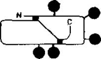
Fig. 2.10. Glycosylation sites (black circles) of the orosomucoid polypeptide chain. The black squares represent two intrachain Disulfide Bonds.
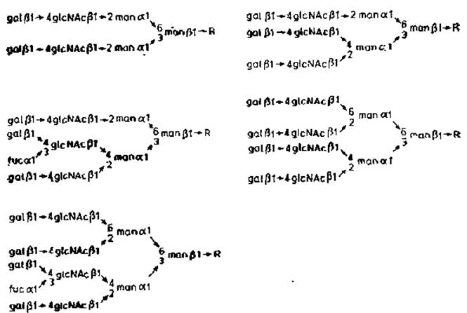
Fig. 2.11. Structure of orosomucoid N-glycans. R — glcNAcß→4glcNAc. Asn.
As a result of the described experiments, some previously unknown properties were discovered in various glycopeptides. In addition, these same experiments confirmed data obtained earlier from the Cleavage of intact Glycoproteins (Section 2.2.1). Sialic acid residues are predominantly attached to N-acetyllactosamine units located at the termini of branched Oligosaccharides. Orosomucoid lacks oligomannose chains, and its carbohydrate components belong to the "complex" type, containing multiple neuNAc.gal.glcNAc units attached to a common core region consisting of two a-mannose residues, one ß-mannose residue, and one chitobiose residue. Similarly to the complex chains of immunoglobulin G, there is also a clear Asymmetry in the structures containing three or more terminal N-acetyllactosamine residues. One of the a-mannose residues linked to C-3 of the ß-mannose residue is substituted at C-2 and C-4, whereas the other is substituted at C-2 and C-6. The reason for this asymmetry is most likely the substrate Specificity of the various N-acetylglucosaminyltransferases responsible for adding each N-acetylglucosamine residue to the precursor chain during chain assembly. Available data on the substrate specificity of two such Enzymes are discussed in Chapter 3.
Another characteristic feature of orosomucoid N-glycans was the presence of fucose attached via an (a1→3)-bond to a specific N-acetylglucosamine residue. The fact that strictly defined N-acetylglucosamine residues in the branched chains are substituted once again points to the very narrow specificity of a1→3-fucosyltransferases. Unlike immunoglobulin G, fucose in orosomucoid is not attached to chitobiose.
From the Brief Overview of our knowledge regarding N-glycan structure, the following Conclusions can be drawn: 1) the Assembly of N-glycans is carried out through the coordinated action of many Glycosyltransferases, each possessing a very narrow substrate specificity; 2) The activity of these enzymes varies noticeably depending on which region of the polypeptide chain undergoes glycosylation. Studies on The Biosynthesis of N-glycans have provided further confirmation of these conclusions. This issue will be discussed in the next chapter.
Last update: 06/08/2026
Editorial and Educational Adaptation: This material has been compiled based on the primary/original source text. The project team performed an editorial review, corrected technical inaccuracies, structured sections, and adapted the content for an educational format.
What was processed:
- elimination of formatting defects (OCR errors, structural breaks, corrupted characters);
- editorial organization of content;
- standardization of terminology in accordance with academic sources;
- verification of factual statements against the original source text.
All mentions of the author, publication year, and origin of the primary text have been preserved in accordance with the source.