BIOCHEMISTRY - L. Stryer - 1984
VOLUME 1
PART I. CONFORMATION AND DYNAMICS
CHAPTER 8. ZYMOGEN ACTIVATION: DIGESTIVE ENZYMES AND COAGULATION FACTORS
8.8. Chymotrypsin has a deep pocket for binding aromatic side chains
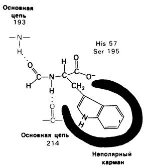
The localization of specific binding sites and the probable orientation of the hydrolyzable peptide bond of an effective substrate were revealed by X-ray crystallographic analysis of Chymotrypsin complexes with substrate analogs. It was established that formyl-L-Tryptophan binds to chymotrypsin via its indole side chain, which fits well in size into a pocket on the enzyme near Serine-195 (Fig. 8.16). The presence of this deep pocket explains the Specificity of chymotrypsin for Amino Acids with aromatic or other large hydrophobic side chains. X-ray crystallographic analysis of chymotrypsin complexes with polypeptide substrate analogs revealed A large number of Hydrogen Bonds between the backbones of the enzyme and the substrate, and these Hydrogen bonds are arranged in the same way as in antiparallel β-pleated sheets.
Class="center">Fig. 8.16. Schematic representation of the binding of formyl-L-tryptophan (a substrate analog) to chymotrypsin
8.9. A transient tetrahedral intermediate is formed during catalysis
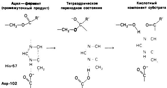
Extensive X-ray crystallographic and chemical studies of chymotrypsin have led to a definite Conclusion regarding its catalytic mechanism. Apparently, Histidine-57 and serine-195 are directly involved in the Cleavage of the substrate's peptide bond. Hydrolysis of this bond begins when the oxygen atom of the serine-195 OH group attacks the carbonyl carbon atom in the hydrolyzable peptide bond of the substrate. As a result, the bond between the Carbon and Oxygen atoms in this carbonyl group becomes a single bond, and the oxygen atom acquires a negative charge. The four atoms bonded to the carbonyl carbon are arranged in a tetrahedron. The formation of this tetrahedral intermediate from the planar amide group is made possible only by the occurrence of hydrogen bonds between the negatively charged oxygen atom of the hydroxyl group (called the oxyanion) and two NH groups of the polypeptide chain itself (Fig. 8.17). A proton transfer from serine-195 to histidine-57 also plays an important role in the Mechanism of formation of this intermediate (Fig. 8.18). This proton transfer is greatly facilitated by the presence of a charge-Relay system. Aspartate-102 strictly orients the imidazole ring of histidine-57 and partially neutralizes the charge that appears on this ring during the Transition State. The proton accumulated by the histidine-aspartate pair is then transferred to the nitrogen atom of the hydrolyzable peptide bond, which consequently cleaves. At this stage, the amine component of the substrate is hydrogen-bonded to histidine-57, and the acid component is ester-linked to serine-195. This completes the acylation step of the hydrolytic reaction.
Fig. 8.17. Tetrahedral intermediate in the acylation and deacylation reactions of chymotrypsin. The Stability of the intermediate is provided by hydrogen bonds formed by the NH groups of the enzyme backbone. This region is called the oxyanion hole

Fig. 8.18. The first step of peptide hydrolysis by chymotrypsin is acylation. A tetrahedral intermediate is formed. Then, the amine component of the substrate rapidly dissociates from the enzyme, and the enzyme is converted into an acyl-enzyme, a catalytic intermediate

The next step is deacylation (Fig. 8.19). The amine component of the substrate diffuses away from the enzyme, and its place in the Active Site is taken by a molecule of
Water. In essence, deacylation is the reverse of acylation, but with the amine component of the substrate replaced by H2O. First, the charge-relay system abstracts a proton from water. The resulting OH- ion immediately attacks the carbonyl carbon atom of the acyl group attached to serine-195. As in acylation, a tetrahedral intermediate is formed. Next, histidine-57 transfers a proton to the oxygen atom of serine-195, leading to the release of the acid component of the substrate. This component diffuses away from the enzyme, which is then ready to enter a new catalytic cycle.
Fig. 8.19. The second step of peptide hydrolysis by chymotrypsin is deacylation. The acyl-enzyme intermediate is hydrolyzed by water. Deacylation is essentially the reverse of acylation, but H2O takes THE PLACE OF the amine component of the substrate
8.10. Mechanism of zymogen activation
Let us now turn to the question of how the cleavage of just a single peptide bond in chymotrypsinogen converts it into an active enzyme. The three-dimensional Structure of chymotrypsinogen was investigated by Joseph Kraut, who established that the enzyme undergoes a series of conformational changes during activation.
1. Hydrolysis of the peptide bond between Arginine-15 and isoleucine-16 generates new C- and N-terminal groups.
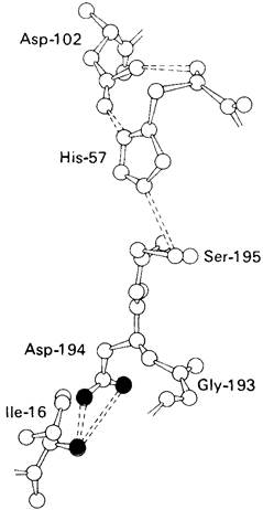
2. The newly formed N-terminal group of isoleucine-16 folds inward and interacts with aspartate-194 inside the chymotrypsin molecule (Fig. 8.20).
Protonation of this amino group stabilizes the active form of chymotrypsin, as evidenced by the pH Dependence of enzymatic activity.
Fig. 8.20. The environment of aspartate-194 and isoleucine-16 in chymotrypsin. The electrostatic interaction between the carboxylate ion of Asp-194 (red) and the α-NH2 group of Ile-16 (blue) plays an important role in the catalytic activity of chymotrypsin. These groups are located in close proximity to the charge-relay system
3. This electrostatic interaction between the positively charged amino group and the negatively charged carboxylate ion, occurring in the nonpolar region of the enzyme, triggers a series of conformational shifts. Methionine-192 moves from the interior of the molecule closer to the surface, while residues 187 and 193 become more extended. As a result, a substrate-specific site for aromatic and large nonpolar groups is formed. One side of this site is made up of residues 189 to 192. In the proenzyme, this substrate-binding pocket is not fully formed.
4. The tetrahedral transition state arising during chymotrypsin catalysis is stabilized by hydrogen bonds between the negatively charged oxygen atom of the carbonyl group and two NH groups of the polypeptide chain itself (Fig. 8.17). In chymotrypsinogen, one of these NH groups is positioned so that it is inaccessible for binding. Consequently, the oxyanion hole in the zymogen is also not fully formed.
5. Conformational shifts in the rest of the molecule are very minor. Thus, the "switching on" of enzymatic activity in the protein is achieved through discrete, strictly localized conformational shifts triggered by the hydrolysis of just a single peptide bond.
Last update: 06/08/2026
Editorial and Educational Adaptation: This material has been compiled based on the primary/original source text. The project team performed an editorial review, corrected technical inaccuracies, structured sections, and adapted the content for an educational format.
What was processed:
- elimination of formatting defects (OCR errors, structural breaks, corrupted characters);
- editorial organization of content;
- standardization of terminology in accordance with academic sources;
- verification of factual statements against the original source text.
All mentions of the author, publication year, and origin of the primary text have been preserved in accordance with the source.