Glycoproteins - Hughes R. 1985
Structure
Structure of N-glycans
Immunoglobulins
All five classes of human IMMUNOGLOBULINS, and presumably all other immunoglobulins as well, contain CARBOHYDRATES [11]. Although the amount and composition of these carbohydrates may vary across different immunoglobulin classes, the molecules of all these Proteins contain N-glycans located within the constant (Fc) region of their heavy chains. Notably, a significant degree of Homology is observed among the segments of the Fc region to which the carbohydrate moieties are attached (Fig. 2.7).
Class="center">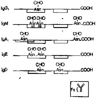
Fig. 2.7. Glycosylation sites and intrachain disulfide bridges in the heavy chains of immunoglobulins. The inset schematically illustrates an intact rabbit immunoglobulin molecule. CHO represents carbohydrate chains. Carbohydrate components (a) and (b) of immunoglobulin M are discussed in the text.
The very fact that homologous carbohydrate chains are localized in similar Regions of the Fc domain across all immunoglobulins strongly suggests that the Amino Acid Sequence surrounding the asparagine residues varies very little. Apparently, this sequence holds considerable Structural and functional importance, most likely serving as the attachment site for functionally vital glycans.
Detailed data are now available on The Structure of two carbohydrate chains from human immunoglobulin M and one chain from human immunoglobulin G. The research utilized immunoglobulins isolated from the serum of two myeloma patients. These proteins should be regarded as secretion products of cellular clones, encoded in each instance by specific genes. Nevertheless, even in these cases, a marked heterogeneity in carbohydrate structure was found, similar to the aforementioned glycan heterogeneity in Ovalbumin.
In the human myeloma immunoglobulin M molecule, each asparagine residue is associated with carbohydrate chains of two main types: chains containing only mannose and N-acetylglucosamine, and chains containing, in addition, sialic acid, galactose, and fucose. The STRUCTURE OF THE second type of chains has not yet been fully elucidated. The structure of the oligomannoside chains is shown in Fig. 2.8. Two important observations should be made. First, all sequences are built by adding a-mannose residues to the core trisaccharide region manß1→HglcNAcß1→4HglcNAc, with the initial addition of one mannose residue to C-3, followed by three mannose residues at C-6 of the central ß-mannosyl residue. This sequence, (man)5R, is structurally identical to the ovalbumin glycopeptide (Fig. 2.6). Structures identical to ovalbumin glycopeptides IIIB and IV were detected among the immunoglobulin carbohydrate chains (minor component (b) and major component (b), respectively). Furthermore, immunoglobulin M was found to contain larger structures with a higher content of a-mannosyl units than ovalbumin glycopeptides: specifically, two (man)8R isomers and one (man)9R sequence. Second, it is crucial to emphasize the presence of glycosylation Asymmetry in regions (a) and (b) of the immunoglobulin M heavy chain. This is best explained by the presence, in regions (a) and (b), of carbohydrate components containing 6 mannose residues each. Five of these residues are identically positioned in both cases, whereas an additional one (a1→2) is attached to different residues of the carbohydrate chains in regions (a) and (b). A similar asymmetry is observed in chains containing 7 and 8 mannose residues; they also differ in the positioning of a single a-mannosyl residue. The reasons for these subtle structural differences among glycans that share a similar overall structure and are attached to adjacent asparagine residues within the same polypeptide chain are not yet fully understood. However, as will be discussed in detail in the next chapter, the described structure of the oligomannose chains in this human immunoglobulin can be rationalized in terms of the Biosynthesis mechanism of N-glycans.
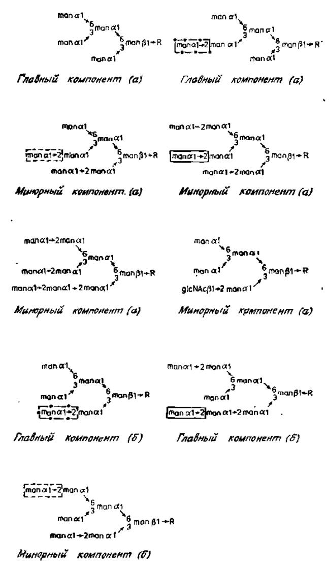
Fig. 2.8. Structure of N-glycans from human immunoglobulin M. R — glсNAcß1→4glcNAc. Asn; (a) and (b) are two glycosylation sites of the Fc region shown in Fig. 2.7. Variable a-mannose residues are enclosed in boxes [13].
Finally, mention should be made of the minor component of region (a), which contains a terminal N-acetylglucosamine residue. As will be discussed below, this carbohydrate serves as a crucial intermediate in the assembly of "complex" glycans containing galactose, fucose, and sialic acid. Such glycans have been identified in immunoglobulins and other Glycoproteins.
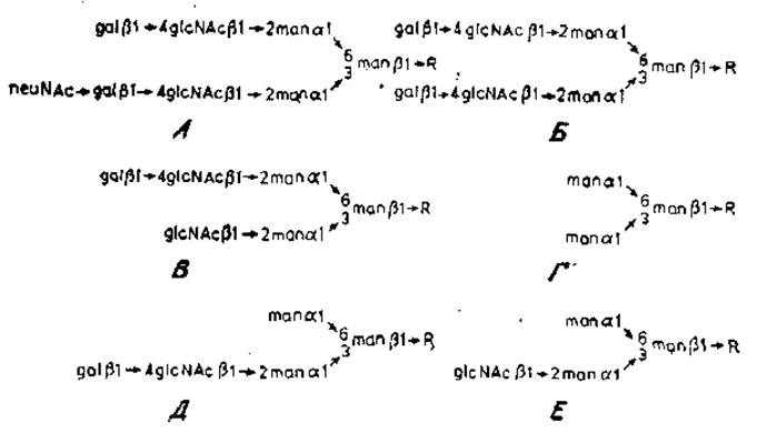
Fig. 2.9. Structure of glycopeptides from human myeloma immunoglobulin G and the products of their Cleavage by exoglycosidases. R — glcNAcß1→4 (fuca1→6) glcNAc. Asn.
Figure 2.9 [10] illustrates the structure of the carbohydrate moieties of glycopeptides isolated from the proteolytic hydrolysate of immunoglobulin G. This protein was obtained from the serum of a patient with multiple myeloma. The glycopeptides were separated using Dowex-50 Column Chromatography. The acidic glycopeptide (A) eluted first, followed by a mixture of neutral glycopeptides (B and C), which in turn could be fractionated for subsequent structural analysis. Sequential Treatment of all these glycopeptides with neuraminidase (for A only), ß-galactosidase, and ß-N-acetylglucosaminidase yielded the same core-attached oligosaccharide (D) containing two a-mannose residues. Evidently, the high-mannose oligosaccharide present in ovalbumin is not a major component of immunoglobulin G. However, N-acetylglucosamine and galactose residues are attached to the two a-mannose residues, in some cases via the same linkages found in ovalbumin glycopeptides (e.g., in fraction I). An interesting feature of immunoglobulin glycopeptide A is its asymmetry. A single N-acetylneuraminic acid residue is linked to the side chain attached via an (a1→3) bond to the central ß-mannose residue of the core region. The existence of such a structure raises questions regarding the Specificity of the Glycosyltransferases that attach sialic acid residues to the terminal galactose of these carbohydrate chains (this topic is addressed in the next chapter). Such an asymmetric structure is of particular significance because it acts as a substrate for specific glycosyltransferases. Treatment of acidic glycopeptide A with ß-galactosidase, followed by ß-N-acetylglucosaminidase and subsequently neuraminidase, yields carbohydrate chain D. Subsequent Treatment of the latter with ß-galactosidase produces chain E. Typically, structures D and E are not found in native immunoglobulins M, although, as noted earlier, a carbohydrate analogous to E has been detected in immunoglobulin M. Another characteristic feature of these N-glycans is the presence of a fucose residue linked via an a1→6 bond to the chitobiose core (to the first residue in the chain). It is important to note that the described glycopeptides are constituents of the immunoglobulins G secreted by the specific myeloma Cell line used in these experiments [10, 11]. Each myeloma is distinct, and they all produce rather diverse repertoires of immunoglobulin glycans.
Last update: 06/08/2026
Editorial and Educational Adaptation: This material has been compiled based on the primary/original source text. The project team performed an editorial review, corrected technical inaccuracies, structured sections, and adapted the content for an educational format.
What was processed:
- elimination of formatting defects (OCR errors, structural breaks, corrupted characters);
- editorial organization of content;
- standardization of terminology in accordance with academic sources;
- verification of factual statements against the original source text.
All mentions of the author, publication year, and origin of the primary text have been preserved in accordance with the source.