BIOCHEMISTRY AND MOLECULAR BIOLOGY - W. ELLIOTT - 2002
CHAPTER 24. GENE CLONING, RECOMBINANT DNA TECHNOLOGY, GENETIC ENGINEERING
While the Structure/179.html">Practical Applications OF biochemical advances are not the primary focus of this book, DNA-based techniques have truly revolutionized biology and biochemistry in particular. Therefore, we deemed it appropriate to dedicate a separate chapter to modern molecular biology Methods.
These techniques have made it possible to isolate individual genes, determine their base sequences, and transfer genes between different species, among other applications. Knowing The base sequence of a coding region allows us to deduce the Amino Acid Sequence of the protein encoded by that Gene.
Elucidating the Principles of Gene function and regulatory mechanisms is impossible without structural data. Much of the information regarding the Introduction/30.html">Regulation of Gene Expression discussed in Chapter 21 was obtained through recombinant DNA experiments.
An isolated gene can be introduced into A wide variety of Cell types to produce virtually unlimited quantities of the protein it encodes. Furthermore, the methods discussed in this chapter have found significant application in the medical Diagnosis of Genetic Disorders.
What were the challenges of gene isolation?
First and foremost was the sheer size of chromosomal DNA molecules. In a diploid Eukaryotic Cell, each homozygous gene is present in two copies—one on each member of a homologous chromosome pair. Among thousands of other genes, they form a fraction of The Cell's total DNA.
The only feature distinguishing any given gene from the rest of the DNA is the information encoded in its base sequence. The task of isolating a single gene is comparable not merely to finding a needle in a haystack, but to finding a specific blade of straw within that haystack! One hardly needs to be overly pessimistic to appreciate the seemingly hopeless nature of this task. Recombinant DNA technology changed everything.
First Step: Cleaving DNA with Restriction Endonucleases
The initial step in gene isolation involves cutting cellular DNA into manageable, sufficiently small fragments for further manipulation. At first, this goal seemed unattainable. The only known enzyme capable of hydrolyzing phosphodiester bonds in DNA (see p. 231) was DNase, originally found among the digestive Enzymes of pancreatic juice. However, incubating DNA with such an enzyme leads to random fragmentation of the molecule.
This explains why the discovery of another Class of endonucleases, or DNases, in Bacteria was revolutionary: endonucleases attack internal bonds within the molecule rather than terminal ones, unlike exonucleases. This new class of enzymes is referred to as restriction enzymes, and the Cleavage events they cause are called restriction. A DNA molecule treated with such enzymes is said to have been restricted. Restriction endonucleases do not hydrolyze DNA randomly; each recognizes a specific short base sequence and makes a cut at a precisely defined site. Different bacteria possess restriction enzymes that recognize distinct DNA sequences and, consequently, cleave at different locations (restriction sites). For example, an enzyme from E. coli cleaves double-stranded DNA at the following sequence:

And the enzyme from Bacillus amyloliquefaciens is specific for another restriction site:

There is no need to memorize these sequences, but notice that they exhibit twofold Symmetry—that is, when both strands are read in the 5' —> 3' direction, the base sequences are identical. Restriction enzymes are named after the bacteria (or bacterial strains) from which they are isolated, and Roman numerals are used when bacterial strains have multiple such enzymes. The aforementioned enzymes are called EcoRI and BamHI, respectively. EcoRI was the first enzyme isolated from the R strain of E. coli. Other restriction enzymes recognize sequences of 4, 5, and 8 bases. Restriction enzymes make it possible to cleave DNA at strictly defined sites determined by base sequences with surgical precision, generating specific fragments.
What is the biological function of restriction enzymes?
In bacterial Cells, restriction enzymes serve to destroy foreign DNA. For example, a bacteriophage, or phage lambda (λ), injects its DNA into an E. coli cell (see p. 314). The restriction enzyme cleaves this DNA at restriction sites, thereby preventing successful phage propagation and blocking foreign DNA from infecting the cell. Why doesn't the enzyme destroy the cell's own DNA? A hexameric base sequence similar to the one recognized by EcoRI should occur in the E. coli chromosome many times—statistically, every 46 (4096) Base Pairs. The cell protects its own DNA from cleavage by adding a methyl group to all sequences of both strands recognized by the restriction enzyme immediately after DNA Synthesis. This does not interfere with base pairing or Gene Expression, but the restriction enzyme fails to recognize the methylated sequence; consequently, the cell's own DNA remains immune to attack by this enzyme.
The Biological Role of restriction Enzymes can be illustrated using phage λ infecting E. coli cells. Different E. coli strains differ in their restriction enzymes; therefore, phage λ that has already replicated in one E. coli strain will infect it with higher efficiency because the phage's own DNA is protected by methylation just like the host E. coli DNA. However, if the phage attempts to infect another E. coli strain containing a different restriction enzyme, the latter will rapidly attack the phage DNA. The success of the infection in this case is determined by the "competition" between the cell's methylation system and the restriction enzyme. The degree of protection against infection is very high, and the phage λ DNA is quite long, potentially containing multiple sites that restriction enzymes can target.
When phage DNA unmethylated at these sites enters the cell, the methylation process must win all "rounds of competition" against the restriction enzyme for successful infection: a single cut in the phage DNA will result in the loss of its ability to reproduce within the cell.
Now let us turn to the technology of gene isolation.
Gene cloning, or how genes are isolated
In this section, we will describe two methods used to produce recombinant DNA. The first of these is based on isolating a gene directly from DNA.
Let us take human DNA as an example, although the methods apply to DNA from any source. Gene clones are DNA fragments identical in base sequence to the segment of cellular DNA containing the gene of interest. The second method involves isolating human cDNA ("c" stands for complementary). cDNA is a double-stranded copy of mRNA. Genomic DNA and cDNA differ in that the former contains introns, whereas the latter does not. This distinction is of great practical importance. The fact is that bacterial cells (such as E. coli) are often used to produce Insulin or human Growth Hormone. Because the primary RNA transcript of E. coli genes does not undergo splicing (see p. 271), cloned eukaryotic genes (containing introns) cannot direct Protein Synthesis in these cells. At the same time, E. coli transcribes cDNA into an mRNA-like transcript (provided that appropriate transcriptional signals are placed on this cDNA). These transcripts will direct protein synthesis in E. coli; this also requires appropriate translational signals to be introduced with the cDNA. Sometimes, the Isolation of the target gene begins with obtaining the corresponding cDNA, which can then be used as a Hybridization probe (see below). Such an approach is justified when preparations enriched in a single type of mRNA, such as that for an inducible enzyme, can be obtained. This mRNA preparation can then be used as a hybridization probe to isolate the cDNA.
Thus, gene clones are required for studying gene structure, while cDNA clones are used for the large-scale Production of Human insulin or other Proteins in E. coli cells.
We chose two methods—genomic cloning and cDNA cloning—because they clearly illustrate the General Principles of gene cloning.
What is cloning?
A clone refers to a set of identical copies derived from a single common ancestor. Gene or cDNA cloning makes it possible to obtain A large number of copies of a DNA fragment.
The Essence of cloning lies in introducing a DNA segment—obtained, for example, by restriction digest of total DNA—into a bacterial cell, where it will replicate. A single cell will give rise to a colony containing a vast number of identical cells: for instance, when grown on Agar overnight, a single E. coli cell will yield ~107 cells in a single colony. Furthermore, each cell may harbor numerous copies of the vector carrying its own inserted DNA fragment. Since this occurs with every DNA fragment in the restriction mixture, cloning ensures both Amplification (an increase in copy number) and the Separation of individual DNA fragments. The latter is possible because E. coli cells containing different vectors are easily separated from one another by growing them as colonies on nutrient plates. If you manage to pick out the colony containing your DNA fragment of interest from thousands of different ones, you can obtain virtually an unlimited number of copies of that fragment by growing a pure culture of the selected colony.
ISOLATION OF A Human Gene Clone
Construction of a Human Gene Library
First, one must select a cloning vector capable of accepting a human DNA fragment and subsequently replicating within E. coli cells. Several options are available, and we will choose the phage λ as our vector (see p. 314). The central region of the phage DNA molecule can be replaced by a human DNA fragment without impairing the ability of such a recombinant phage to replicate in E. coli cells. Genes essential for the phage's own Replication will flank the inserted fragment. Phage λ can accept a foreign DNA fragment 15–20 kilobase pairs (kbp) in length without compromising its replication. This fragment size is well-suited for most genomic cloning applications.
How are recombinant molecules constructed?
This method relies on The Use of DNA "sticky ends." Many restriction enzymes, such as EcoRI, do not make a straight cut across both strands of double-stranded DNA, but rather a "staggered" one.
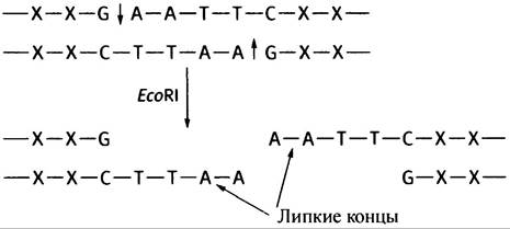
This results in The formation of sticky ends, where the "overhanging" complementary sequences can spontaneously pair with each other (in this case, A with T). If two DNA fragments with identical sticky ends are mixed together, they will join (Fig. 24.1). If pre-digested human and phage λ DNAs are mixed, the sticky ends will facilitate the formation of recombinant phage DNA molecules, as described above. To covalently seal the nicks, the enzyme ligase is used, which catalyzes bond formation between a 3'-OH- group and a 5'-phosphoryl group.
Fig. 24.1. Main steps in producing recombinant DNAs used to construct a λ phage-based DNA library. Note that while Other types of DNA fragment associations may occur due to sticky ends, only recombinant molecules containing a human DNA fragment (15–20 kbp) in the center and phage DNA fragments on the sides will be packaged into the phage HEAD.

Upon adding the recombinant molecules to the protein components of phage λ, phage self-assembly takes place, and the recombinant DNA is automatically packaged into its head to form a mature phage.
Packaging of recombinant DNA into infectious phage particles will occur only if the human DNA fragment, no larger than 15–20 kbp, has phage λ DNA at its ends. This is how recombinant molecules are selected (certain precautions are taken during the preparation of human DNA fragments to ensure their sizes meet these requirements, but we will not discuss them here). The phage λ carrying the recombinant DNA molecule is used to infect E. coli cells via a procedure designed to infect only a small fraction of the cells. This is necessary to maximize the probability that a single phage particle enters a given cell. It is also important that the E. coli strain is mutant for the restriction enzyme gene. Ultimately, the entire fragmented Human Genome will be distributed throughout an E. coli cell culture (one fragment per cell). This "complete collection" of The Human Genome, distributed across "individual volumes" of recombinant phage particle molecules enclosed within the "dust jacket" of a bacterial cell, is termed a genomic library. The cell culture is then plated onto a solid nutrient medium and incubated. Uninfected cells grow, forming an opaque "lawn" across the entire surface of the plate. Interspersed among them are phage-infected cells, whose colonies appear as spots, or plaques. The formation of a plaque is due to the fact that after phage multiplication within a cell, the cell lyses, and the phage particles infect neighboring cells, disrupting the "lawn" formed by the initially uninfected bacteria. Each plaque contains multiple copies of the phage that have multiplied from a single infectious particle and carries an identical copy of the human DNA fragment (Fig. 24.2).
If we can identify the plaque containing the phage that carries the desired gene, it will enable us to grow an unlimited amount of that phage in E. coli and isolate its DNA. How do we identify the correct plaque?
Screening Plaques for a Selected Human Gene
To identify a phage carrying the target human gene, DNA–DNA hybridization is the most widely used approach. This requires a hybridization probe consisting of a short DNA fragment (about 20 NUCLEOTIDES long) whose base sequence is complementary to a known sequence within the gene. The method involves transferring phage from each plaque onto a DNA-adsorbing membrane. When a membrane disk is pressed against The surface of the culture plate (keeping in mind to mark it so THE POSITION OF each plaque can later be identified), phage from each plaque adheres to the membrane. The membrane is then treated with alkali to disrupt the protein–DNA complexes (phage heads) and to release and separate the DNA strands. The single-stranded DNA is fixed to the membrane, which is subsequently treated with non-specific DNA to prevent non-specific binding of the probe to the membrane. To hybridize with any complementary DNA, the hybridization probe is incubated with the membrane (having previously been radiolabeled via enzymatic phosphorylation using labeled ATP). Under these conditions, only the complementary DNA—the target gene—will "stick" to the probe. X-ray film is then used to determine the position of the hybridized probe (see Fig. 24.2). Once the plaque containing the target gene is identified, an unlimited amount of phage can be grown from that plaque by infecting E. coli, after which the human DNA inserts can be retrieved from the phage DNA using an appropriate restriction endonuclease.
Fig. 24.2. Steps in gene cloning using bacteriophage λ as a vector

How can one obtain a suitable probe—with a nucleotide sequence complementary to the gene sequence—if The structure of the latter is unknown? In this case, one can work backward, relying on METABOLISM/28.html">The Genetic Code (see Table 22.1). If The amino acid sequence of the protein encoded by the gene of interest is fully or partially known, we can deduce the base sequence of that gene. Of course, due to the degeneracy of the genetic code, such a reverse approach is not always straightforward. Where an amino acid has multiple codons, we cannot deduce which one
was actually used by the cell for protein synthesis (gene cloners always take pleasure in finding methionines or tryptophans encoded by a single triplet within sequenced fragments). In cases where the degeneracy of the genetic code must be accounted for, methods involving the synthesis of a probe mixture are employed. Specialized instruments can readily synthesize short DNA probes with a specific base sequence. An enzymatically added radioactive phosphate group is then attached to the probe. If such a probe is unavailable, alternative screening methods are used, which are beyond The Scope of this book.
Let us now consider the isolation of a cDNA clone.
Cloning Human cDNA
Construction of a Human cDNA Library
The Principle of the method is that mRNA is isolated from human cells expressing the gene of interest; it is then copied in vitro into double-stranded DNA, which is subsequently cloned. A significant advantage of this approach lies in choosing a tissue where the target gene is highly active, meaning the corresponding mRNA is abundant.
Among all cellular RNA, the proportion of mRNA is small, yet in human cells, all mRNAs (with rare exceptions) feature poly-A tails (see p. 270). If an RNA preparation is passed through a Column containing an inert matrix with a synthetic oligo-dT Ligand (a short single-stranded DNA composed exclusively of T bases), ribosomal and Transfer RNAs pass through the column unhindered, whereas mRNA binds to it via A = T hydrogen bonding between the tail and the ligand. The mRNA can then be eluted using low Ionic strength solutions.
The isolated mRNA mixture is copied in vitro into DNA using the viral Reverse Transcriptase polymerase domain (see p. 313), yielding an RNA-DNA duplex. The RNA is degraded by Treatment with NaOH or RNase, and the single-stranded DNA is converted into double-stranded DNA using exonuclease-free DNA polymerase I isolated from bacteriophage.
For cloning cDNA molecules, phage λ can be used as a cloning vector, as previously described for Genomic Libraries. cDNA molecules lack sticky ends; however, methods exist for enzymatically attaching sticky ends to so-called blunt-ended molecules. Other blunt-end ligation methods are also employed.
What can be done with cloned DNA?
Thus, we have phage λ containing the desired genomic DNA insert (also referred to as an insert) or cDNA insert. We can obtain it in any desired quantities
by infecting E. coli cells, subsequently isolating the DNA, and excising the inserts via restriction Digestion.
Further application of these inserts for various purposes almost invariably involves inserting the DNA of interest into another cloning vector—most frequently a bacterial plasmid. Phage λ is convenient for constructing libraries because it can accept large DNA fragments (around 15 kb) and infect cells efficiently, enabling the generation of a complete genomic library or a comprehensive cDNA library. However, when it comes to studying a cloned genomic DNA or cDNA fragment, the disadvantage of the phage is its own large DNA flanking the insert (about 30 kb), which is essential for its replication but entirely irrelevant to the tasks at hand. Plasmids are much smaller and easier to handle. They cannot accept large fragments of foreign DNA like phage λ can, but if, for example, the goal is to sequence a gene isolated using phage λ, that gene is cut into fragments, which are then inserted into plasmids (see below), and each fragment is sequenced. cDNA clones can be processed in a similar manner. They are small enough to be inserted into expression vectors, including E. coli plasmids, provided that appropriate transcriptional and translational signals are added. Upon infecting E. coli cells with these vectors, the protein encoded by the cDNA can be produced in the required amounts. We will now examine bacterial plasmids.
Bacterial plasmids
An E. coli cell contains a single main circular chromosome carrying thousands of genes that constitute the bulk of the cell's genetic material. However, The Cytoplasm also harbors separate small minichromosomes, or plasmids: circular DNA molecules carrying a small number of genes that play a protective role in the cell. Typically, they harbor genes that confer Antibiotic Resistance to the cell by encoding, for example, an enzyme that degrades the antibiotic. Each plasmid is capable of replication, ensuring its duplication within the cell.
Plasmids are non-infectious in the sense that Bacteriophages are infectious. However, if E. coli cells are treated with, for example, CaCl2 at 0 °C and then subjected to a Temperature Shock at 42 °C, they become competent—capable of taking up plasmids.
Suppose we are dealing with a cloned cDNA molecule isolated via the method described above. First, our chosen plasmid is cleaved
with a restriction enzyme, and appropriate sticky ends are added to the cDNA molecules (we will not describe how this is done). When the cleaved plasmids and modified cDNA are mixed and ligated (i.e., their ends are joined enzymatically), recombinant plasmids are formed (Fig. 24.3). To avoid unnecessary complication of the process, we do not describe here the measures taken to prevent the regeneration of original circular plasmids and to promote the formation of recombinant plasmids. E. coli cells are infected with the recombinant plasmids and grown on agar plates such that each colony arises from a single cell. As a result of plasmid transformation, only a small fraction of the cells are infected, and the probability of a single cell taking up two plasmids is minimal. Since non-transformed cells vastly outnumber transformed ones, a rapid method for selecting the latter is required.
Fig. 24.3. Transfer of a DNA insert from phage λ into a plasmid. A DNA fragment cloned in a phage or in an expression-ready cDNA is transferred into E. coli within a specially constructed plasmid in order to produce the desired protein

One of the engineered plasmids used for cloning is pBR322; it carries inserted genes for ampicillin and tetracycline resistance. Each of these genes contains a distinct restriction site. Suppose a foreign DNA fragment is inserted into the tetracycline resistance gene; the latter can no longer direct the Synthesis of the protein that confers resistance to this antibiotic. A cell carrying such a plasmid will be resistant to ampicillin but sensitive to tetracycline. A cell containing a plasmid without a DNA insert will be resistant to both Antibiotics, whereas a cell lacking the plasmid will be sensitive to both. This serves as the basis for selecting exclusively those cells that contain the plasmid of interest.
Determining the base sequence of a cloned DNA fragment
All information within a gene is encoded in its base sequence, the determination of which is of paramount importance. There are two methods of DNA Sequencing (as this process is called). One is the direct chemical Maxam-Gilbert method. The other, most commonly used, is based on DNA replication and is known as the Sanger method, or dideoxy method.
Scheme of the dideoxy DNA sequencing method
Suppose you have a cloned DNA fragment that you wish to sequence. It is inserted into a specially constructed plasmid that replicates along with the host E. coli cell.
Once the plasmids are isolated, you have at your disposal a large number of recombinant plasmids containing the selected DNA fragment. The sequencing procedure (see below) requires that the fragment targeted for sequencing be copied in vitro by DNA polymerase. In addition to the four deoxynucleoside triphosphates (dNTPs), this requires: 1) that the DNA template be single-stranded; 2) that a primer hybridizes to the starting region (recall that DNA polymerase cannot initiate a de novo chain). Plasmids designed for sequencing are constructed such that a specific nucleotide sequence (plasmid DNA) is always located at the 3' end of the inserted cloned fragment. Knowing this sequence, one can synthesize a complementary (oligo)nucleotide to serve as a primer.
Incubation of the single-stranded DNA to be sequenced with a primer, polymerase I (exonuclease-free), dATP, dGTP, dCTP, and dTTP allows the DNA fragment to be copied. One of the triphosphates (or the primer) must be radioactive so that all newly synthesized DNA strands are labeled.
Two aspects are critical when performing sequencing. The first involves the electrophoretic method for separating DNA molecules, which migrate in an electric field through an acrylamide gel plate at a rate determined by chain length: the shorter the DNA chain, the faster it moves. Each nucleotide addition alters the mobility of the chain. On the gel, chains with different mobilities correspond to bands, where these bands represent chains differing by a single nucleotide. They are identified using autoradiography, which involves exposing an X-ray film sensitive to radioactivity to the gel. The second aspect concerns dideoxy derivatives of nucleoside triphosphates (ddNTPs). DNA polymerase adds a 3'-nucleoside to the 3'-OH end of a growing DNA chain. DideoxyNTPs lack the 3'-OH group (Fig. 24.4); they can be incorporated into the chain via their 5'-P group, but once incorporated, chain elongation cannot continue.
Fig. 24.4. Structures of deoxyATP (dATP) and dideoxyATP (ddATP). The absence of a 3'-OH group means that upon incorporation of a ddNTP into the growing chain, the latter is terminated
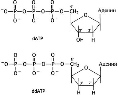
It is important to emphasize that when we speak of a sequenced DNA "fragment," the experiment actually involves many copies of this "fragment." Even a negligible amount of DNA contains a large number of molecules (remember that 1 mole of any chemical substance contains an astronomical 6.03-1023 molecules). Suppose that for copying we have all four dNTPs and a small amount of a single dideoxyNTP; for example, let us take dideoxyATP (A). The quantity of completed chains is sufficient to detect them as distinct bands on a gel using Electrophoresis. Chain elongation will continue until A is added; the lack of a 3'-OH group makes the attachment of the next nucleotide impossible.
How is all this interpreted in terms of base sequence?
Suppose that the sequenced DNA fragment has the sequence shown below, which includes T bases:

If copying occurs in the presence of dideoxyATP (along with an excess of deoxyATP), The addition of each A will terminate the growth of a portion of the chains, ultimately forming the following chains (attached to the primer).
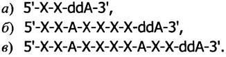
On the gel (referred to as a sequencing gel), these chains will be visible as bands (Fig. 24.5, left column).
Fig. 24.5. Autoradiogram of a sequencing gel. The polynucleotide sequence is read from bottom to top. a, b, c - Bands corresponding to polynucleotides formed in the presence of dideoxyATP
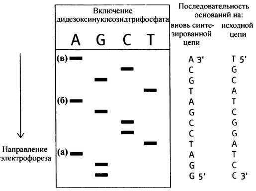
If the second incubation mixture contains dideoxyTTP instead of dideoxyATP, a set of chains ending in T is produced. Likewise, if dideoxyCTP or GTP is present in the medium, the chains will end in C or G, respectively. When all four incubations are performed, the reaction products align on the gel one after another in what is called a sequencing ladder (see Fig. 24.5). It allows for the easy Determination of the DNA fragment's base sequence, which is read from the bottom of the gel upwards: this is The sequence of the copy strand (i.e., the template's partner), rather than the template itself. Consequently, the sequence runs in the 5' -> 3' direction, since synthesis always proceeds in this direction.
Each sequencing ladder allows for the determination of approximately 200–300 bases. If overlapping fragments of the original cDNA are obtained using various restriction enzymes, its complete sequence can be mapped. A photograph of a sequencing ladder obtained in such an experiment is shown in Fig. 24.6.
Fig. 24.6. Photograph of a sequencing ladder. A, C, G, and T designate the dideoxynucleoside triphosphates present in the incubation mixture. Kindly provided by Dr. Cris Hahn, Department of Biochemistry, University of Adelaide
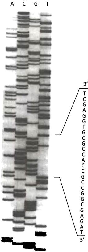
If the cDNA is sequenced to determine the amino acid sequence of a protein, the correct reading frame (out of 6 possible across the 2 strands) is identified using the genetic code.
The technique described above is used in many laboratories. However, sequencing technology has advanced to the point where the process has become automated. THE PRINCIPLE OF the new method does not differ from that used by Sanger, but it employs fluorescently labeled nucleoside triphosphates, with each of the 4 types having its own fluorescence emission maximum. DNA is incubated in a medium containing all four dideoxy compounds and, of course, the four deoxynucleoside triphosphates; the products are analyzed in a single electrophoretic lane. The gel is scanned automatically, and a computer prints out the base sequence.
Using the Polymerase Chain Reaction (PCR) to Amplify DNA Segments
The polymerase chain reaction (PCR) has become one of the most popular methods for working with DNA in recent years. It allows for the amplification of a DNA segment by synthesizing a massive number of its copies. Negligible amounts of DNA (from just a few cells) are sufficient to perform PCR. The principle of the method is simple and elegant. To carry out PCR, one must know The nucleotide sequence of the DNA of interest and synthesize two oligonucleotides, each complementary to a region on one of the two DNA strands flanking the segment chosen for amplification. These oligonucleotides serve as primers for DNA synthesis in vitro. The strands of the DNA duplex are separated by heating, and the chosen segment is copied using synthetic primers (Fig. 24.7). At the end of each synthesis cycle, the DNA duplexes formed during copying are separated by heating to prepare for the next round of priming and copying. The use of thermostable DNA polymerases (from thermophilic bacteria) eliminates the need to add the enzyme after each copying cycle and subsequent thermal strand separation.
Fig. 24.7. Schematic diagram illustrating the basic principle of DNA segment amplification during the polymerase chain reaction (PCR). a - In the center is the region selected for amplification using appropriate primers (—> <—); each is complementary to the 3'-end of the amplified strand;
b - heating causes strand separation (from this point on, for the sake of simplicity, the diagram shows the amplification of only one strand, although in reality both are amplified); c - the incubation mixtures contain the 4 dNTPs, a thermostable DNA polymerase, and the appropriate primers; d - the DNA is replicated; e - strands are separated by heating; f - a new strand is primed; g - the new strand is replicated; h - the desired fragment is isolated. Over 25 cycles of replication, heating, priming, and synthesis, the selected region can be amplified millions of times. Note that priming and copying occur in every cycle.
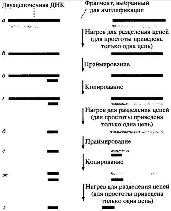
Until recently, it was only possible to amplify DNA segments no longer than 2 kb, but newer technologies allow for the amplification of fragments up to 35 kb in length. DNA copies obtained in this way can be used for a wide variety of purposes. This method is vital in forensic science and in the Prenatal Diagnosis of genetic abnormalities in utero (see below). It is also widely used in biochemical research.
Applications of Recombinant DNA Amplification Technology
As noted previously, the primary role of gene cloning is to determine base sequences. This is a prerequisite for studying gene function and its regulation. A DNA base sequence provides a direct "window" into the evolutionary relationships among organisms. Recombinant DNA amplification has also found practical applications in A number of other areas, which we will briefly discuss here.
GENE EXPRESSION IN Bacterial and Eukaryotic cells
An isolated cDNA clone or bacterial gene can be inserted into a specially constructed vector, such as a plasmid containing appropriate bacterial DNA promoters and Translation initiation signals. The cDNA molecule inserted into each plasmid is expressed; the cell synthesizes mRNA from the cDNA and translates it into protein. Thus, inserted prokaryotic genes or eukaryotic cDNAs (remember that cDNA lacks introns) can be efficiently transcribed, and the corresponding mRNAs translated, yielding the desired protein.
Using the methods described above, bacteria such as E. coli are capable of producing unlimited quantities of human proteins used as therapeutic drugs. In addition to yielding proteins that are present in The Human Body in vanishingly small amounts, the use of E. coli eliminates the risk of contaminating the protein product with infectious agents. This is how human insulin and several other proteins, such as growth hormone, are produced. As a safe, infection-free vaccine, an immunogenic hepatitis B virus protein produced in Yeast is employed. In some cases, E. coli can produce a foreign protein in such high amounts that it precipitates as inclusion bodies and fails to fold into its proper conformation. Sometimes, correct protein folding can only be achieved in vitro. Bacteria are unable to glycosylate proteins; however, if animal cells (such as insect cells) are used as expression hosts, glycosylated Proteins can be generated in large quantities.
This is a remarkably powerful method for analyzing The Role of individual amino acid residues within a protein. Consider a scenario where we have an enzyme whose amino acid sequence is known, determined either through direct sequencing or deduced from the base sequence of its gene or cDNA. We now wish to investigate THE CONTRIBUTION OF a specific amino acid side chain to the function of this protein. If the bacterial gene or eukaryotic cDNA is cloned into an expression vector, such as a plasmid, E. coli cells will produce the protein in quantities sufficient for research. For example, in the case of an enzyme, we might be interested in its catalytic activity or substrate Specificity. Site-directed mutagenesis enables the specific replacement of one or more selected Amino Acids in a protein. This is achieved by substituting the corresponding fragment of the isolated gene with a synthetic oligonucleotide whose base sequence encodes the altered amino acid. Once the mutant protein is obtained, its catalytic activity can then be determined.
Transgenesis
Transgenesis refers to the introduction of foreign genes into animals or plants. The introduction of normal genes into patients to correct defective genes (Gene Therapy) can be used to treat genetic disorders. Retroviruses, genetically crippled to prevent their replication, could potentially be used as vectors for introducing genes into Human Chromosomes. However, this approach does not allow for control over the insertion site, and random insertion carries inherent risks. Methods for targeted gene insertion are currently under development. The insertion of the human adenosine deaminase gene into patient leukocytes has been successfully used to treat children with immunodeficiency (see Fig. 18.6). For introducing foreign genes into plant chromosomes, naturally occurring (yet appropriately modified) Ti plasmids (tumor-inducing plasmids) derived from the pathogenic soil bacterium Agrobacterium tumefaciens are utilized as cloning vectors. The gene gun method (or biolistics) involves “shooting” genes into plant cells using a specialized device. This technique is used to engineer herbicide-resistant crops, allowing weeds to be destroyed with herbicides without hindering crop growth. Transgenic Animals can also serve as “expression vectors.” For instance, a human protein can be produced by linking its gene to the signal sequence of a milk protein gene, ultimately resulting in the secretion of the target protein into the milk. This approach has been successfully applied to produce human protein in the milk of sheep.
Detection of Genetic Anomalies via Restriction Analysis or Southern Blotting
Once gene structures are known, hybridization probes can be designed to detect genetic anomalies. This analysis requires a minimal amount of DNA, making prenatal fetal diagnosis feasible (when combined with the polymerase chain reaction, even minute quantities of DNA are sufficient). Access to information regarding potential hereditary disorders allows for informed decisions regarding Pregnancy termination. For instance, in muscular dystrophy, the gene anomaly typically stems from a deletion in a portion of the coding DNA, which can be identified via Southern blot analysis. Named after its inventor, this procedure involves digesting DNA with one or more restriction enzymes, separating the resulting fragments by gel electrophoresis, transferring the separated fragments onto a membrane, and probing the membrane with a gene-specific radioactive hybridization probe. This process reveals specific DNA segments as distinct bands. Gene anomalies can yield varying band patterns because Mutations may create new restriction sites or disrupt and eliminate previously existing ones. Even though the gel may contain numerous distinct DNA fragments, only those few that hybridize with the specific probe will be visualized.
If the chromosomal Location OF THE gene responsible for a genetic disease is not precisely known or has not yet been isolated, RFLP analysis (restriction fragment length polymorphism analysis) can be employed. The methodology is not discussed here, but it can be used to identify family members who are carriers of the abnormal gene.
The principle of RFLP analysis is most easily understood through the example of DNA fingerprinting. Repetitive DNA sequences scattered throughout The Genome exhibit a high degree of restriction site polymorphism, resulting in restriction patterns that are unique to each individual. Although historically used in forensic medicine, this technique is gradually being supplanted by newer PCR-based technologies. A large number of repetitive sequences have been identified in the human genome, and the number of repeats varies widely among individuals. The method involves selecting primer binding sites on either side of a repetitive sequence and amplifying it via PCR.
During electrophoresis, the mobility of the analyzed DNA samples depends on the number of repeats within the amplified segment. By selecting a panel of such loci for amplification, a unique banding pattern characteristic of each individual is obtained.
Chapter 24 Questions
1. How does a restriction endonuclease differ from pancreatic DNase?
2. The E. coli restriction enzyme EcoRI cleaves a hexameric base sequence. Such a sequence should occur frequently within the E. coli genome. Why doesn't the enzyme degrade its own host DNA?
3. What is meant by the term "sticky ends" in the context of DNA molecules?
4. WHAT IS A gene clone? What is a cDNA clone? How do they differ in eukaryotes?
5. What are the key steps in preparing a genomic library using bacteriophage λ?
6. What are the steps involved in isolating a specific clone from a genomic library harbored in phage λ?
7. What is a dideoxynucleoside triphosphate? What is its role in Sanger DNA sequencing?
8. Briefly outline the principles of the polymerase chain reaction and the conditions required for its performance.
9. What is a bacterial expression plasmid vector?
10. What is restriction fragment analysis?
Last update: 06/08/2026
Editorial and Educational Adaptation: This material has been compiled based on the primary/original source text. The project team performed an editorial review, corrected technical inaccuracies, structured sections, and adapted the content for an educational format.
What was processed:
- elimination of formatting defects (OCR errors, structural breaks, corrupted characters);
- editorial organization of content;
- standardization of terminology in accordance with academic sources;
- verification of factual statements against the original source text.
All mentions of the author, publication year, and origin of the primary text have been preserved in accordance with the source.