Genetics - A. V. Sivolob 2008
Genetic Engineering and Methods of Molecular Genetics
Methods of Genetic Engineering
Genomic Libraries
The principles of DNA fragment cloning discussed so far leave open the question of where a given DNA fragment actually comes from. To work with a specific region of DNA (such as a particular Gene), researchers first clone A large number of diverse genomic fragments to create a clone library, from which the gene of interest is subsequently isolated.
The standard Procedure for constructing a library—known as the shotgun method—begins with partial Digestion (for a limited time) of the total genomic DNA using a specific restriction enzyme. This yields a set of overlapping, diverse fragments of a defined average length (Fig. 9.4). All these fragments are inserted into vectors, followed by cellular transformation. Bacteria are then plated onto a solid medium so that each colony originates from a single Cell. Consequently, the collection of colonies forms a set of diverse clones, each carrying a single genomic fragment.
cDNA clone libraries are generated in a similar fashion to genomic libraries. First, total mRNA is isolated from a specific cell type. Using Reverse Transcriptase and oligo(dT) primers complementary to the 3'-terminal poly(A) tails of the mRNA, complementary DNA strands are synthesized. The resulting cDNA molecules are then cloned. Unlike genomic libraries, a cDNA clone library contains only the coding sequences (exons) of genes, and specifically those genes that are active in the given cell type.
Class="center">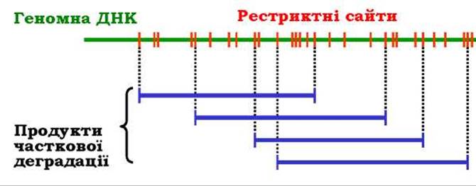
Fig. 9.4. The shotgun method
The target nucleotide sequence within a clone library can be identified by hybridizing the cloned DNA with a radiolabeled DNA probe (Fig. 9.5). Bacterial colonies from a Petri dish are transferred onto a nitrocellulose filter to create a replica. The Cells are then lysed, and the DNA is deproteinized and denatured in an alkaline solution. After drying the filter, the single-stranded DNA is irreversibly fixed to it. The filter is subsequently incubated with a radiolabeled DNA probe of a specific sequence. If the probe is complementary to the clone's DNA, Hybridization—the renaturation of double-stranded DNA—takes place. Detecting this event via autoradiography allows the desired clone to be identified.
Probes are most frequently synthesized using the EST (Expressed Sequence Tag) database. This public database is an extensive collection of short (200–400 bp) cDNA sequence fragments from numerous organisms. To prepare a probe, one simply searches the EST database for a short stretch corresponding to the fragment of the protein sequence whose gene is being sought, and then synthesizes it.
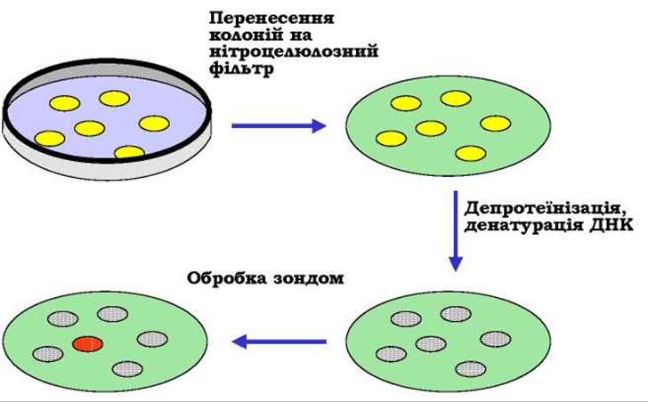
Fig. 9.5. Hybridization of cloned DNA with a radioactive probe on a nitrocellulose filter
Sequencing (see below) each fragment contained in a genomic clone library, followed by comparing the overlapping sequence fragments (Fig. 9.4), makes it possible to order the cloned fragments according to their natural arrangement in The Genome, thereby establishing the complete genome sequence.
The vast amount of sequence data (including complete genome sequences) that has already been generated and continues to accumulate poses significant challenges for data storage and analysis. Naturally, such a task—which arises as soon as one begins comparing individual cloned fragments to determine a genome's nucleotide sequence—can only be accomplished using computers. Bioinformatics, the field dedicated to solving these problems, is today an indispensable component of genetics. The largest publicly accessible internet Databases for nucleotide and Amino acid sequences were established by the European Molecular Biology Laboratory (EMBL Sequence Data Base) and the U.S. National Institutes of Health (GenBank). Respective portals also host software tools for sequence comparison, promoter identification, finding METABOLISM/31.html">Transcription start sites, introns, and exons, as well as deducing protein sequences from nucleotide sequences. Based on bioinformatic analysis, one can determine, for instance, the Functional Significance of a newly cloned, unknown gene through its Homology with a known gene from another Organism; elucidate gene Structure (locating promoters, exons, and transcription termination sites); reveal the Structural and functional features of novel Proteins; map functional relationships between various proteins and genes; and trace Phylogenetic relationships among different taxa. Thanks to the advancement of bioinformatics, it has become possible to address various biological problems simply via computer—in silico—complementing results obtained in vivo and in vitro.
Last update: 11/08/2026
Editorial and Educational Adaptation: This material has been compiled based on the primary/original source text. The project team performed an editorial review, corrected technical inaccuracies, structured sections, and adapted the content for an educational format.
What was processed:
- elimination of formatting defects (OCR errors, structural breaks, corrupted characters);
- editorial organization of content;
- standardization of terminology in accordance with academic sources;
- verification of factual statements against the original source text.
All mentions of the author, publication year, and origin of the primary text have been preserved in accordance with the source.