Pediatric Medical Genetics - S.I. Smiian 2003
Chromosomal diseases
Human chromosomes
The primary carrier of the hereditary traits in living organisms is the chromosomal apparatus of the Cell Nucleus. Each biological species is characterized by a constant chromosomal Complement consisting of a specific number of Chromosomes. For instance, a species of roundworm (parasitic roundworm) typically has 2 chromosomes, whereas some crustaceans feature up to 200 chromosomes.
In most higher organisms, every cell contains a diploid (2n) set of chromosomes. Chromosomes vary in shape and size. The collection of quantitative and qualitative characteristics of chromosomes, as observed microscopically in a single cell, is termed the karyotype.
The normal diploid chromosome number in humans is 46. Due to the limitations of early cytological techniques, the total human chromosome number was long believed to be 48 (from 1912 to 1956). It was only in 1956 that Swedish cytologists H. Tjio and A. Levan, utilizing refined cytological Methods, demonstrated that the standard human chromosome count is 46. These findings were confirmed later that year by British scientists C.E. Ford and J.L. Hamerton, marking the beginning of the rapid development of cytogenetics.
The human chromosomal complement consists of 23 pairs. Of these, 22 pairs are autosomes, and the 23rd pair comprises the sex chromosomes, whose primary role is Sex Determination. Specifically, the female sex is represented by two X chromosomes (XX), and the male sex by an XY chromosome pair. It is worth noting that about 3/4 of all hereditary disorders—both Gene-based and chromosomal—are associated with abnormalities in the sex chromosomes (X and Y), while only 1/4 stem from autosomal changes.
Somatic Cells contain 46 chromosomes, whereas Germ Cells possess half that number—23 chromosomes. Autosomes are identical in both males and females.
Depending on size, centromere position, and arm length, human chromosomes are classified into 3 main types.
1. Median, or metacentric — the centromere is located in the middle of the chromosome, resulting in two equal arms.
2. Submedian, or submetacentric — the centromere is positioned closer to one end, making the chromosome arms unequal in length.
3. Subterminal, or acrocentric — the centromere is situated near the very end of the chromosome.
In addition to the primary constriction (centromere), certain chromosomes exhibit a secondary constriction, most frequently observed in chromosomes 1, 9, and 16.
Among other structural features visible in human chromosomes using routine staining techniques, small chromatic satellite bodies can be observed on the short arms of large and small acrocentric chromosomes; these vary among individuals and largely depend on preparation techniques. Based on size and centromere Location, all chromosomes are divided into 7 groups (Fig. 5), designated by the letters A, B, C, D (K), E, F, and G. This Classification relies on two fundamental principles: chromosome size and structural features associated with THE POSITION OF the centromere (the primary constriction dividing the chromosome into two parts, or arms, connected by the chromatid). Group A (pairs 1–3) comprises large chromosomes with approximately median centromeres; chromosomes 1 and 3 are metacentric, while 2 is submetacentric. They are clearly distinguishable and easily differentiated. Group B (pairs 4–5) consists of relatively large chromosomes with submedian centromeres. Morphologically they are difficult to distinguish, though the 4th pair is slightly longer. Group C (pairs 6–12) includes medium-sized chromosomes with submedian centromeres that show poor differentiation. The sex X chromosome, in terms of size and centromere position, falls between chromosome pairs 6 and 7. Chromosomes 6, 7, 8, and 11 can be classified as metacentric, whereas chromosomes 9, 10, and 12 are more submetacentric. Group D (K) (pairs 13–15) features medium-sized chromosomes with nearly terminal centromeres (acrocentric). Chromosomes 13 and 14 frequently bear satellites on the ends of their short arms, while chromosome 15 does so less often. Group E (pairs 16–18) comprises relatively short chromosomes with nearly median to submedian centromeres. Chromosome 16 is more metacentric and often features a secondary constriction near the proximal end of the long arm, while chromosomes 17 and 18 have submedian centromeres. Group F (pairs 19–20) consists of short chromosomes with near-median centromeres that are extremely difficult to distinguish from one another. Group G (pairs 21, 22, and the Y chromosome) contains very short acrocentric chromosomes. The first two typically bear well-defined satellites on their short arms, whereas the Y chromosome lacks satellites and has long-arm chromatids that lie closely parallel to each other.
The determination and development of human sex are governed by the sex chromosome system. During female Meiosis, the ovum receives a haploid set consisting of 22 autosomes (one from each pair) and a single X chromosome; consequently, all egg cells share an identical karyotype. During male meiosis, one gamete receives a haploid set with an X chromosome, while the other receives a Y chromosome, thus producing Two Types of sperm. Upon Fertilization, the sex of the offspring is determined by whether an X- or Y-bearing sperm fertilizes the egg.
Class="center">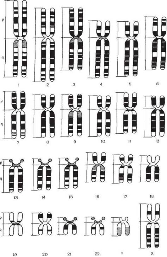
Fig. 5. Schematic maps of transverse banding patterns in human chromosomes using Q-, G-, and R-banding techniques (Paris Conference, 1972).
1 - R-bands; 2 - G- and Q-bands; 3 - variable bands.
Each chromosome contains a vast number of genes arranged in a linear sequence, with each gene occupying a strictly defined locus. Genes are the carriers of hereditary traits. While a single gene may influence The Development of one specific trait, it is more common for a single gene to affect multiple traits. Conversely, certain traits are controlled by the combined action of multiple genes.
Chromosomes store hereditary information that dictates all aspects of an Organism's development, Structure, and vital Functions. Furthermore, chromosomes act as a complex cellular apparatus regulating METABOLISM/35.html">Protein Biosynthesis. Damage to this system disrupts the synthesis of Proteins, Enzymes, Hormones, and other vital molecules. Chromosomal aberrations can take various forms. They may arise at the molecular (submicroscopic) level—without visible changes to Chromosome structure—due to the loss of one or more bases in a DNA molecule. These are known as gene or point Mutations; they cause complex Metabolic Disorders and trigger various pathological syndromes. Additionally, chromosomal and genomic mutations involve alterations in chromosome structure or number, and are collectively referred to as chromosomal aberrations.
Specific chromosomal aberrations cause pathological conditions, manifesting as chromosomal and polychromosomal disorders. Polychromosomal disorders include leukemias and tumors.
Last update: 11/08/2026
Editorial and Educational Adaptation: This material has been compiled based on the primary/original source text. The project team performed an editorial review, corrected technical inaccuracies, structured sections, and adapted the content for an educational format.
What was processed:
- elimination of formatting defects (OCR errors, structural breaks, corrupted characters);
- editorial organization of content;
- standardization of terminology in accordance with academic sources;
- verification of factual statements against the original source text.
All mentions of the author, publication year, and origin of the primary text have been preserved in accordance with the source.