BIOCHEMISTRY AND MOLECULAR BIOLOGY - W. ELLIOTT - 2002
CHAPTER 23. VIRUSES AND VIROIDS
Aside from their immense biological and medical importance, Viruses have found widespread application in biochemical research. They are used as simple models to study the complex processes of Gene function and serve as valuable tools for A wide variety of purposes, including the generation of recombinant DNA (see Chapter 24).
With the exception of specialized survival strategies such as spore formation, in which chemical transformations are reduced to a minimum, living Cells are always metabolically active: they synthesize ATP, perform osmotic work, synthesize cellular components, and so on. Furthermore, cells grow and divide. This requires thousands of genes encoding the necessary Proteins.
Viruses are distinct from cells. They are much smaller, requiring an Electron microscope to be visualized.
Viruses lack METABOLISM. A viral particle—a virion—is in itself completely inert. It generates no energy and carries out no Chemical Reactions; it is simply an organized, inert complex of molecules.
Viruses multiply only after infecting living cells. Various viruses infect animal and plant cells, as well as Bacteria (bacterial viruses are called Bacteriophages). The primary function of a virus is to deliver its genetic material into a Cell and hijack the cellular machinery for its own Replication.
A viral particle consists of a nucleic acid surrounded by one or more protective coats. Although these protective coats contain many copies of protein molecules, in most viruses they are formed from just a few types of proteins. The total number of viral genes can range from hundreds (such as in the vaccinia virus) down to three or four.
The protein shell surrounding The Genome is called the capsid, and the viral genome enclosed within this shell is termed the nucleocapsid. Some viruses possess an additional lipid bilayer envelope embedded with protein molecules exposed On the surface, which participate in the Initial Stages of the infection process.
Viral Life Cycle
A virus must enter The Cell it infects by introducing its genetic material (RNA or DNA). Viral genes direct the synthesis of virus-specific Enzymes required for viral reproduction, as well as proteins needed for the assembly of new Viral Particles. The virus must produce numerous copies of its genome and assemble progeny virions from the synthesized components, which must then exit the cell.
Below, we outline the Stages of the viral life cycle and then examine several specific viruses in greater detail to illustrate these General Principles.
A living cell is surrounded by a Lipid Bilayer Membrane that prevents The entry of viruses. However, mechanisms exist to overcome this barrier, such as receptor-mediated endocytosis in animal cells (p. 204). Certain viruses exploit this cellular import mechanism to enter the cell. In the first step, the virion binds to the cell surface via a coat protein complementary to a specific receptor located on the exterior of the host cell. The presence of such a receptor is a key prerequisite for viral infection of a given cell. Following attachment, the virus-receptor complex migrates along the membrane to a depression known as a coated pit, the inner surface of which is lined with the protein clathrin (Fig. 23.1). As in normal receptor-mediated endocytosis, the pit invaginates until the virion, enclosed within a coated vesicle, is internalized into the cell. The clathrin then dissociates and returns to The Plasma Membrane, while the virus-containing vesicle fuses with a cytoplasmic vesicle called an endosome, leaving the virus inside. In normal (non-viral) endocytosis, endosomes deliver their contents to vesicles formed by the Golgi apparatus (Lysosomes), which contain a battery of hydrolytic enzymes that degrade the captured particle (see Fig. 16.2). The action of a proton pump maintains an acidic pH within the endosome. This acidic environment causes the virus to dissociate from the host cell membrane receptor. If the virus is enveloped, the viral membrane fuses with the endosomal membrane, releasing the viral contents into the Cytoplasm. Here, the virus uncoats, freeing its genetic material into the cytoplasm. The mechanism by which non-enveloped viruses escape from the endosome into the cytoplasm is not yet fully understood.
Class="center">Fig. 23.1. Process of invasion of an animal cell by an enveloped virus via receptor-mediated endocytosis

The second pathway, available exclusively to lipid-enveloped viruses, involves direct fusion with the outer membrane of the host cell. In this case, the attachment of the virus to the cell surface results from the interaction between a viral protein and a specific cellular protein. This interaction triggers the fusion of the two membranes (viral and cellular), creating a pore through which the viral contents enter the cell. This is the pathway utilized by the AIDS virus.
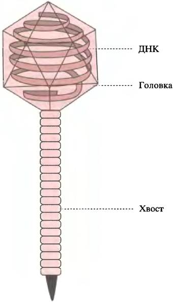
Bacterial viruses—bacteriophages—penetrate cells by a different mechanism. Bacteria possess a rigid Cell wall. Lambda phage (λ) resembles a tadpole (Fig. 23.2); its HEAD is a protein capsule containing a DNA molecule. The phage attaches to The cell wall by a tail-like fiber and, acting much like a hypodermic syringe, injects its DNA into the bacterial cell.
Types of Genetic Material in Various Viruses
The genetic material of viruses may consist of double-stranded or single-stranded DNA, or double-stranded or single-stranded RNA. Up to this point in the book, we have focused on double-stranded DNA as genetic material. As noted previously (see p. 232), DNA is more stable than RNA: the presence of two complementary strands means that damage to one strand can be repaired using the opposite strand as a template. The replication machinery of The Nucleus includes proofreading mechanisms that greatly reduce the frequency of errors. No such machinery exists for RNA Synthesis; nevertheless, RNA serves as the genetic material for many viruses. Why? Probable reasons include: 1) viral genomes are extremely small compared to cellular genomes, thereby lowering the probability of deleterious Mutations during viral RNA replication; 2) although errors occur more frequently during RNA replication than during DNA replication—and some may be lethal—this disadvantage is offset by a high rate of reproduction. (Under intense selective pressure, this actually becomes an advantage, as rapid mutation helps the virus evade host immune responses.)
Fig. 23.2 Lambda phage. The phage head consists of a chromosome containing approximately 50,000 Base Pairs of DNA surrounded by a protein coat
Upon entering the host cell, The Fate of Different types of viral genetic material varies. Double-stranded viral DNA is transcribed by host cell RNA polymerase to produce mRNA. (Among this class of viruses, the vaccinia virus is unusual in possessing its own RNA polymerase.) The minus (template) strand of the double-stranded genome is copied to form mRNA, which in turn directs the synthesis of virus-specific proteins. In the case of viruses containing single-stranded DNA—either the (+) or (-) strand—host cell enzymes synthesize a double-stranded DNA intermediate during replication, from which mRNA is subsequently transcribed (for strand Structure/97.html">Definitions, see below).
Cells lack the machinery to replicate viral RNA. Viruses with double-stranded RNA carry RNA-dependent RNA polymerase molecules within their virions. Inside the host cell, this enzyme transcribes mRNA from the viral RNA template. The mRNA is translated by the host cell machinery to produce the virus-specific proteins required for viral replication. Single-stranded RNA viral genomes exist in two forms. Recall (see p. 263) that within a cell, one of the two DNA strands is called the sense (non-template) strand, whose base sequence is identical (except for the substitution of U for T) to the mRNA transcribed from the complementary, antisense (template) strand. In various single-stranded RNA viruses, the RNA strand may be equivalent to either the sense or the antisense strand, referred to as plus [(+)] or minus [(-)] strands, respectively. In other words, the RNA of a (+)-strand virus Functions directly as mRNA, whereas that of a (-)-strand virus does not. When (+)-RNA enters a cell, the host protein-synthesizing machinery can immediately translate the RNA to produce viral proteins—including the enzymes necessary for viral reproduction—since it is effectively an mRNA. Such a virus requires only an RNA genome encoding all the proteins needed for reproduction post-infection. The situation is more complex for a virus containing (-)-RNA: because it is not an mRNA, the cell can neither translate nor replicate it. Copying (-)-RNA into (+)-RNA (mRNA) requires an additional RNA-replicating enzyme provided by the virus.
Another class of (+)-strand RNA viruses is the Retroviruses, of which the most notable is the HUMAN IMMUNODEFICIENCY VIRUS (HIV), the CAUSATIVE AGENT OF Acquired Immunodeficiency Syndrome (AIDS). Its virion carries an enzyme that converts single-stranded RNA into double-stranded DNA, which can then integrate into the host cell chromosome.
How Do Viruses Release from Cells?
Some viruses, such as the poliovirus, are released through simple cell lysis. In the case of bacteriophage λ, the tough Introduction/37.html">Bacterial cell wall would otherwise prevent the release of new phage particles. However, one of the phage genes encodes an enzyme called Lysozyme, which degrades the cell wall, causing cell lysis. This gene is transcribed only at a late stage of infection, ensuring that the cell does not rupture until new phage particles have been formed. Although bacteriophage λ is merely a small "piece" of DNA—48,502 NUCLEOTIDES long with 63 genes—these genes are regulated by a sophisticated control system.
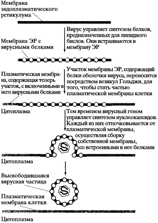
The release mechanism of animal viruses surrounded by a lipid bilayer membrane is more complex. Here, too, the virus hijacks the host cell it infects. Recall that in a normal cell, a new cell membrane containing the required set of proteins is assembled in the rough Endoplasmic reticulum (see p. 301). These proteins are glycosylated and transported via Golgi vesicles to the plasma membrane; the vesicles fuse with it, forming a new cell membrane complete with the necessary Membrane Proteins.
Certain enveloped viruses adapt this process for their own purposes. The mRNA for envelope proteins encodes a leader sequence
and an anchor sequence (see p. 303), so the positioning of these proteins in the rough ER—from which they are delivered—is identical to that described above for the normal cell membrane. There, the viral membrane proteins accumulate. The newly formed viral nucleocapsid (genome + capsid envelope proteins) buds off from the membrane, acquiring its lipid envelope along with the incorporated membrane proteins. Influenza and HIV use precisely this method of particle assembly (Fig. 23.3).
Fig. 23.3. Release of viral particles from the cell by budding from the cell surface
Mechanisms of Replication in Certain Viruses
Viruses successfully overcome the challenges associated with diverse replication pathways. Reviewing all of them would take up too much space, so we will focus
on just a few viruses whose reproduction illustrates the principal modes of replication. Furthermore, these particular viruses are of independent interest.
Vaccinia Virus
The virus that causes cowpox was famously used to develop the smallpox vaccine. It is one of the largest and most complex viruses, possessing a double-stranded DNA genome. This DNA is transcribed into mRNA, which directs the host cell Ribosomes to synthesize the protein components required for The formation of new virions (including the viral DNA REPLICATION ENZYMES).
The genomes of most double-stranded DNA viruses find their way into the nucleus, where host RNA polymerase transcribes the viral genes into mRNA. Vaccinia is unusual in that its complete replication takes place in the cytoplasm, where host RNA polymerase is absent. Consequently, the virus must carry its own RNA polymerase molecules within its particles, thereby ensuring mRNA production and the synthesis of all necessary proteins, including RNA polymerase itself.
Poliovirus
This is a "naked" virion containing only a protein capsid shell. It binds to a specific receptor found exclusively on the epithelial cells of humans and other primates. Because its single-stranded (+)-RNA acts directly as Messenger RNA (mRNA), it is translated upon entering the cytoplasm. This Translation yields RNA replicase, which synthesizes RNA using the viral RNA as a template. The replicase copies the initial (+)-strands into (-)-strands, which in turn serve as templates for the synthesis of numerous additional (+)-strands. Among RNA-synthesizing enzymes, poliovirus RNA replicase is unique in that it cannot initiate RNA strand synthesis on its own. Instead of an oligonucleotide primer, it utilizes a protein primer to which the enzyme adds the first nucleotide before elongating the chain.
Translation of the mRNA produces a large "polyprotein," which is subsequently cleaved into individual proteins. The rationale behind forming a polyprotein is not entirely clear: translation yields an equal number of capsid protein and replicase molecules, yet viral particle assembly requires far more capsid protein molecules than The amount of replicase needed for replication.
Influenza Virus
This virus possesses a (-)-sense RNA chain. Its genome is divided into eight segments, each enclosed within a helical nucleocapsid surrounded by a lipid membrane embedded with molecules of two distinct Glycoproteins (Fig. 23.4). One of these is the surface protein hemagglutinin. It earned this name because mixing it with red Blood Cells causes them to agglutinate. The carbohydrate component on The surface of erythrocytes, attached to molecules of the transmembrane protein Glycophorin, terminates in neuraminic (sialic) acid (see p. 56). When mixed with red blood cells in vitro, hemagglutinin binds to neuraminic acid and cross-links the cells into aggregates. The virus gains entry into the host cell by attaching to its receptor (which also bears a terminal neuraminic acid residue) and subsequently penetrating via endocytosis.
Fig. 23.4. Cytology/cytology/92.html">SCHEMATIC STRUCTURE OF the influenza virus. The outer membrane matrix protein layer is not shown
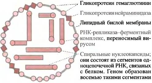
The surface of the virion bears 700 hemagglutinin molecules. Additionally, there is another surface protein—the enzyme neuraminidase, which hydrolyzes the terminal neuraminic acid groups of glycoproteins. The exact Functional Significance of this enzyme remains debated. At first glance, it seems paradoxical that a virus would possess a surface enzyme designed to destroy the very cellular receptors required for its cell entry. Perhaps this enzyme thins sialic acid-containing mucin (see p. 56), facilitating access to the cell surface for infection and the release of new viral progeny. The thinning of mucins may also aid in the more efficient airborne transmission of the virus during sneezing. Finally, it is possible that newly formed virions remain tethered to the host cell via neuraminic acid receptors. Neuraminidase counteracts this by cleaving the receptors and releasing the viruses. Evidence suggests this enzyme is crucial for efficient viral propagation during an infection.
As noted previously, the viral genome is organized as a helical nucleocapsid distributed across 8 distinct (-)-RNA segments (see Fig. 23.4). Because a (-)-sense RNA strand cannot function as a template for translation, RNA replicase is essential for its replication.
The virus carries RNA replicase molecules within its nucleocapsids, which copy the (-)-strands into (+)-RNA strands (the mRNA equivalent). Subsequent translation produces all required viral proteins, including abundant molecules of RNA replicase.
The Immune Response is directed primarily against the hemagglutinin of the influenza virus. It is neutralized in such a way that an individual who has recovered from the disease becomes immune to reinfection by the identical viral strain. However, due to frequent mutations (characteristic of RNA viruses), the Amino Acid Composition of viral hemagglutinin constantly drifts, gradually eroding immune protection. This phenomenon is known as antigenic variation. Minor Amino Acid Substitutions do not trigger major epidemics because the antigen-binding sites on the protein surface are largely conserved during gradual structural changes. Only their number decreases, resulting in partial population-level Immunity and relatively mild infections. Nevertheless, if a virus acquires a completely novel hemagglutinin via antigenic shift, a pandemic can erupt because pre-existing residual immunity is entirely absent. Such a scenario can arise when a host cell is co-infected by two different viral strains. Due to the segmented genome, reassortment of RNA segments can occur during the assembly of new viral nucleocapsids. The 1918 pandemic, believed to have originated from such a reassortment between human and avian influenza strains, claimed 20 million lives.
Novel approaches to influenza Treatment focus on targeting structural viral proteins. X-ray crystallography has revealed the precise 3D STRUCTURE OF THE neuraminic acid-binding pocket in crystalline influenza neuraminidase. This breakthrough enabled the design of synthetic neuraminic acid analogues that bind with high affinity to the enzyme, inhibiting its action. Clinical trials of these substrate analogues as anti-influenza drugs are currently underway. Because these compounds do not block the hemagglutinin region responsible for host receptor binding, viral cell entry still occurs. Inhibiting neuraminidase activity presumably arrests the further spread of the infection by preventing the active enzyme-dependent release of newly formed viruses from the host cell.
Retroviruses
As already noted, There is a fourth class of single-stranded RNA viruses that is currently generating a great deal of interest. These are retroviruses, a group that includes the AIDS virus. They belong to the category of viruses containing a (+)-RNA strand. However, upon cell infection, retroviruses do not follow The pathway of the poliovirus. A retrovirus particle carries several molecules of a remarkable enzyme. Its discovery initially met with skepticism, but ultimately culminated in the Nobel Prize being awarded to its discoverers, Howard Temin and David Baltimore. This enzyme is called Reverse Transcriptase. Prior to its discovery, it was of course known that DNA could direct the synthesis of RNA, but the reverse process seemed unthinkable!
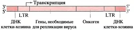
Reverse transcriptase is a remarkable multifunctional enzyme. When a retrovirus infects a cell, the viral reverse transcriptase copies the (+)-RNA strand into DNA. As in all replication processes, DNA Synthesis requires a primer. In retroviruses, this function is performed by a tRNA molecule that enters the virion from the host cell and interacts with the viral DNA through complementary base pairing. The resulting RNA/DNA hybrid is converted into single-stranded DNA following the Hydrolysis of the RNA. This RNase reaction is also catalyzed by reverse transcriptase. Next, the single-stranded DNA is copied by the same enzyme to form double-stranded DNA. The viral genome now exists as a standard DNA duplex (known as proviral DNA) and integrates into the host cell chromosome (Fig. 23.5). Another vital viral enzyme plays a key role in this process—integrase, which integrates the proviral DNA into the host chromosome. At each end of the double-stranded viral genome DNA (the proviral DNA) lies a base sequence called a Long Terminal Repeat (LTR) (Fig. 23.6), which participates in forming the "junctions" with the cellular DNA. As a result, the double-stranded viral DNA essentially constitutes an additional set of cellular genes that replicates alongside the host DNA during division. To generate new retroviral particles, the proviral genes (the viral genes within the host Chromosomes) are transcribed into (+)-RNA transcripts. Some of these serve as the genome for new retroviral progeny, while others undergo Processing into mRNA to direct the translation of proteins required for viral particle assembly.
Fig. 23.5. Replication of a hypothetical retrovirus

Fig. 23.6. Proviral genome of an oncogenic retrovirus integrated into the host cell genome. LTR stands for long terminal repeat. An oncogene may encode an abnormal regulatory protein or promote The production of excessive amounts of such a protein
The drug AZT (azidothymidine), whose structure is shown below, is used in the treatment of AIDS because it inhibits reverse transcriptase. To exert its effect, AZT must be converted into a triphosphate, which acts as a dTTP analogue. AZT triphosphate inhibits reverse transcriptase and also causes DNA chain termination due to the absence of the 3'-OH group. DNA polymerase is inhibited to a much lesser extent.
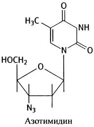
Retroviruses can be utilized in Gene Therapy to deliver a "healthy" gene capable of correcting a defect to the cells of a patient carrying a defective gene. The underlying principle involves disrupting the reproductive capacity of a retrovirus by deleting a gene and replacing a segment of its RNA with a DNA fragment equivalent to the gene intended for integration into the host chromosome. The drawback of this method is the random nature of integration, which carries inherent potential risks. Methods for site-specific gene insertion are currently under development.
Oncogenic Retroviruses
The integration of an oncogenic retrovirus's genetic material into a chromosome (in the form of its DNA, see Fig. 23.6) can lead to tumor formation. The first oncogenic virus to be described was Rous Sarcoma virus, which causes tumors in chicken Muscle tissue. Tumor development is driven by a single retroviral gene—an oncogene.
Since then, 18 to 20 oncogenes have been discovered in mammals. As discussed earlier in the context of DNA Hybridization, a small DNA fragment rendered single-stranded by heating will unerringly locate a complementary sequence among thousands of bases and bind to it via
Hydrogen Bonds. Methods have been developed to detect hybridization complexes (see p. 233). Remarkably, in every case, the retroviral oncogene responsible for causing a tumor has a "counterpart" in normal cells. This applies only to the exons of the normal gene, because RNA splicing ensures that the viral gene RNA lacks introns. During evolution, the viral oncogene apparently originated from its cellular counterpart. The only difference between them may be a divergence of a single base. The viral gene is designated by the prefix "v", while the corresponding cellular gene carries the prefix "c" (for cellular). These genes are given intriguing Abbreviations (see name explanations on p. 356): for example, c-myc and v-myc, c-ras and v-ras, etc. The cellular gene is referred to as a proto-oncogene. Proto-oncogenes have been found to encode proteins involved in crucial regulatory cellular mechanisms (see Chapter 26), the disruption of which can lead to uncontrolled Cell Division. Some of these encode abnormal Transcription factors (see p. 355). Evolutionarily, retroviral oncogenes may have originated from retroviruses that accidentally "picked up" an mRNA molecule transcribed from a cellular regulatory gene. The oncogenic viral form of such a regulatory gene could have arisen through mutation of the normal cellular counterpart either during the "capture" stage or afterwards. Upon reinsertion of this gene (via retroviral infection) into the host cell chromosome using the mechanism already described, the presence of the oncogene will lead either to the synthesis of an abnormal regulatory protein or to the overproduction of such a protein, resulting in disrupted cellular regulation (for details, see Chapter 26).
Bacteriophage Lambda
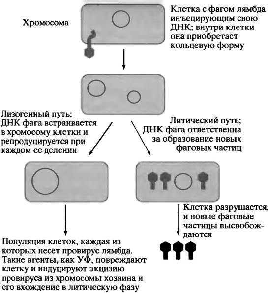
This bacterial virus has been intensively studied and is widely used in molecular biology and Gene cloning (see Chapter 24 and Fig. 23.2). Phage λ contains double-stranded DNA, and its life cycle exhibits several distinctive features. Upon entering a cell, the viral DNA faces a choice of two pathways. It can immediately replicate and produce new viral particles, which are released from the cell with the help of a phage-encoded enzyme called lysozyme; this enzyme breaks down the cell wall, leading to cell lysis. This is known as the virulent or lytic pathway (Fig. 23.7). The alternative is the lysogenic pathway, in which the phage genome integrates into the E. coli chromosome, where it can remain dormant indefinitely, replicating along with the host cell DNA. Consequently, a large population of otherwise normal E. coli cells may carry the phage DNA. A cell containing lambda DNA in its chromosome is called a lysogenic cell, and the integrated phage DNA is termed a prophage. If E. coli cell DNA is damaged by ultraviolet light, chemical agents, or ionizing radiation, the so-called SOS responses, designed to repair such damage, are activated. In a lysogenic cell, this type of damage triggers the excision of the prophage from the DNA via a complex mechanism that we will not discuss here. The excised prophage then enters the lytic replication cycle. Bacteriophage lambda provides a striking example of self-assembly. If the viral protein components are mixed with Viral DNA molecules in a test tube, a fully functional virus will spontaneously assemble, with the DNA packaged inside its head. Phage lambda is a powerful tool for gene cloning.
Fig. 23.7. Injection of bacteriophage λ DNA into a bacterial cell. Lytic and lysogenic replication pathways
What are Viroids?
Viroids are the smallest known infectious agents, being smaller and simpler than viruses. They consist of naked RNA molecules
devoid of any protein coat or other envelope (Fig. 23.8). Infecting plant cells with viroids requires mechanical damage to the cells (in experimental settings, plants are infected with viroid RNA by rubbing it onto leaves using an abrasive material). The effects of viroid infection can vary, manifesting as mild plant debilitation or ultimately leading to plant death, as is the case with palm trees. It remains unknown how viroids cause disease, and The Mechanism of viroid RNA replication is likewise an enigma. Another surprising fact is that viroids lack genes encoding proteins. To identify a protein-coding gene, one must find an Open Reading Frame, i.e., a sequence of triplets encoding a long stretch of Amino Acids. Although sequences responsible for Translation initiation have been identified in viroids, subsequent reading frames are so short (due to stop codons) that Protein Synthesis is precluded.
Fig. 23.8. Structure of the coconut cadang-cadang viroid. Loops represent unpaired regions, and cross-bars indicate base pairs. This is the smallest known viroid (246 nucleotides). Palm trees die 5–10 years after infection. Kindly provided by Professor R. Symons, Department of Plant Science, Waite Institute, University of Adelaide

Chapter 23 Questions
1. Explain how a virus can gain entry into a Eukaryotic Cell.
2. How must a single-stranded (+)-RNA virus (other than a retrovirus) differ from a single-stranded (-)-RNA virus?
3. Why does the vaccinia virus carry its own DNA-dependent RNA polymerase within its virion, even though the host cell has its own enzyme?
4. A retrovirus is a virus containing a (+)-strand RNA. How does it differ from a non-retrovirus containing a single-stranded (-)-RNA?
5. Explain how a virus with a lipid membrane acquires it along with its complete set of integral proteins.
6. Influenza epidemics sweep across the globe regularly. Most of them are relatively mild, but occasionally, as in 1918, epidemics occur with a high mortality rate. Why?
7. Why does the influenza virus cause hemagglutination when viruses and erythrocytes are mixed together?
8. The surface of the influenza virus exhibits neuraminidase activity. Why is this fact unusual? What is the function of the neuraminidase activity?
9. Describe the "choice" that bacteriophage λ must make upon infecting an E. coli cell.
10. A. WHAT IS A viroid? B. It is known that viroids infect plants and replicate there; therefore, they must have genes for protein synthesis. Is this true?
Last update: 06/08/2026
Editorial and Educational Adaptation: This material has been compiled based on the primary/original source text. The project team performed an editorial review, corrected technical inaccuracies, structured sections, and adapted the content for an educational format.
What was processed:
- elimination of formatting defects (OCR errors, structural breaks, corrupted characters);
- editorial organization of content;
- standardization of terminology in accordance with academic sources;
- verification of factual statements against the original source text.
All mentions of the author, publication year, and origin of the primary text have been preserved in accordance with the source.