BIOCHEMISTRY AND MOLECULAR BIOLOGY - W. ELLIOTT - 2002
CHAPTER 22. PROTEIN SYNTHESIS, INTRACELLULAR TRANSPORT, AND DEGRADATION
In the previous chapter, we examined The process of Gene METABOLISM/31.html">Transcription, which leads to The formation of mRNA. In this chapter, we will discuss the processes of Protein Synthesis, targeted delivery, and selective degradation. You will learn how mRNA is translated into protein. Most Introduction/5.html">Eukaryotic Cell Proteins are synthesized in the Cytoplasm (except for Mitochondrial and Chloroplast proteins), and specific mechanisms exist for their targeted delivery to The Nucleus, Plasma Membrane, mitochondrial membranes, and other cellular Organelles. Finally, different proteins are programmed for either short or long lifespans.
Basic principles of Protein Biosynthesis
Let us first consider the problem in general terms. mRNA is a relatively long molecule containing 4 types of nitrogenous bases. Proteins are synthesized from 20 different Amino Acids, and The base sequence in the messenger (mRNA) determines the Amino Acid Sequence in the protein. A code consisting of 1 nitrogenous base could encode only 4 amino acids; a code comprising 2 nitrogenous bases could encode 16 amino acids, which is still clearly insufficient for 20. Thus, at least 3 nitrogenous bases are required to encode 20 amino acids. Each amino acid in mRNA is "spelled out" as a triplet of nitrogenous bases called a codon. 4 different bases can form 64 triplets, or codons (4 • 4 • 4).
At first glance, evolution could well have limited itself to using 20 codons, ignoring the rest. However, random Mutations would then inevitably generate triplets that do not encode amino acids. This would lead to gene inactivation, since protein synthesis halts upon encountering an mRNA triplet that does not encode an amino acid.
Evolution chose an alternative strategy. Three codons were reserved as stop signals, indicating to the protein-synthesizing machinery that Protein synthesis is complete. These 3 triplets (one of them being UAA) do not encode amino acids.
If a mutation introduces a stop signal into the coding region of an mRNA, the protein product of that gene will not be produced. If there were more such stop triplets (44), mutations causing the cessation of protein synthesis would occur much more frequently (although in Bacteria, stop mutations can be overcome via suppressor mutations, but we shall not dwell on this).
All remaining triplets (61 in total) encode amino acids, meaning that a single amino acid can be encoded by several different triplets. This phenomenon is known as the degeneracy of The Genetic Code. As shown in Table 22.1, only Two amino acids — Methionine and Tryptophan — have a single codon each (AUG for methionine).
Class="center">Table 22.1. The Genetic Code
5′-End base |
Middle |
base |
3'-End base |
||
U |
C |
A |
G |
||
U |
UUU Phe UUC Phe UUA Leu UUG Leu |
UCU Ser UCC Ser UCA Ser UCG Ser |
UAUTyr UAC Tyr UAA Stop* UAG Stop* |
UGU Cys UGC Cys UGA Stop* UGG Trp |
U С А G |
С |
CUU Phe CUC Phe CUA Leu CUG Leu |
CCU Pro CCC Pro CCA Pro CCG Pro |
CAU His CAC His CAA Gln CAG Gln |
CGU Arg CGC Arg CGA Arg CGG Arg |
U С А G |
А |
AUU Ile AUC Ile AUA Ile AUG Met+ |
ACUThr ACCThr АСА Thr ACG Thr |
AAU Asn AAC Asn AAA Lys AAG Lys |
AGU Ser AGC Ser AGA Arg AGG Arg |
U С А G |
G |
GUU Phe GUC Phe GUA Leu GUG Leu |
GCU Ala GCC Ala GCA Ala GCG Ala |
GAU Asp GAC Asp GAA Glu GAG Glu |
GGU Gly GGC Gly GGA Gly GGG Gly |
U С А G |
* Stop triplet does not encode amino acids + The AUG triplet also serves as a Translation initiation codon
The remaining Amino acids have 2 or more codons; leucine even has 6. Codon distribution is not random. When multiple codons specify a single amino acid, they tend to be similar. For example, the isoleucine codons — AUU, AUC, and AUA — differ only at the third base position. Furthermore,
similar triplets encode amino acids with structural similarities: for instance, the aliphatic amino acids isoleucine and leucine (see p. 38) have the codons CUU (leucine) and AUU (isoleucine). Because of these Features of the genetic code, many mutations that alter only a single base often do not affect The Structure of the synthesized protein (changing CUU to CUC does not affect the encoded amino acid, as both codons specify leucine) or result in substitution with a structurally similar amino acid (changing CUU to AUU leads to the substitution of leucine with isoleucine). Isoleucine and leucine are so similar in size and hydrophobic properties that such a substitution may not affect protein function. Thus, the Organization OF THE genetic code creates a kind of genetic "buffer" that minimizes The impact of many point mutations on the synthesized protein.
How Codons Are Translated
Protein synthesis, known as translation, takes place on Ribosomes. We will discuss their Structure and function later; for now, we will focus on the translation of codons into amino acids.
There is no physical or chemical resemblance between an Amino Acid and its codon that would drive their direct association. It was therefore hypothesized that adaptor molecules exist to ensure the linkage of amino acids to their corresponding codons. It was later discovered that small RNA molecules act as these adaptors, and they were named Transfer RNAs, or tRNAs.
Transfer RNA, or tRNA
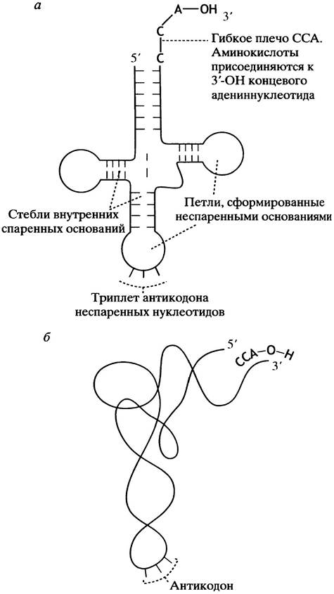
These are small RNA molecules. Their structure is schematically depicted as a cloverleaf (Fig. 22.1, a). Base pairing creates a double-helical stem with a loop of unpaired bases. The most crucial regions are the 3 unpaired bases that form the anticodon, and the flexible 3'-CCA arm where The amino acid attaches.
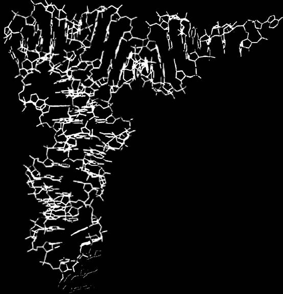
During biosynthesis, 2 tRNA molecules must fit side by side on the ribosome, and their anticodons must pair with adjacent codons on the mRNA. In reality, tRNA molecules are compactly folded (Fig. 22.1, b). Figure 22.2 illustrates a three-dimensional model of tRNA.
Fig. 22.1. Transfer tRNA. a - Structure of Transfer tRNA (cloverleaf model); b - schematic representation of the folded tRNA molecule
The structure of tRNA molecules is somewhat of a "black box". Although tRNAs contain modified, unusual bases, their core components are the anticodon and the amino acid attachment site. The anticodon is a base triplet complementary to the codon (Fig. 22.3). Thus, if the mRNA codon is UUU (which encodes phenylalanine), the corresponding anticodon on the tRNA molecule will be AAA. Importantly, this specific tRNA molecule will bind only phenylalanine. We will examine the amino acid attachment process shortly. As already noted, 61 codons specify amino acids, suggesting that an equal number of different tRNA molecules is required for translation. In reality, there are fewer varieties of tRNA. Naturally, there must be at least one for each of the 20 amino acids. Some tRNA molecules can recognize multiple codons, which, of course, must specify the same amino acid. This is achieved through a mechanism of wobble base pairing.
Fig. 22.2. Three-dimensional structural model of Yeast tRNAPhe. The CCA terminus and the anticodon loop are highlighted in color
The Wobble Hypothesis, or Wobble Mechanism
So far, in our Discussion of Replication and transcription, we have seen that the Watson-Crick principle of complementary base pairing is inviolable. Therefore, a slight departure from this rule during codon-anticodon base pairing might seem to introduce some confusion. However, this departure applies only to the first Base of the anticodon (with one exception, discussed later). This "improper" pairing of the first base results from The flexibility of the neighboring tRNA Structure (see Fig. 22.3), allowing U at this position to interact with either A or G on the codon, and G with either C or U. This is precisely what is known as wobble base pairing (see Fig. 20.17).
Fig. 22.3. Schematic representation of base pairing between a tRNA anticodon and an mRNA codon. To achieve antiparallel pairing, the tRNA molecule is flipped over. Therefore, while the tRNA structure shown in Fig. 22.1 (with the 5'-end on the left) is in its standard orientation, in the paired form it appears inverted (with the 3'-end on the left); mRNA is always depicted with the 5'-end on the left.

THE CONCEPT OF the "first base of the anticodon" requires some clarification. When an anticodon sequence is written by itself, just like any other nucleotide sequence, it is presented in the 5' -> 3' direction. However, during codon-anticodon interaction, the triplets are aligned in an antiparallel orientation. Therefore, to illustrate the same anticodon interacting with a codon, the codon is always written in the 5' -> 3' direction, while the "first" base of the anticodon must consequently be written in the opposite direction—on the right. This is why in diagrams the tRNA molecule alone is shown (in Fig. 22.1) with its 5'-end on the left, but when its base pairing with the codon is illustrated, the 5'-end is positioned on the right (in Fig. 22.3). Thus, the GGC anticodon will interact with the codon as shown below:
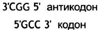
The wobble mechanism allows for alternative pairing of the same anticodon:
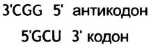
Since both GCC and GCU encode the same amino acid—Alanine—such pairing does not alter the amino acid sequence of the synthesized protein, but it allows a single tRNA to translate multiple codons. This enables The Cell to synthesize a smaller number of tRNA molecules.
Evolution has also provided other pathways that allow for more flexible codon-anticodon interactions without compromising translational fidelity. One such mechanism involves the incorporation of the non-standard base hypoxanthine (see Fig. 18.7) into the anticodon to enable its pairing with C, U, or A in codons.
How Amino acids are Attached to tRNA Molecules
A tRNA molecule attaches an amino acid in accordance with the mRNA codon, which is complementary to its anticodon. Thus, a tRNA molecule with the AAA anticodon is "responsible" exclusively for phenylalanine, since UUU is the complementary mRNA codon. The attachment of any other amino acid to this tRNA would lead to an error in the protein molecule's structure, as phenylalanine would then be replaced by a different amino acid. Phenylalanine-specific tRNA is designated as tRNAPhe. tRNAs for Other Amino Acids are denoted similarly, using three-letter Abbreviations for the amino acids (note that tRNAPhe simply specifies the type of tRNA, not whether Phe is currently attached to it; the latter is denoted as Phe-tRNAPhe). The attachment of amino acids to tRNAs is catalyzed by Enzymes called Aminoacyl-tRNA synthetases. Every cell must contain at least 20 Different types of these enzymes, each attaching a specific amino acid to its corresponding tRNA. This means that each enzyme recognizes one or more specific tRNAs and the appropriate amino acid, joining them together. Synthetase recognition of the correct tRNA can occur in two ways: in some cases, the anticodon is recognized; in others, several bases located in different PARTS OF THE molecule are identified. The overall reaction, utilizing ATP as an energy source, proceeds as follows:
Amino acid + tRNA + ATP <-> Aminoacyl-tRNA + PPi + AMP
This process is sometimes referred to as Amino Acid Activation.
Inorganic pyrophosphate is hydrolyzed to Pi, which drives the reaction forward from left to right. The accuracy of translation depends on the correct choice of a specific amino acid by synthetases. Once tRNA binds to its cognate amino acid, the aminoacyl-tRNA enters the protein-synthesizing machinery, which no longer verifies whether the correct tRNA was attached to the amino acid. Therefore, it is crucial that the amino acid-activating enzyme makes very few mistakes during operation. The Active Site of the enzyme is usually highly specific for its substrate, yet limits of precision do exist. The enzyme easily distinguishes between amino acids with vastly different properties, but it has a much harder time recognizing structurally similar amino acids, such as valine and isoleucine. Let us recall their structure.
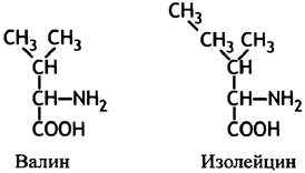
The difference in binding energy resulting from a single CH2 group is insufficient to provide high selectivity for isoleucine and valine. Without additional safeguards, the error rate would be unacceptably high, and leucine-specific aminoacyl-tRNA synthetase would attach valine to tRNAIle, leading to intolerable errors in mRNA Translation. Fortunately, this does not happen due to a proofreading mechanism, which can be described as a two-step overall reaction.
Amino acid + ATP Aminoacyl-AMP + PPi (1)
Aminoacyl-AMP + tRNA Aminoacyl-tRNA + AMP (2)
Aminoacyl-AMP does not leave the enzyme. The binding of tRNA induces a conformational change in the enzyme molecule, leading to the formation of
an additional catalytic site capable of hydrolyzing the incorrect aminoacyl adenylate. In the event of erroneous adenylation, the isoleucine-specific enzyme hydrolyzes valyl-AMP, whereas isoleucyl-AMP (presumably due to its much larger size) is excluded from this site. Such a mechanism reduces the error rate of isoleucine incorporation to 1 in 60,000 molecules. Other aminoacyl synthetases "proofread" in a similar manner by hydrolyzing incorrect aminoacyl-tRNAs. The hydrolytic site of the respective aminoacyl synthetase recognizes tRNAs carrying amino acids that are smaller (than the correct substrate) or of a similar size but possess different side chains. For example, Threonine and valine are similar in size; however, the hydrolytic site of the valine-specific enzyme preferentially binds the hydrophilic OH group of threonine rather than the hydrophobic CH3 group of valine. This dual selectivity—preferential binding of the correct amino acid to tRNA and preferential Hydrolysis of aminoacyl-tRNAs bearing an incorrect amino acid—reduces the error rate to about 1 in several thousand molecules. Not all aminoacyl-tRNA synthetases possess such proofreading mechanisms; they are required only for distinguishing structurally similar amino acids. A random error in protein biosynthesis does not have severe consequences because multiple protein molecules are produced, all of which eventually undergo degradation (with defective molecules presumably degraded more rapidly). Such errors are not as critical as errors in DNA replication; nevertheless, a high degree of fidelity remains essential.
The tRNA molecule features a terminal 3'-trinuclueotide sequence—CCA. The amino acid is linked via an ester bond to the free 3'-OH group of the ribose moiety of the terminal nucleotide (A) (Fig. 22.4). This trinucleotide forms a flexible arm that can position the aminoacyl group at the appropriate catalytic center of the ribosome (see below). The ester formed by the aminoacyl group has the same energy level as a peptide. Thus, there is no thermodynamic barrier to transferring the aminoacyl-ester group to the -NH2 group of another aminoacyl moiety during peptide bond formation. In other words, the energy required for peptide bond formation is provided by the aminoacyl-tRNA synthetase utilizing ATP. Next, we will examine how amino acids are organized by tRNA molecules into a polypeptide chain.
Fig. 22.4. Attachment of an amino acid to tRNA. a - A tRNA molecule with CCA bases at the 3'-end, covalently linked to an amino acid via an ester bond; b - STRUCTURE OF THE terminal nucleotide with the attached amino acid. In solution, the aminoacyl group rapidly migrates between the 2'- and 3'-OH groups of the ribose ring. In the text, we consider it in the 3'-position

Ribosomes
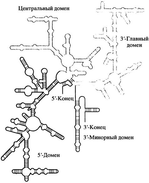
Ribosomes are small particles present in large numbers in Cells (with the exception of highly differentiated cells, such as erythrocytes, which do not synthesize proteins). They derive their name from ribonucleic acid (RNA), which accounts for about 60% of their dry weight. A ribosome consists of two subunits: in E. coli, the large subunit contains 2 RNA molecules, while the small subunit contains one. We have already examined mRNA and tRNA. Ribosomal RNA (rRNA) also has unique features. Due to internal base-pairing, rRNA forms highly twisted, compact structures. The diagram in Fig. 22.5 illustrates the complex shape of one of the RNA molecules from an E. coli ribosome—specifically, the 16S rRNA of the small subunit. The large subunit contains two distinct rRNAs, sedimenting at 23S and 5S. Numerous proteins are associated with rRNA to form dense particles; 34 proteins are part of the large subunit and 21 belong to the small subunit.
Fig. 22.5. Structure of 16S rRNA (schematic diagram)
When dealing with very large structures such as ribosomes, their size is estimated by their sedimentation rate during ultracentrifugation, expressed in Svedberg units, denoted as S. The larger the molecules, the faster they sediment. The size of the E. coli ribosome is 70S, and its subunits are 50S and 30S (the sum of these values does not equal the arithmetic addition of the subunits because the S value depends not only on size, but also on shape).
The general principle of protein synthesis, in highly simplified terms, is as follows. The ribosome attaches near the 5' end of the mRNA and then moves along the mRNA toward the 3' end, linking the aminoacyl groups of charged tRNA molecules (i.e., tRNA molecules with attached amino acid residues) into a polypeptide chain According to the codon sequence. At the end of the mRNA, the ribosome encounters a stop codon: at this point, the protein is released, and the ribosome detaches and dissociates into its subunits. Note that there are no "specialized" ribosomes. Within a given cell, any ribosome can use any mRNA, much like a tape player can play any recorded tape; restrictions exist only for mitochondrial and chloroplast ribosomes, which differ from cytoplasmic ribosomes (see p. 296).
However, this is merely the basic principle. To truly understand the process, we need to examine the details. Protein biosynthesis can be divided into three stages: initiation, elongation, and termination.
Translation Initiation
Translation begins at a strictly fixed point on the mRNA chain, which features 5' and 3' untranslated regions flanking a central coding region. The ribosome must recognize the first triplet of the coding sequence and initiate translation from it. Absolute precision in initiation is critical, as the correct translation of mRNA by the ribosome relies on the exact Setting of the reading frame. Suppose the coding region of an mRNA begins with the sequence:
5' AUGUUUAAACCCCUG................. 3'.
The first 5 amino acids are specified by the codons AUG, UUU, AAA, and so on. There are no punctuation marks to indicate which bases constitute a codon, with the exception of the first three bases of the coding region, which form constitutive codon 1, the next triplet forming codon 2, and so forth. There are neither commas nor periods between them. Consequently, the correct reading of information is determined solely by the starting point. Suppose a single-base error occurs and reading begins at base 2. In this case, the codons would be read as:
(A) UGU, UUA, AAC, CCC, UG ...
In other words, the translated codons, as well as their corresponding amino acids, would be completely different. This type of error is known as a frameshift mutation. Mutations caused by the deletion or insertion of one or two bases in the mRNA result in a reading frameshift. This causes the amino acid sequence of the peptide synthesized after the frameshift to be incorrect, rendering the resulting product incapable of
performing the Functions of a protein sculpted by millions of years of evolution and encoded by the given gene. While the General scheme of PROTEIN SYNTHESIS AND its participating components is largely applicable to both PROKARYOTES AND EUKARYOTES, significant differences exist in the actual mechanisms, so we will examine them separately. Let us begin with prokaryotes.
Translation Initiation in E. coli
As already noted, upstream of the start point in the mRNA, There is a sequence located near the 5' end of the chain that is recognized by the ribosome. Since initiation does not simply start at the 5' end of the mRNA, the first codon must be specifically identified.
The starting point is the AUG codon (more rarely GUG); however, paradoxically, the AUG triplet can be located anywhere within the mRNA because it also encodes the amino acid methionine. For a long time, it remained unclear: how do ribosomes initiate translation specifically at the first AUG rather than any other AUG within the mRNA? Why not use a specialized codon for initiation? As it turns out, There are two distinct tRNAs specific for methionine. Both tRNAs share the same anticodon, but one tRNA is used exclusively for initiation, while the other is used solely for adding methionine during elongation.
The translation-initiating tRNA can pair with AUG or GUG codons through "wobble" base-pairing (see p. 287). Typically, wobble involves the 5' base of the anticodon. In the tRNA used for translation initiation, this corresponds to the 3'-terminal base of the anticodon (Fig. 22.6).
Fig. 22.6. The anticodon loop, illustrating the "wobble" position. a – Conventional tRNAs involved in elongation; b – initiator tRNAfMet. The wobble base is determined by the adjacent structure of the tRNA. The Significance of tRNAfMet is explained in the text

What determines the distinct Functions of the two methionine-specific tRNAs? The initiator aminoacyl-tRNA possesses structural features that are recognized by an initiation protein, or initiation factor IF2, which delivers the initiator aminoacyl-tRNA to the assembling initiation complex. The elongation-participating aminoacyl-tRNA recognizes a different cytoplasmic factor that delivers it to the ribosome; this factor does not bind to the initiator tRNA. In E. coli, the methionine attached to the initiator tRNA undergoes formylation at its NH2 group. This process is catalyzed by transformylase, which uses N10-formyltetrahydrofolate (see p. 219) as the formyl group donor, meaning that prokaryotic protein synthesis—for reasons still unclear—begins with N-formylmethionine. The formyl group (and frequently the methionine as well) is removed before synthesis is complete. The initiator tRNA is generally designated as tRNAfMet ("f" stands for formyl), and its amino acid-charged counterpart as fMet-tRNAfMet (often abbreviated as fMet-tRNAf). The foregoing clarifies how the correct utilization of methionine-specific tRNAs for initiation and elongation is achieved: specific protein factors involved in initiation and elongation recognize exclusively Met-tRNAfMet and fMet-tRNAfMet, respectively.
In the cytoplasm, there is a pool of free 30S and 50S ribosomal subunits in equilibrium with 70S ribosomes. Initiation factor IF3 binds to the 30S subunit, preventing its premature reassociation with the 50S subunit at this stage (Fig. 22.7, a). In addition, other factors are required,
namely IF1 and IF2. These protein factors bind to the 30S subunits, participate in the initiation process, and are subsequently released back into the cytoplasm to function in a new round of initiation. The function of IF1 remains unknown, whereas IF2 is essential for binding fMet-tRNAfMet.
Fig. 22.7. The Effect of translation initiation factor IF3 and the Shine-Dalgarno sequence on 70S ribosomal subunits. a – Dissociation of the 70S ribosome into 50S and 30S subunits. Initiation factor IF3 prevents the premature reassociation of the 50S and 30S subunits. During translation initiation, IF3 must be released to allow 50S joining;
b – diagram illustrating The Role of the Shine-Dalgarno sequence in positioning the E. coli 30S ribosomal subunit on the mRNA during initiation. P – peptidyl site, A – aminoacyl site. The P and A sites are fully formed only upon the attachment of the 50S subunit

mRNA, fMet-tRNAfMet, and GTP form a complex with the 30S subunit, and IF3 is released into the cytoplasm. As shown in Fig. 22.7, b, the mRNA contains a base sequence known as the Shine-Dalgarno sequence, which is complementary to a region of the 16S rRNA. Upon their binding, the mRNA is properly positioned on the small ribosomal subunit, the tRNA approaches the P-site of the subunit, and its anticodon pairs with the AUG codon (Fig. 22.8).
Following the release of IF3, this complex associates with the 50S subunit, a process accompanied by the hydrolysis of GTP and the release of GDP and Pi, as well as IF1 and IF2 (see Fig. 22.8). We now have a complete 70S ribosome with the mRNA positioned in the P-site holding fMet-tRNAf (whose anticodon is paired with the initiation codon AUG) and an empty A-site awaiting the delivery of the second amino acid corresponding to the next tRNA. Initiation is complete.
Fig. 22.8. Translation initiation in E. coli. The initiator tRNA, tRNAfMet, is shown as a grey line; the anticodon is represented by a short horizontal line. Factor IF2 delivers fMet-tRNAfMet to the 30S subunit. NNN represents any codon (N denotes any nucleotide). The ribosome also features an exit site, which is not shown in the diagram

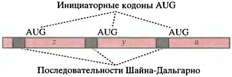
Some bacterial mRNAs are polycistronic; for example, the lac mRNA. A single mRNA molecule contains three regions that encode 3 different proteins. In this situation, each coding region is adjacent to a Shine-Dalgarno sequence, and translation initiation occurs independently in each case (Fig. 22.9).
Fig. 22.9. Structure of a prokaryotic polycistronic mRNA. The coding Regions of the lac mRNA are designated as z, y, and a (see Fig. 21.9)
Elongation is the stage of translation that follows initiation
Cytoplasmic elongation factors
The cytoplasm contains two soluble protein elongation factors. These are G-proteins that can bind to ribosomes when complexed with a GTP molecule. Both proteins function as latent GTPases. On the ribosome, they hydrolyze the bound GTP molecule to GDP and Pi; the release of inorganic phosphate triggers Conformational Changes in these proteins. Complexed with GDP, they dissociate from the ribosome. In the cytoplasm, GDP is exchanged for GTP, and the factors are ready to participate in the next round of elongation.
These two factors are EF-Tu (elongation factor Temperature unstable) and EF-G. The role of EF-Tu is to deliver aminoacyl-tRNA to the ribosome. Once the aminoacyl group is added to the growing peptide chain, EF-G promotes the translocation of the ribosome along the mRNA in the 5' -> 3' direction toward the next codon.
These two factors alternately move across the ribosome in their GTP-bound state, perform their respective functions, and dissociate in their GDP-bound state to participate again in subsequent rounds of elongation. We can now proceed to a detailed examination of peptide bond synthesis.
Mechanism of elongation
Let us begin with the initiation complex shown in Fig. 22.10, a. This complex contains fMet-tRNAfMet in the P-site and an empty A-site. Only during initiation does the P-site accept a tRNA charged with the amino acid N-formylmethionine; all subsequent aminoacyl-tRNAs enter the A-site. Aminoacyl-tRNAs (other than the initiator) form a complex with elongation factor EF-Tu bound to a GTP molecule. The EF-Tu-GTP-aminoacyl-tRNA complex binds to the ribosome such that the aminoacyl-tRNA occupies the A-site, and its anticodon aligns with the mRNA codon (Fig. 22.10, b). It is important to note that EF-Tu-GTP does not bind to fMet-tRNAf (the initiator tRNA), but interacts exclusively with aminoacyl-tRNAs involved in elongation. Thus, initiation and elongation occur separately. The concentration of EF-Tu in E. coli cells is fully sufficient to bind all aminoacyl-tRNAs. On the ribosome, EF-Tu, possessing GTPase activity, hydrolyzes the bound GTP molecule to GDP and Pi, releasing the latter. This serves as a signal for the dissociation of EF-Tu from the aminoacyl-tRNA, presumably As a result of allosteric changes, and EF-Tu-GDP leaves the ribosome to be regenerated via GDP-GTP exchange in the cytoplasm (Fig. 22.10, c).
Fig. 22.10. The elongation process. tRNAs are depicted as grey lines; AA represents the aminoacyl group. THE POSITION OF EF-Tu-GTP on the tRNA and the ribosome is shown schematically

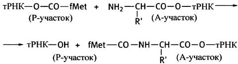
The aminoacyl groups on the two tRNA molecules located in the P- and A-sites are brought into close proximity to ribosomal peptidyl transferase; the latter catalyzes The transfer of fMet from the tRNA in the P-site to the free amino group of the aminoacyl-tRNA in the A-site, thereby forming a dipeptide attached to the tRNA (Fig. 22.10, d). We have described peptidyl transferase as an enzyme, but evidence indicates that this catalytic activity persists in ribosomes even after the extraction of 95% of the ribosomal proteins. This provides strong argument that this activity is an intrinsic property of ribosomal RNA.
The scheme of the peptidyl transferase reaction is as follows:
Following the Synthesis of the first peptide bond, the A-site is occupied by peptidyl-tRNA, while the P-site contains uncharged tRNA. The ribosome moves by one codon along the mRNA—a process known as translocation. The deacylated tRNA moves to the "exit" site (E-site) located on the 50S subunit, from which it is released for subsequent reutilization (Fig. 22.10, e). The movement of the ribosome along the mRNA requires factor EF-G, also known as translocase. The translocation process requires the hydrolysis of GTP. The binding of EF-Tu-GTP and EF-G to the ribosome occurs in such a way that only one of these factors can be bound to the ribosome at any given moment. Consequently, Peptide Synthesis and translocation take place alternately (in succession).
Figure 22.10 does not detail the mechanism by which the peptidyl transferase reaction and translocation progress from stage (c) to (d). Recent studies have shown that during this process, each tRNA occupies two sites on the ribosome simultaneously (Fig. 22.11). Once released from its peptide group, the tRNA occupies the P and E sites (E standing for exit); the anticodon end of the molecule remains in the P-site, while the other end occupies the E-site. Similarly, the tRNA located in the A-site (carrying the peptide) spans the A and P sites.
Both models are based on the observation that tRNAs occupy the P/E and A/E sites on the ribosome. According to model I, the aminoacyl-tRNA rocks in order to take a step; model II involves the formation of hybrid sites on the ribosome. It is hypothesized that synthesis proceeds in such a way that the peptide always remains in a fixed position relative to the large subunit.
Two alternative models have been proposed to explain The Mechanism of this process. Model I (see Fig. 22.11) assumes that one end of the aminoacyl-tRNA rocks as shown in the figure, peptide transfer occurs, and the deacylated tRNA swings one of its ends toward the E-site. The subsequent translocation then returns the system to the state shown in Fig. 22.10, d, where everything is ready for the next round of elongation.
Fig. 22.11. Alternative models of translocation and peptide synthesis on the ribosome. In this diagram, the synthesis of the first peptide bond is chosen as an example, so the grey circle denotes fMet attached to the tRNA in the P-site, which is positioned at the first codon of the mRNA; at the end of this round, the P-site is located at the second codon. Rectangles indicate binding sites: E, exit site; P, peptidyl site; A, aminoacyl (acceptor) site. E/P and A/P are hybrid centers formed by the movement of the large subunit relative to the small one

According to alternative model II, the large ribosomal subunit moves while the small subunit remains stationary. This results in the formation of hybrid P/E and A/P binding sites. Following the peptidyl transferase reaction, these hybrid sites become occupied (see Fig. 22.11). Subsequent translocation of the small subunit leads to the state predicted by model I, where everything is set for the incorporation of the next amino acid.
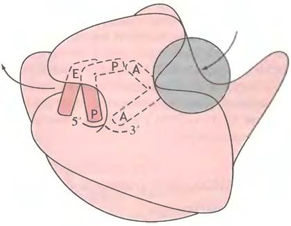
In both models, the nascent peptide remains in a fixed position relative to the large subunit. This overcomes the physical challenge of moving a tRNA molecule burdened with a relatively large peptide. Model II also provides an explanation for why ribosomes invariably consist of two subunits. The ribosome features a highly complex, tunnel-like structure (Fig. 22.12).
Fig. 22.12. The ribosome with the probable arrangement of ribosome-bound tRNAs in the A/A, P/P, and E sites (schematic). The gray area on the right indicates the approximate region of EF-Tu interaction. The diagram shows the polarity of the mRNA fragment containing codons in the A and P sites. Arrows indicate the putative pathway of tRNA through the ribosome.
Thus, the ribosome is now in the state shown in Fig. 22.10, e. The P site is occupied by peptidyl-tRNA, whereas the A site, positioned at the third codon, is vacant. Polypeptide chain elongation involves repeated cycles of the same process. Aminoacyl-tRNA, forming a complex with EF-Tu-GTP, is delivered to the A site; the peptidyl group from the P site is transferred to it; the deacylated tRNA moves toward the "exit" (E site); the peptidyl-tRNA shifts from the A site to the P site; and the ribosome advances by one codon—whereupon the entire cycle begins anew. When a protein comprising 200 amino acids is synthesized, the final round involves transferring a 199-amino-acid peptide to the terminal amino acid and its tRNA, yielding a protein-tRNA complex. Metaphorically speaking, the dog is added to the tail, not the other way around. The N-terminus of the polypeptide chain is synthesized first, and the last added amino acid forms the C-terminus.
Translation fidelity is achieved through codon-anticodon interaction, which dictates the Selection of the correct tRNA; however, the precise mechanism of this selection remains unclear. The issue is that EF-Tu cannot "recognize" which aminoacyl-tRNA should come next, and it appears to deliver them to the A site indiscriminately. One hypothesis suggests that unrecognized aminoacyl-tRNA-EF-Tu-GTP complexes undergo dissociation prior to the reaction because they are not held as firmly in the A site as they would be through proper hydrogen bonding during accurate codon-anticodon recognition. Peptide synthesis does not proceed until EF-Tu-GDP is released, which can only occur following GTP hydrolysis. It is hypothesized that this brief delay is necessary for "proofreading," thereby ensuring process efficiency. Furthermore, ribosomes are thought to participate in some yet undetermined manner in recognizing the correct aminoacyl-tRNA. At present, however, this remains merely a hypothesis, and no definitive mechanism has yet been proposed to explain it.
Termination of Protein Biosynthesis
The mRNA molecule terminates with at least one of three stop codons (also known as termination codons) for which no corresponding tRNA exists: the triplets UAG, UAA, and UGA. When the ribosome encounters a stop codon, a specialized cytoplasmic release factor (termination factor) promotes the release of the completed polypeptide from the tRNA. It induces a conformational change in peptidyl transferase, causing the enzyme to hydrolyze the ester bond between the C-terminal carboxyl group of the protein and the hydroxyl group of the 3'-terminal nucleotide. The ribosome then detaches from the mRNA and dissociates into subunits ready for the next initiation event. Two distinct factors are known to recognize different stop codons.
WHAT IS A Polysome?
The synthesis of a single E. coli protein on a ribosome—which incorporates about 15 amino acids per second—takes roughly 20 seconds. However, as soon as an initiated ribosome
has advanced by approximately 30 codons, another translation initiation event can occur. Consequently, multiple ribosomes move sequentially along the same mRNA molecule, each independently synthesizing a protein copy. Typically, a single mRNA molecule accommodates about five ribosomes, although this number varies depending on the length of the mRNA. Such a multi-ribosome structure is referred to as a polysome.
How does protein synthesis differ in eukaryotes?
In eukaryotes, the translation process is essentially similar to that in prokaryotes, albeit with a few distinct differences. Eukaryotic ribosomes are larger (80S, consisting of 40S and 60S subunits) and contain a greater number of rRNA and protein molecules. Methionine is always the first amino acid in the synthesized protein; however, the methionyl-tRNA used for initiation is not formylated (the Biological Significance of prokaryotic formylation remains unclear). Just like prokaryotes, eukaryotes employ a specialized initiator methionyl-tRNA that is distinct from the elongation tRNAs. This does not imply that all eukaryotic proteins start with methionine, as this amino acid is frequently cleaved off from the polypeptide chain later on. The assembly of the 40S initiation complex mirrors the Formation of the 30S initiation complex in prokaryotes. The resulting complex then associates with the large 60S ribosomal subunit to finally establish the complete initiation complex. The protein biosynthesis process is likewise accompanied by GTP hydrolysis. However, in eukaryotes, the correct positioning of mRNA—specifically the precise alignment of the P-site with the AUG initiation codon—relies on a completely different mechanism. Recall that the 5' ends of eukaryotic mRNAs are capped with a methylated guanine nucleotide (p. 270). Eukaryotic mRNAs lack a Shine-Dalgarno sequence; instead, a group of protein factors binds to the cap and associates with the 40S ribosomal subunit (Fig. 22.13). Thus, at each 5' end of the mRNA, at a certain distance from the AUG codon, we find the 40S preinitiation complex. Driven by an ATP-dependent mechanism, the 40S subunit complex moves along the mRNA until it encounters the first AUG triplet, which triggers the joining of the 60S subunit and the completion of initiation. This process is accompanied by GTP hydrolysis. Unlike prokaryotic mRNAs, which are frequently polycistronic and contain multiple Shine-Dalgarno sequences to ensure the initiation of each Cistron, eukaryotic mRNAs are monocistronic (coding for a single polypeptide) and contain only a single initiation site per molecule. The functions of prokaryotic EF-Tu and EF-G are performed by EF1α and EF2 in eukaryotes, respectively.
Fig. 22.13. Simplified diagram of eukaryotic initiation. In addition to eIF2, several other initiation factors are involved in this process. tRNAMet stands for initiator RNA; eIF2 stands for the eukaryotic initiation factor corresponding to the prokaryotic factor IF2

How does protein synthesis occur in Mitochondria?
Mitochondria contain DNA and possess their own protein-synthesizing machinery. A widely accepted hypothesis suggests that mitochondria evolved from Prokaryotic Cells that invaded ancestral Eukaryotic cells (p. 282).
Similar to prokaryotes, mitochondrial ribosomes utilize fMet-tRNAfMet for initiation. They also exhibit other fascinating features: a slightly modified genetic code and simpler codon-anticodon interactions, allowing mammalian mitochondria to function with just 22 types of tRNA. Such simplifications are likely possible because mitochondria synthesize a limited repertoire of different proteins. A mitochondrion is not an autonomous entity; the vast majority of mitochondrial proteins are encoded by nuclear genes and subsequently imported into these organelles. It is also believed that Chloroplasts originated from photosynthetic prokaryotes that established an endosymbiotic relationship with eukaryotic cells.
Effects of Antibiotics and toxins on protein synthesis
Antibiotics act as the chemical "missiles" of microorganisms, deployed against competitors in the struggle for survival. They target vital components of cellular metabolism, and several of them interfere directly with translation.
In prokaryotes, streptomycin disrupts initiation, kirromycin prevents the release of EF-Tu, while erythromycin and chloramphenicol inhibit peptidyl transferase (the latter is also true for mitochondrial ribosomes). Fusidic acid impedes translocation by blocking the release of the EF-G-GDP complex.
Furthermore, diphtheria toxin inhibits eukaryotic eEF2 translocase (which is the functional equivalent of bacterial EF-G). Ricin, a toxin derived from the castor bean plant, is an N-glycosidase that removes an adenine base from a specific eukaryotic ribosomal RNA, thereby inactivating the large ribosomal subunit. A single molecule of ricin can destroy a cell containing tens of thousands of ribosomes.
How does the polypeptide chain synthesized on the ribosome fold?
It was long believed that once a polypeptide chain was synthesized on the ribosome, the protein spontaneously folded into its three-dimensional structure solely through the interactions of its amino acid side chains. Of course, this concept is fundamental, as we know that Protein Structure is dictated entirely by a single type of information—the base sequence in the gene, which is translated into the amino acid sequence of the polypeptide. Furthermore, experimental evidence Supports this notion. The enzyme Ribonuclease was denatured by Treatment with urea (which disrupts Hydrogen Bonds) and by reducing its Disulfide Bonds. Upon removing the urea via dialysis and reoxidizing the protein, a fraction of the enzyme spontaneously refolded into its correct conformation and even recovered its catalytic activity. Thus, the amino acid sequence of a protein undoubtedly dictates its native conformation. However, a polypeptide chain offers virtually limitless possibilities for intramolecular associations among amino acid residues. As the protein is being synthesized, a hydrophobic stretch in one part of the chain may interact with another hydrophobic segment; yet this contact might be completely "inappropriate" and absent in the native protein. Theoretically, one could imagine that the polypeptide "samples" every conceivable internal association until it reaches the Free energy minimum of the native protein. This trial-and-error approach to folding has been calculated to take millions of years, whereas in a living cell, the entire process takes mere minutes.
Following the successful refolding of ribonuclease, similar in vitro experiments were conducted with other small, single-domain proteins. Attempts to refold larger proteins, particularly multi-domain ones, have met with far less success. Since the majority of proteins fall into the latter category, it is evident that the folding of large molecules poses a formidable challenge.
The in vivo protein folding mechanism—by which newly synthesized Peptides acquire their correct three-dimensional configuration—is not yet fully understood. It is believed that certain polypeptide fragments can rapidly adopt a Secondary structure, which somehow facilitates the correct folding of the entire molecule. This latter assumption forms The basis of the modular folding hypothesis, in which folded modules form rapidly and assist in the folding of the rest of the molecule. Thus, protein folding appears to occur in several stages, although the details of this process remain elusive.
Up to this point, we have discussed protein folding as an autonomous, unaided process. However, there are two classes of proteins
involved in folding. The first comprises traditional enzymes, and the second consists of molecular chaperones. We will examine them in this order.
Enzymes Involved in Protein Folding
The first enzyme is protein disulfide isomerase (PDI), which "reshuffles" disulfide (S–S) bonds within the polypeptide chain. If an erroneous S–S bond forms, its covalent nature prevents it from breaking spontaneously, locking the polypeptide into an incorrect configuration. By breaking and reforming S–S bonds between various Cysteine residues, PDI helps correct the folding process. High concentrations of PDI are found in The Endoplasmic reticulum, where secretory proteins undergo folding, many of which contain disulfide bonds.
Another enzyme, peptidyl-prolyl isomerase (PPI), catalyzes the cis-trans isomerization of Proline peptide bonds, helping the protein adopt the correct configuration. A whole class of remarkable proteins called cyclophilins has been discovered that possess prolyl isomerase activity. They bind the immunosuppressant drug cyclosporine (used in organ transplantation). The connection between proline isomerase and the Biological Role of cyclophilins remains poorly understood.
Molecular Chaperones and Folding
Chaperones (derived from the French term for an elderly woman who chaperones a young lady at balls) are a family of specialized intracellular proteins that ensure the rapid acquisition of the correct Spatial Structure. They can recognize and bind to partially folded (or unfolded) proteins. As a protein emerges from the ribosome, "erroneous" interactions can occur—for instance, between hydrophobic patches—which would hinder the proper folding of the molecule. The binding of chaperones to such regions stabilizes the partially folded molecule until correct protein folding can take place. This means that chaperones must eventually dissociate from the polypeptide; following dissociation, favorable conditions are created for the completion of proper folding. Although the details of this mechanism remain unclear, the essential role of chaperones in folding polypeptide chains is undeniable. The complexity of this system is further evidenced by the requirement for ATP to release chaperones from the polypeptide chain. Note that chaperones affect The kinetics of the folding process rather than the final Tertiary Structure of the protein, which is determined entirely by its amino acid sequence.
Prion Diseases and Protein Folding
There is a group of fatal neurological diseases affecting humans and animals. It includes Creutzfeldt-Jakob disease and kuru in humans. The latter is known as "laughing death" due to the facial grimaces it causes. It is believed that kuru is contracted through cannibalism in certain tribes of New Guinea. In sheep, this disease is called scrapie because afflicted animals scrape their wool against fence posts. In cattle, it is known as bovine spongiform encephalopathy (BSE), or mad cow disease. These diseases can be transmitted by consuming infected tissue. Hereditary forms are much rarer. It was previously thought that all these conditions were caused by "slow Viruses" because they are infectious and develop insidiously over a span of years. However, all attempts to detect Nucleic Acids in infectious material isolated from the Brain have failed, ruling out the involvement of a conventional infectious agent (such as a virus) in the disease's Pathogenesis. Nevertheless, the infectious agent appears to replicate.
It turned out that the infection is caused by a protease-resistant form of a normal protein—the prion protein (PrP)—found in the brain. The disease-causing protein is named a prion, an acronym for "proteinaceous infectious particle."
The protein that infects mice with scrapie is designated PrPsc, and its normal counterpart is PrPc ("c" stands for constitutive). Both represent the exact same polypeptide and are encoded by the same gene, yet their Conformations differ: PrPsc is characterized by a high content of β-sheets, which are virtually absent in PrPc. Unlike PrPc, the PrPsc protein readily forms aggregates that lead to the amyloid plaques characteristic of prion diseases. This raises the question: how can a misfolded protein become infectious and self-replicate? After all, none of the Biochemical Mechanisms of protein synthesis we have examined allow a protein to direct its own replication. Nevertheless, there is compelling evidence
that PrPsc somehow converts PrPc (the normal protein) into the abnormal conformation. This was confirmed by the co-incubation of the two proteins.
Models for this conversion mechanism involve the participation of a chaperone and an energy source to unfold the PrPc protein and refold it under the Influence of the PrPsc molecule. According to an alternative "nucleation-polymerization" model, PrPc molecules are captured by a PrPsc aggregate, after which the conformational reorganization of PrPc takes place. Due to experimental limitations, there is still no direct in vitro proof that a protein acquires infectious properties through the events described above.
In the absence of infection, The conversion of PrPc to PrPsc is a rare event, which is why spontaneous cases of the disease are extremely uncommon. It is believed that mutations in the gene encoding normal PrP can increase the likelihood of this post-translational conversion, leading to hereditary forms of the disease. Thus, once formed, PrPsc is capable of triggering its own further autocatalytic production.
Other functions of Molecular Chaperones
We have examined the role of chaperones in protein folding, but this is only part of their repertoire. As already noted, chaperones can bind to unfolded or partially folded proteins—such as a polypeptide just released from ribosomes.
However, Proteins can also partially denature, unfolding to varying degrees under The Influence of various factors: heat, excessive oxidation, ionizing radiation, ultraviolet light, and so on. Protein Denaturation can likewise be triggered by various cellular stresses. For a long time, it was believed that heat Shock induces the synthesis of specialized proteins whose role was thought to be cellular protection. If the temperature of E. coli is raised to 42°C, there is a surge in the synthesis of heat shock proteins (p. 267). It turned out that these are actually chaperones, which is why they are sometimes referred to as stress proteins.
In addition to participating in the folding of Newly synthesized proteins, chaperones play a vital role in unfolding and stabilizing denatured proteins, as well as tagging Aging proteins for subsequent degradation by Lysosomes. Chaperones are also essential for transporting proteins across mitochondrial membranes, reorganizing subunits within complexes, and assembling oligomeric Protein Complexes. A particularly apt definition of this class of proteins was provided by Hendrick and Hartl (see the "Further Reading" section): "We now define a molecular chaperone as any protein that interacts with, stabilizes, or helps another protein to acquire its conformation without being bound to the final product. By controlling the binding and release of this protein substrate, the chaperone facilitates its correct folding in vivo—whether it be folding, oligomeric assembly, transport into a specific cellular compartment, or the controlled switching of active/inactive conformations."
The final point concerning conformational switching relates to intracellular glucocorticoid Hormone Receptors (see p. 346).
Carrying out such diverse functions requires a variety of many different chaperones, A large number of which have already been identified.
How Are Synthesized Proteins Delivered to Their Destinations?
There is only one type of ribosome in the cell, and all of them are located in the cytoplasm (with the exception of prokaryote-type ribosomes found in Mitochondria and chloroplasts). Which protein is synthesized by a ribosome at any given moment depends entirely on the mRNA currently being translated. Newly synthesized proteins have diverse final destinations. Many are cytoplasmic: upon completion of synthesis, they are released from the ribosomes and remain in the cytoplasm. At the same time, many proteins reside in The cell membrane. How do they get there? Consider a Liver cell, which produces Blood Plasma Proteins. These are large proteins, yet cellular membranes are structured in a way that prevents protein "leakage." How, then, does the selective release of blood proteins occur? A similar question arises regarding the release of any extracellular protein, such as digestive enzymes or Insulin from the Pancreas. There are other challenges as well. Most mitochondrial proteins are synthesized in the cytoplasm. How are they selectively transported into mitochondria? Lysosomes and Peroxisomes (see Chapter 16) are membrane-bound vesicles filled with enzymes, yet they cannot synthesize proteins themselves. How are these proteins transported into such vesicles?
We will now explore the various types of protein translocation. Let us begin with those that are first transported into the endoplasmic reticulum (ER).
What is the Endoplasmic Reticulum (ER)?
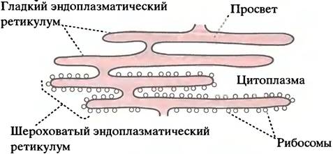
If you examine a section of the liver or pancreas under an Electron microscope, you will see a large number of membranes inside the cell enclosing a vast network of flattened cavities. This intricate system of interconnected cavities is called the endoplasmic reticulum (ER). The cytoplasm lies outside the ER, whereas the ER lumen forms a distinct compartment bounded by the ER membrane (see the electron micrograph in Fig. 3.15). Part of the ER is studded with ribosomes, giving it a rough appearance under the electron microscope; this is known as the rough endoplasmic reticulum. Other regions lack ribosomes and are therefore referred to as the smooth endoplasmic reticulum (Fig. 22.14). Proteins destined, for instance, for the nucleus or mitochondria are first transported into the lumen of the rough endoplasmic reticulum, which brings us to the next question.
Fig. 22.14. Structure of the endoplasmic reticulum
How do proteins cross the ER membrane?
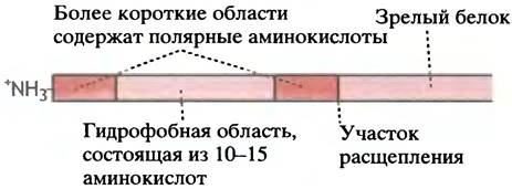
A protein destined to cross The Plasma Membrane, enter lysosomes, or reach the ER lumen contains a leader sequence at its N-terminus, typically consisting of 25 ± 11 amino acids. This leader amino acid sequence defines a common structural feature of many proteins transported into various compartments (Fig. 22.15). The mature protein is released following proteolytic Cleavage at a site with a specific sequence.
Fig. 22.15. Attachment of a typical leader sequence to the N-terminus of a protein transported across the ER membrane. Such leader sequences are formed by a specific type of amino acids but do not possess a strict consensus amino acid sequence; they lack acidic amino acids
Note that in the cytoplasm, a free ribosome first synthesizes the leader sequence of the protein, which is immediately recognized by the signal recognition particle (SRP). The latter is a cytoplasmic ribonucleoprotein complex. This particle binds to the ribosome-nascent peptide complex, halting further elongation of the polypeptide chain. The mechanism that drives the translocation of the "arrested" ribosome with its growing peptide across the ER membrane is quite complex and not yet fully understood; however, several key aspects have already been elucidated (Fig. 22.16).
Fig. 22.16. Translocation of a polypeptide across the ER membrane mediated by a membrane translocon
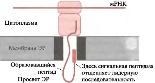
The ER membrane contains SRP receptors, also known as docking proteins, to which the cytoplasmic ribosome-SRP complex attaches. Through a series of sequential steps involving the hydrolysis of one GTP molecule to GDP, the ribosome binds to a membrane protein that forms a polypeptide-conducting channel, sometimes referred to as the translocon (see Fig. 22.16), while the SRP is released back into the cytoplasm for recycling. The growing polypeptide is translocated through the protein pore in the membrane as a loop, with the signal peptide anchored to the pore (see Fig. 22.16). A nearby signal peptidase cleaves off the leader sequence, and the polypeptide enters the ER lumen.
It remains unclear what precisely drives the translocation of the nascent peptide. Molecular chaperones located within the ER lumen are thought to assist in polypeptide folding.
Protein glycosylation in the ER lumen
In Chapter 3 (see p. 62), we noted that certain proteins (especially membrane and secretory proteins) are linked to Oligosaccharides. The attachment sites are either the NH2 side chains of asparagine (N-glycosylation) or the OH groups of Serine and threonine residues (O-glycosylation). This attachment occurs in several stages. N-glycosylation takes place within the ER. The first step of this process is particularly noteworthy: an oligosaccharide "core" consisting of 14 sugar monomers is assembled in the cytoplasm and transported across the membrane attached to a long hydrophobic chain. This chain contains over 100 carbon atoms and is called dolichol phosphate. An enzyme called transferase, located on the inner leaflet of the ER membrane, transfers the oligosaccharide group to the nascent peptide as soon as it emerges into the ER lumen. O-glycosylation occurs in the Golgi apparatus (see the next section).
What happens to the polypeptide translocated into the lumen of the rough ER?
The ER is essentially a "dead-end pouch." Within the lumen of the rough ER, the protein becomes surrounded by a membrane, and the resulting vesicles bud off from the ribosome-free regions of the ER, delivering the luminal contents to another organelle—the Golgi apparatus—via membrane fusion.
The structure of the Golgi apparatus is simple: about half a dozen flattened membrane-bound sacs with neither an entry nor an exit. Using the transport vesicles discussed above, it receives proteins from the ER and dispatches them to their final destinations by budding off vesicles, as shown in Fig. 22.17. An electron micrograph of the Golgi apparatus is shown in Fig. 3.15.
Fig. 22.17. Schematic representation of The Central Role of the Golgi apparatus in post-translational protein sorting and tagging. Newly synthesized Membrane Lipids are tagged to ensure transport vesicles are delivered precisely to their destinations. Vesicle formation for transport between Golgi cisternae relies on a remarkable mechanism: a GTP-protein complex interacts with the membrane, leading to the recruitment of "coat" proteins. Budding from the binding sites results in the formation of coated vesicles. They "uncoat" upon contact with the target membrane as a result of GTP hydrolysis to GDP; uncoating triggers the fusion of the vesicle with the target membrane. Target molecules on the vesicle direct it precisely to its destination. This "coating" mechanism appears to be widespread
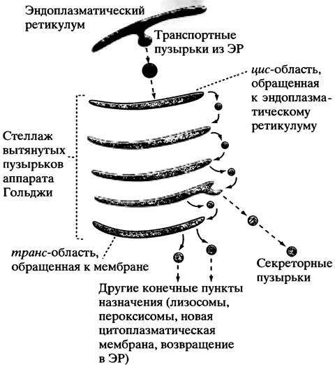
Not all proteins entering the Golgi apparatus are destined for secretion. The final destination of some is lysosomes or peroxisomes. Others, such as protein disulfide isomerase (p. 298) or glycosylation enzymes, must return to the rough ER because they were accidentally "swept away" to the Golgi apparatus along with other proteins. The Golgi apparatus sorts these proteins and packages them into vesicles that deliver their contents to the correct Location. Therefore, the Golgi apparatus is arguably the most remarkable functional "department" of the cell, capable of sorting proteins according to their final destinations and dispatching them in "packages" to precise addresses.
Proteins destined for secretion are packaged by the Golgi apparatus into vesicles that migrate toward the plasma membrane. There are Two Types of secretion. Some proteins are released continuously as they are synthesized; these include blood proteins secreted from liver cells without The Need for additional signals. In this case, vesicles fuse with the cell plasma membrane upon arrival, and their contents are released via exocytosis. The release of digestive enzymes, for example from the pancreas, is required only when food enters the intestine. In this case, the vesicles are larger. They store enzymes until a neuronal or hormonal stimulus triggers their massive release through the exocytosis of secretory vesicles (the signal and mechanism for this process are described on p. 358). The budding of transport vesicles from Golgi cisternae occurs after their membranes are surrounded by a coat protein complexed with a G-protein and bound GTP. GTP hydrolysis acts as a trigger for uncoating and the fusion of vesicles with the target membrane. Specific proteins located on the vesicles and target membranes play a crucial role in this recognition process.
What are the "address tags" on proteins involved in sorting within the Golgi apparatus?
As described below, in the case of enzymes targeted to lysosomes, the signal is carried by the carbohydrate portion of the glycoprotein. In most cases, however, the sorting mechanism must involve receptors that recognize specific structural features or Sequence Motifs on the proteins being sorted. These have not yet been fully identified, but one category of receptors is already known. For proteins (such as protein disulfide isomerase) that must be returned to the ER, the "tag" consists of a four-amino-acid sequence: Lys-Asp-Glu-Leu. In the single-letter amino acid code (Table 22.2), this sequence is written as KDEL.
Table 22.2. Single-letter amino acid symbols
Alanine |
A |
Leucine |
L |
R |
K |
||
Asparagine |
N |
Methionine |
M |
Aspartic acid |
D |
Phenylalanine |
F |
Cysteine |
C |
Proline |
P |
Glutamine |
Q |
Serine |
S |
Glutamic acid |
E |
Threonine |
T |
G |
Tryptophan |
W |
|
H |
Y |
||
Isoleucine |
I |
Valine |
V |
Packaging of Lysosomal Proteins
Up to this point, we have focused on proteins destined for secretion. The intracellular pathway of secretory proteins is quite similar to that of lysosomal proteins. As described in Chapter 16, lysosomes are small, membrane-bounded intracellular organelles containing a group of highly active hydrolytic enzymes. These enzymes are synthesized on the ER in the same manner as secretory proteins and undergo N-glycosylation within the ER lumen.
In the Golgi apparatus, the enzyme system recognizes lysosomal enzymes and phosphorylates the mannose residues of their oligosaccharide chains. The Golgi membrane contains internal receptors that bind the phosphorylated mannose; subsequently, vesicles containing the lysosomal enzymes bud off. These vesicles fuse with other "sorting vesicles," inside of which a low pH is maintained (driven by proton pumps in their membranes). The acidic pH of these vesicles promotes the dissociation of mannose-6-phosphate receptors from the Glycoproteins, allowing the receptors to return (via budding vesicles) back to the Golgi apparatus, while the enzymes are delivered to the lysosome. In a genetic disorder known as I-cell disease ("I" stands for inclusion of mucopolysaccharides), the mannose-phosphorylating system is defective, leading to a failure in delivering a group of lysosomal enzymes to lysosomes. This, in turn, disrupts mucopolysaccharide degradation, and their accumulation in lysosomes leads to severe clinical symptoms.
When it was discovered that the phosphorylation of mannose residues serves as a specific "tag" for lysosomal enzymes, it was hypothesized that carbohydrate tagging might be a widespread phenomenon. However, in most cases, the sorting machinery of the Golgi apparatus recognizes specific Structural Features of the proteins themselves.
How are integral plasma Membrane Proteins tagged?
Integral membrane proteins are also synthesized on the rough ER, inserted into the ER membrane, transported to the Golgi apparatus in vesicles, and from there directed (also via vesicles) to the Plasma Membranes. Membrane lipid synthesis likewise occurs in the ER, making this organelle the primary site for the generation of new membranes.
A fascinating question arises: why does a secretory polypeptide pass straight through the membrane of the rough ER, whereas an integral protein becomes anchored within it? All the necessary information determining whether—and if so, how—a given peptide will be integrated into the Membrane Structure is encoded within the amino acid sequence of the protein itself.
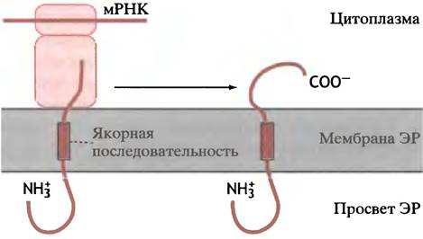
The simplest scenario occurs when a polypeptide contains an anchor sequence (also known as a stop-transfer sequence) (Fig. 22.18). This sequence becomes fixed in the Hydrophobic core of the bilayer and halts further translocation. The ribosome then completes the synthesis of the C-terminal portion of the polypeptide, resulting in a protein whose N-terminus is located on the exterior of the ER and whose C-terminus protrudes into the ER lumen. When this region of the ER eventually becomes part of the cell membrane, this protein retains the same orientation, with its N-terminus exposed on the cell surface. Membrane proteins can also adopt the opposite orientation by "spanning" the membrane multiple times. The mechanism for inserting such proteins involves the participation of several leader and stop-transfer signals.
Fig. 22.18. Illustration of the role of anchor signals in the insertion of integral membrane proteins into the ER membrane. The N-terminus of the polypeptide is oriented toward the exterior, and the C-terminus toward the interior. Proteins involved in translocation are not shown. Alternative arrangements of anchor signals can lead to proteins adopting the opposite orientation.
Is the translocation of all proteins co-translational?
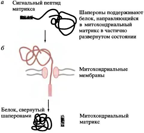
So far, we have examined the transport (translocation) of proteins across or into membranes during their synthesis. However, eukaryotic cells utilize another type of protein transport—into mitochondria or plant chloroplasts. As noted previously, mitochondria possess their own chromosome and protein-synthesizing machinery, but these account for only a small fraction of the organelle's proteins. Several hundred different proteins or subunits are imported into mitochondria, making up more than 95% of the proteins found there. These are synthesized in the cytoplasm. By analogy with the ER, one might expect protein synthesis to occur on mitochondria-associated ribosomes, but this is not the case. Mitochondrial proteins are synthesized on free ribosomes. Nuclear-encoded mitochondrial proteins (i.e., those encoded by nuclear genes) are synthesized and released into the cytoplasm as pre-proteins, after which chaperones bind to them to keep them in an unfolded conformation. Once released from the ribosome, these proteins cross the membrane in an extended (unfolded) state, a process driven by the Membrane Potential. After the protein crosses the membrane, other chaperones inside the mitochondrial matrix facilitate its folding.
The leader sequence of a mitochondrial protein typically consists of roughly 12–70 amino acids. While these sequences vary from one pre-protein to another, a common structural hallmark is an amphipathic α-Helix, in which one face is positively charged and the other is largely hydrophobic. This helix binds to receptors on the outer mitochondrial membrane.
In the simplest case, the N-terminal leader sequence directs the protein into the mitochondrial matrix through a pore located at contact sites between the outer and inner membranes. The leader sequence is subsequently cleaved off (Fig. 22.19). When the final destination is the intermembrane space, the pre-protein carries two leader sequences. As described above, the first sequence directs it into the matrix. However, its removal exposes the second sequence, which guides the polypeptide from the matrix, across the inner membrane, and into the intermembrane space, where the second leader sequence is then cleaved off.
Fig. 22.19. Transport of mitochondrial pre-proteins. (a) Cytology/cytology/92.html">SCHEMATIC STRUCTURE OF a mitochondrial pre-protein synthesized in the cytoplasm, destined for the matrix; (b) the polypeptide crosses the mitochondrial membrane through a proteinaceous pore, and the leader sequence is removed by a matrix signal peptidase.
The initial attachment of the polypeptide to the mitochondria is mediated by a receptor protein in the membrane (not shown). If the intermembrane space is the final destination, a second leader sequence directs the movement of the protein from the matrix across The inner mitochondrial membrane.
The Cell Nucleus is enclosed by a double nuclear membrane. Proteins synthesized in the cytoplasm are transported into the nucleus through specialized pores. In many cases, passing through these pores requires a signal sequence containing a short amino acid stretch; this is not required for the Transport of Small histone proteins. For transcription factors delivered to the nucleus, the signal sequence is located within the polypeptide chain.
Pre-proteins and pro-proteins should not be confused! Insulin is secreted into the ER lumen as a single polypeptide chain known as proinsulin. The removal of its central segment converts proinsulin into two polypeptide chains linked by a disulfide bond. The insulin mRNA encodes the leader sequence of proinsulin, which led to the coinage of the term pre-proinsulin—although, strictly speaking, because the leader (pre-sequence) is cleaved off as the protein crosses the ER membrane, pre-proinsulin as such does not exist in the cell.
Protein Degradation
We have explored protein synthesis and translocation. Now let us turn to protein degradation.
It is important to note two key points: 1) protein degradation occurs in all cells as older protein molecules are continually replaced by new ones; 2) the destruction of protein molecules is highly selective. While some proteins have half-lives exceeding 20 hours (and liver proteins can last several days), others survive for only 10 or even 2 minutes.
Several factors trigger protein degradation. Because absolute translational fidelity is seemingly impossible, protein molecules containing erroneous amino acids are inevitably produced, which can lead to improper folding. Proteins damaged by chemical insults, such as oxidation, must also be eliminated.
However, this process is not merely about "waste disposal." Some proteins appear to be intentionally designed for rapid turnover, as evidenced by
the presence of specific Amino acid sequences. Why would a cell bother with the rapid degradation of highly efficient enzymes? In many cases, it serves Metabolic Regulation. If an enzyme is degraded rapidly, its cellular levels can be finely and continuously controlled by its rate of synthesis. Such enzymes typically have relatively short half-lives. Structural proteins and Hemoglobin, by contrast, belong to the "long-lived" class.
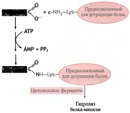
One of the selective protein destruction mechanisms involves ubiquitin—a small protein found in all eukaryotes but absent in bacteria, so its name (from the English "ubiquitous") is somewhat of an overstatement. In an ATP-dependent reaction, its terminal carboxyl group binds to the ε-amino group of a side-chain lysine residue in the target protein, thereby tagging the latter for degradation (Fig. 22.20). The selection of proteins destined for destruction by ubiquitin appears to be determined by their N-terminal amino acid. Chaperones may also participate in tagging proteins designated for destruction.
Fig. 22.20. Ubiquitin-dependent tagging of proteins destined for degradation. The mechanism by which proteins are selected for destruction remains unknown, although it has been observed that the terminal amino acid influences the protein's half-life.
Lysosomes play a specific role in the degradation of long-lived structural proteins (see Fig. 16.2): they are responsible for the autolytic destruction of cells during development (for example, the resorption of the tadpole's tail).
Overall, we know much less about protein degradation than about the far more complex process of its biosynthesis. Yet this area is of vital importance. Recall (see p. 149) that during starvation, The breakdown of Muscle Proteins supplies amino acids for Gluconeogenesis; without this, a person could not survive for more than 24 hours due to the complete depletion of Glycogen stores. Intracellular protein degradation is currently an active area of research.
Questions for Chapter 22
1. Why are 61 codons used to encode 20 amino acids instead of 64?
2. There are fewer types of tRNA than 61. Why?
3. Why are tRNA molecules depicted in diagrams in an inverted orientation when base-paired with a codon compared to how the same tRNA is shown independently?
4. What mechanisms ensure The fidelity of translation?
5. Describe the involvement and (where known) the role of GTP in protein synthesis.
6. Studies have shown that in E. coli, tRNA molecules (with attached aminoacyl or peptidyl moieties) occupy the A, P, and E sites on the ribosome. Explain the cause and mechanism of this phenomenon.
7. The mechanism of translation initiation in eukaryotes is incompatible with polycistronic mRNA. Explain why.
8. Explain the role of chaperones in protein synthesis.
9. What disease can be caused by improper protein folding?
10. Explain how proteins are transported across the endoplasmic reticulum.
Last update: 06/08/2026
Editorial and Educational Adaptation: This material has been compiled based on the primary/original source text. The project team performed an editorial review, corrected technical inaccuracies, structured sections, and adapted the content for an educational format.
What was processed:
- elimination of formatting defects (OCR errors, structural breaks, corrupted characters);
- editorial organization of content;
- standardization of terminology in accordance with academic sources;
- verification of factual statements against the original source text.
All mentions of the author, publication year, and origin of the primary text have been preserved in accordance with the source.