Review of Medical Physiology - William F. Ganong 2002
Respiration
Pulmonary Functions
Other Functions of the Respiratory System
Protective Mechanisms in the Lungs
The Airways, extending from the external environment to the alveoli, perform many Functions In addition to gas transport. They humidify and either cool or warm the inhaled air so that it is neither too hot nor too cold, closely approaching body Temperature by the time it reaches the alveoli. Bronchial secretions contain secretory IMMUNOGLOBULINS (IgA; see Chapter 27) and other substances that help combat infection and maintain mucosal integrity. Furthermore, the epithelium of the Paranasal Sinuses produces NO, which is bacteriostatic and helps prevent infection.
The pulmonary epithelium contains an interesting group of protease-activated receptors (PARs) that, when activated, release PGE2, which in turn protects the epithelial Cells. Such receptors are also present in the gastrointestinal tract; they are activated when Thrombin or Trypsin partially digests the ligands attached to them. PAR2 isoforms represent one form of these receptors in the airways.
Alveolar macrophages (AMs; dust cells) are another vital component of the lung's defense mechanisms. Like other macrophages (see Chapter 27), these cells originate from the Bone Marrow. They are active phagocytes that engulf and digest inhaled Bacteria and small particles. AMs also facilitate antigen presentation for immune responses and secrete substances that attract granulocytes to the lungs, as well as factors that stimulate The production of granulocytes and monocytes in the bone marrow. Their role in the Pathogenesis of emphysema is described in Chapter 27. Macrophages ingest large quantities of substances contained in cigarette smoke and can release lysosomal products into the extracellular space, thereby triggering an inflammatory response. Silica and asbestos particles also induce the release of lysosomal Enzymes.
Various mechanisms prevent foreign bodies from entering the alveoli. Nasal hairs filter out particles greater than 10 µm in diameter. Most particles of this size remain on the mucous membranes of the Nose and Pharynx because they fail to follow the airstream flowing down into the lungs. They become trapped on or near the Tonsils and adenoids, which are large aggregates of immunologically active lymphoid tissue located on the pharyngeal wall. Particles with a diameter of 2–10 µm mostly settle on the walls of the Bronchi as the air slows down in the smaller airways, eliciting a reflex bronchoconstriction and cough (see Chapter 14). Such particles are also cleared upward and out of the lungs by the ciliary "escalator." The airway epithelium from the anterior third of the nose to the beginning of the respiratory bronchioles is ciliated, and the cilia covering the mucosa beat rhythmically and in a coordinated fashion at a frequency of 1,000–1,500 cycles/min. The ciliary mechanism is capable of transporting particles at a rate of 16 mm/min. Particles smaller than 2 µm in diameter typically reach the alveoli, where they are engulfed by macrophages. Structure/19.html">The Importance of these protective mechanisms becomes obvious when we recall that in modern cities, every liter of air may contain several million dust particles and irritants.
When ciliary motility is impaired, effective mucus transport ceases. This leads to chronic sinusitis, recurrent pulmonary infections, and Bronchiectasis. Ciliary immotility can be caused by various air pollutants or may be congenital. One congenital form is Kartagener's syndrome, which lacks axonemal dynein, the ATPase molecular motor that drives Ciliary movement (see Chapter 1). Patients with this condition are also sterile because their spermatozoa lack motility, and they frequently exhibit situs inversus (complete reversal of Internal Organs), suggesting that the cilia required for organ rotation fail to function properly during embryonic development.
Metabolic and Endocrine Functions of the Lungs
In addition to gas exchange, the lungs perform several metabolic functions. They produce surfactant for local use, as noted above. The lungs also harbor a fibrinolytic system that dissolves thrombi within the pulmonary Blood Vessels. They release several substances into the systemic arterial blood (Table 34-6) and remove other substances reaching them via the pulmonary Arteries. Prostaglandins are cleared from the Circulation, but they are also synthesized by the lungs and released into the blood when lung tissue is stretched.
Class="center">Table 34-6. BIOLOGICALLY ACTIVE SUBSTANCES metabolized by the lungs
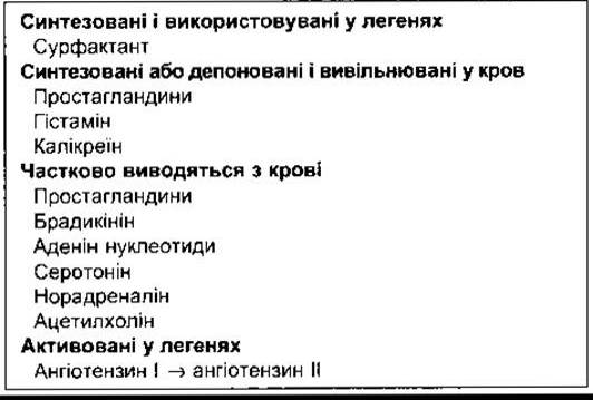
The lungs activate one key hormone. The physiologically inactive decapeptide angiotensin I is converted into the vasoconstrictor and aldosterone-stimulating octapeptide angiotensin II within the Pulmonary Circulation (see Chapter 24). Large amounts of angiotensin-converting enzyme (ACE), responsible for this activation, are located On the surface of the endothelial cells in the pulmonary capillaries. The converting enzyme also inactivates bradykinin. The transit time of blood through the pulmonary capillaries is less than 1 s, yet 70% of the angiotensin I reaching the lungs is converted to angiotensin II during a single pass through the capillaries. Four other peptidases have been identified on The surface of pulmonary endothelial cells, although their physiological roles remain unclear.
The removal of serotonin or norepinephrine reduces The amount of these vasoactive substances entering the systemic circulation. Many other vasoactive Hormones pass through the lungs without being metabolized, including epinephrine, dopamine, oxytocin, vasopressin, and angiotensin II. Furthermore, as noted in Chapter 26, various amines and Polypeptides are secreted by neuroendocrine cells in the lungs.
Last update: 10/08/2026
Editorial and Educational Adaptation: This material has been compiled based on the primary/original source text. The project team performed an editorial review, corrected technical inaccuracies, structured sections, and adapted the content for an educational format.
What was processed:
- elimination of formatting defects (OCR errors, structural breaks, corrupted characters);
- editorial organization of content;
- standardization of terminology in accordance with academic sources;
- verification of factual statements against the original source text.
All mentions of the author, publication year, and origin of the primary text have been preserved in accordance with the source.