Metabolism and Energy Transformations in Body Cells - Renata Armenakovna Petrosova 2004
Protein Biosynthesis
Structure of Transfer RNA and Amino Acid Coding
Transfer RNAs (tRNAs) are small molecules consisting of 70 to 90 NUCLEOTIDES. They account for approximately 15% of all cellular RNA. The function of tRNA depends directly on its Structure. Studies of tRNA molecules have shown that they are folded in a specific manner to resemble a cloverleaf (Fig. 20). The molecule features loops and double-stranded regions held together by complementary base pairing. The most crucial part is the central loop, which contains the anticodon—a nucleotide triplet corresponding to the code for a specific amino acid. Through its anticodon, a tRNA molecule is able to bind to the corresponding codon on an mRNA molecule via THE PRINCIPLE OF complementarity.
Class="center">METABOLISM/metabolism.files/image029.jpg" width="342"/>
Fig. 20. Structure of a tRNA molecule: 1 — anticodon; 2 — amino acid attachment site
Each tRNA can carry only one of the 20 Amino Acids. Consequently, there is at least one tRNA for each amino acid. Since an amino acid may be specified by multiple triplets, the number of tRNA types corresponds to the number of amino acid triplets. Thus, the total number of tRNA types matches the number of codons, equaling 61. None of the three stop codons correspond to any tRNA.
One end of the tRNA molecule always terminates in a guanine nucleotide (the 5'-end), while the other end (the 3'-end) always features three nucleotides, CCA. It is to this exact end that The amino acid attaches (Fig. 21). Each amino acid is attached to its specific tRNA carrying the appropriate anticodon. The Mechanism of this attachment relies on specific Enzymes known as Aminoacyl-tRNA synthetases, which link each amino acid to its corresponding tRNA. There is a distinct synthetase for each amino acid. The conjugation of an amino acid to tRNA is driven by ATP energy, whereby the high-energy bond is transferred to the bond between the tRNA and the amino acid. This process accomplishes the activation and coding of amino acids.
Stages of Protein Biosynthesis. The process of Polypeptide chain synthesis carried out on a ribosome is called Translation. Messenger RNA (mRNA) acts as an intermediary in transmitting information regarding the Introduction/19.html">Primary Structure of a protein, tRNA delivers the encoded amino acids to the site of synthesis and ensures their correct sequential assembly, and Ribosomes facilitate the assembly of the polypeptide chain.
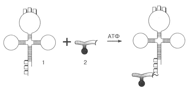
Fig. 21. Activation and coding of an amino acid: 1 — tRNA; 2 — amino acid
The ribosome contains three binding sites for RNA molecules: one site for mRNA and two for tRNA. One aminoacyl-tRNA is held in the aminoacyl center, while the other is held in the peptidyl center, where the polypeptide chain elongates.
The First stage is initiation. The mRNA synthesized during Transcription leaves The Nucleus and travels to the Cytoplasm, heading to the site of Protein Synthesis—the ribosome. Driven by various protein factors and ATP energy, the mRNA binds with the two ribosomal subunits, which until this moment remained dissociated. Before the ribosome can begin synthesizing the polypeptide chain, a specialized molecule—the initiator tRNA carrying an amino acid—must attach to the mRNA. Protein synthesis always begins with this step. Following the principle of complementarity, the tRNA uses its anticodon to bind to the start codon on the mRNA and enters the ribosome. This codon on the mRNA is referred to as the start codon.
As a result of the interaction among all these components, a complex is formed: ribosome — mRNA — initiator tRNA — amino acid.
The Second Stage is elongation. This is the phase of polypeptide chain growth. The assembly of the polypeptide chain then begins. The next aminoacyl-tRNA binds to the mRNA via complementary base pairing between its anticodon and the codon and enters the ribosome. The first tRNA is anchored in the peptidyl center, while the second tRNA carrying its amino acid is positioned in the aminoacyl center. The Amino acids are brought into close proximity, a peptide bond forms between them, and a dipeptide is produced. Meanwhile, the first tRNA is released and, as it leaves the ribosome, pulls the mRNA along with it by one triplet.
The second tRNA, now carrying the dipeptide, shifts to the peptidyl center, and a third aminoacyl-tRNA enters the ribosome (Fig. 22). This entire process then repeats over and over. As the mRNA moves sequentially through the ribosome, it continually brings in a new aminoacyl-tRNA and expels the discharged tRNA. The polypeptide chain progressively grows longer. The entire process is sustained by enzymatic activity and the energy of high-energy compounds (such as ATP).

Fig. 22. Scheme of polypeptide chain biosynthesis. The arrow indicates the direction of mRNA movement
The final stage is termination. As soon as a stop codon enters the aminoacyl center, synthesis halts. At this point, the tRNA is replaced by a specific protein factor (enzyme) that catalyzes the Hydrolysis of the bond between the final tRNA and the synthesized protein. The ribosome dissociates from the mRNA and splits back into its two subunits, while the last tRNA is also released back into the cytoplasm. The synthesized protein molecule enters The Endoplasmic reticulum (ER) or cytoplasm, where it undergoes modification and acquires its functional structure.
The translation process typically occurs repeatedly. A single mRNA molecule can bind to multiple ribosomes, forming a polyribosome or polysome, on which many molecules of the same protein are synthesized simultaneously (Fig. 23).
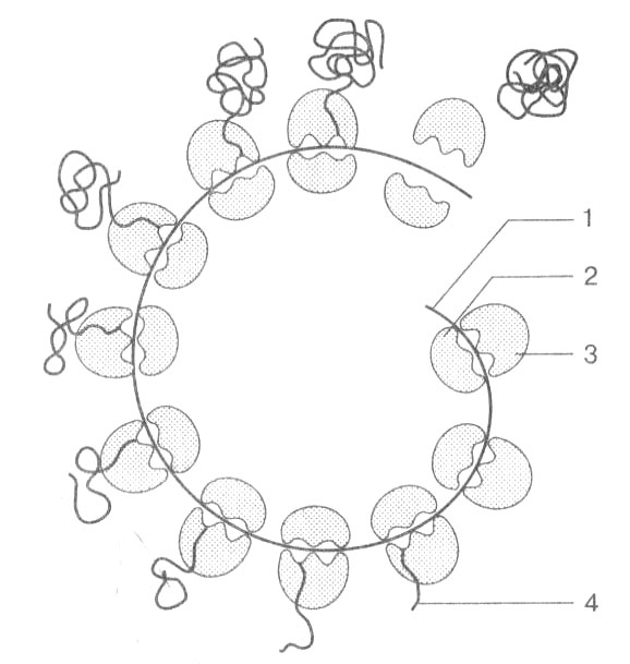
Fig. 23. Polysome: 1 — mRNA; 2 — small ribosomal subunit; 3 — large ribosomal subunit; 4 — synthesized polypeptide chain
If synthesis occurs on the rough endoplasmic reticulum, the polypeptide chain enters the ER lumen. Here, it achieves its final conformation and transforms into a mature protein molecule. Subsequently, the protein is transported to the Golgi apparatus and secreted from The Cell. If synthesis takes place on free ribosomes in the cytoplasm, the synthesized molecules remain there and are utilized by the cell.
The entire synthesis of a single protein takes anywhere from 20 to 500 seconds, depending on the length of the polypeptide. For instance, in an E. coli ribosome, a protein consisting of 300 amino acids is synthesized in approximately 20 seconds.
Last update: 13/08/2026
Editorial and Educational Adaptation: This material has been compiled based on the primary/original source text. The project team performed an editorial review, corrected technical inaccuracies, structured sections, and adapted the content for an educational format.
What was processed:
- elimination of formatting defects (OCR errors, structural breaks, corrupted characters);
- editorial organization of content;
- standardization of terminology in accordance with academic sources;
- verification of factual statements against the original source text.
All mentions of the author, publication year, and origin of the primary text have been preserved in accordance with the source.