BIOCHEMISTRY - L. Stryer - 1984
VOLUME 1
PART I. CONFORMATION AND DYNAMICS
CHAPTER 3. OXYGEN CARRIERS—MYOGLOBIN AND HEMOGLOBIN
3.11. Hemoglobin Is Composed of Four Polypeptide Chains
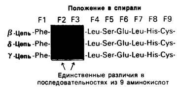
Let us now turn to Hemoglobin, a protein related to Myoglobin. Whereas myoglobin consists of a single polypeptide chain, hemoglobin is made up of four. These four chains are held together by noncovalent interactions. Each chain contains a single heme group, meaning that the hemoglobin molecule possesses four oxygen-binding sites. Hemoglobin A—the major hemoglobin of adult humans—consists of two identical chains called α chains and two identical chains called β chains. Overall, the subunit Structure of hemoglobin A is described by the formula α2β2. Adults also have a minor component, hemoglobin A2, which accounts for roughly 2% of the total hemoglobin; its subunit structure is α2δ2. Embryos contain different Hemoglobins. In early embryonic development, embryonic hemoglobin α2ε2 is expressed. This is subsequently replaced by fetal hemoglobin F, which has the subunit structure α2γ2. The Biological Significance of these different hemoglobins presents a fascinating problem that we will discuss in the next chapter. The α chain, common to all these hemoglobins, contains 141 amino acid residues, while the β, δ, and γ chains each contain 146 residues with highly homologous Amino acid sequences (Fig. 3.23).
Class="center">Fig. 3.23. The β, γ, and δ chains of human hemoglobins share similar amino acid sequences. Regions spanning residues F1 through F9 in three of the chains are shown as an example.
3.12. X-Ray Diffraction Analysis of Hemoglobin
As mentioned previously, the three-dimensional structure of hemoglobin A was solved by Max Perutz and his coworkers. This monumental endeavor began in 1936 when Perutz left Austria for Cambridge, England, to pursue his graduate studies and joined the laboratory of J. D. Bernal, where the first X-Ray Diffraction patterns of protein crystals had been obtained just two years earlier. Bernal and a graduate student, Dorothy Crowfoot Hodgkin, had obtained excellent diffraction patterns of Pepsin, thereby demonstrating that Proteins possess a highly ordered, definite structure. As early as 1934, they foresaw the potential of X-ray crystallography as a method "capable of yielding far more detailed information about Cell/13.html">Protein Structure than previous Physical and Chemical approaches could provide." More than 20 years passed, however, before this prediction was fully realized. When Perutz first chose hemoglobin as his research subject, the largest molecule whose structure had been solved was the dye phthalocyanine, which contains 58 atoms. Perutz, by contrast, tackled a molecule hundreds of times larger. Not surprisingly, "my colleagues looked at me with a pitying smile... Fortunately, the examiners of my thesis did not insist on the final structure being solved, or I would have remained a graduate student for 23 years." However, Lawrence Bragg—who, along with his father, had pioneered X-ray analysis in 1912—became the HEAD of the Cavendish Laboratory around this time and supported Perutz's work. He wrote: "I was under no illusions about the Prospects. It looked like multiplying a zero probability of success by an infinite importance of the result; the outcome of this mathematical operation was entirely unknown." Success finally came in 1959, when Perutz obtained a low-resolution electron density map of horse oxyhemoglobin. High-resolution maps were subsequently produced for both oxy- and deoxyhemoglobin from horse and human sources. The hemoglobins of these two species are remarkably similar in structure.
3.13. Quaternary Structure of Hemoglobin
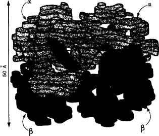
The hemoglobin molecule is nearly spherical, with a diameter of 55 Å. The four chains that make up the molecule are arranged in a tetrahedral geometry (Fig. 3.24). The four heme groups, one per subunit, reside in crevices on the exterior of the molecule. These four oxygen-binding sites are situated far apart from one another, the distance between the two closest iron atoms being 25 Å. Each α chain contacts both β chains, whereas interactions between the two α chains or between the two β chains are minimal.
Fig. 3.24. Low-resolution model of hemoglobin. The α chain is shown in yellow, the β chain in blue, and the heme group in red.
3.14. The α and β Chains of Hemoglobin Bear a Striking Resemblance to Myoglobin
The three-dimensional structures of myoglobin and the α and β chains of hemoglobin show a striking degree of similarity (Fig. 3.25). This close correspondence in backbone conformation was unexpected because there are numerous differences in The amino acid sequences of these three polypeptide chains. In fact, only 24 out of 141 positions are identical across all three chains, demonstrating that very similar tertiary structures can be built from completely different amino acid sequences (Fig. 3.26).
Fig. 3.25. Comparison of the backbone Conformations of myoglobin and the β chain of hemoglobin. The similarity in conformation is readily apparent.

Fig. 3.26. Comparison of the amino acid sequences of sperm whale myoglobin and the α and β chains of human hemoglobin, illustrated for the region spanning residues F1 through F9. The amino acid sequences share far less Homology than do the three-dimensional structures of these three polypeptide chains.

It is abundantly clear that the three-dimensional fold shared by sperm whale myoglobin and the α and β chains of human hemoglobin possesses profound general biological significance. Indeed, this structural motif is characteristic of all vertebrate myoglobins and hemoglobins. The intricate folding pattern of the polypeptide chain, first revealed in myoglobin, represents the fundamental architecture selected by nature for Oxygen transport: its physiological purpose is to provide a specialized microenvironment around the heme group that ensures the reversibility of oxygen binding.
Last update: 06/08/2026
Editorial and Educational Adaptation: This material has been compiled based on the primary/original source text. The project team performed an editorial review, corrected technical inaccuracies, structured sections, and adapted the content for an educational format.
What was processed:
- elimination of formatting defects (OCR errors, structural breaks, corrupted characters);
- editorial organization of content;
- standardization of terminology in accordance with academic sources;
- verification of factual statements against the original source text.
All mentions of the author, publication year, and origin of the primary text have been preserved in accordance with the source.