BIOCHEMISTRY AND MOLECULAR BIOLOGY - W. ELLIOTT - 2002
CHAPTER 28. MUSCLE CONTRACTION
Muscles are highly specialized Organs in which adaptations for mechanical movement are most prominently developed. In this chapter, we examine the Molecular Basis of Muscle contraction, while the next chapter covers the motor systems of non-muscle Cells.
A reminder on protein conformational changes
Muscle contraction is brought about by coordinated movements of protein molecules resulting from changes in their conformation. These conformational changes are driven by the interaction of protein molecules with various ligands.
For a "molecular motor" to generate movement via protein conformational changes, a counterbalancing antagonistic Structure is required: if the motor molecule is anchored, the partner molecule will move, and vice versa. Before discussing the Mechanism of muscle contraction, we should note that Myosin acts as the anchored "motor", whereas the Actin filament serves as the moving antagonistic structure.
Types of muscle cells and their energy supply
There are two MAIN TYPES OF muscle cells: smooth and striated. The former are found in the intestines and Blood Vessels; they contract involuntarily and are under the control of the Autonomic Nervous system. Smooth muscles contract slowly, but can sustain contraction for long periods. They are so named because microscopic examination reveals no transverse striations (unlike the other muscle type). Striated muscles contract rapidly and form The basis of skeletal musculature, whose voluntary contractions are regulated by motor nerves. Cardiac muscle also exhibits striations; however, unlike Skeletal Muscle, it contracts involuntarily, and The structure of myocardial muscle cells has its own distinctive features.
In voluntary Cytology/practical/58.html">Striated skeletal muscle, two main fiber types are distinguished: fast-twitch and slow-twitch. White striated muscles in animals consist of fast-twitch fibers with an inadequate blood supply, where ATP is generated via Glycolysis. These muscles mediate immediate survival responses (flight or fight), but due to the low ATP yield of glycolysis and the accumulation of lactic acid, they fatigue rather quickly.
Slow-twitch red fibers are capable of more sustained contraction. They are richly supplied with blood and characterized by a high content of Cell/35.html">Mitochondria and Myoglobin-bound oxygen. ATP in these fibers is produced through Oxidative Phosphorylation, which is much more efficient for ATP generation than glycolysis. However, in critical situations, red fibers require time and an increased Heart rate to deliver adequate amounts of oxygen. Consequently, the response of red muscles is slower compared to white ones, yet they can function for long periods without showing signs of fatigue. In humans, voluntary striated muscles contain both fiber types, with their ratio depending on the specific role of the given muscle in the body. The back muscles must sustain the tension required to maintain posture over long periods, and therefore they predominantly contain slow-twitch fibers. The extraocular muscles involved in eye movement are a prime example of fast-twitch fibers.
In all muscle types, the ATP reserve—upon which contraction largely depends—lasts for only a very short period of intense contraction. This is where the high-energy reserve compound creatine phosphate plays a vital role; the Standard Free energy of its Hydrolysis (ΔG°′) is -43.0 kJ • mol-1 (cf. -30.5 kJ • mol-1 for the hydrolysis of ATP to ADP and Pi). When ATP is converted to ADP and Pi during contraction, ATP is regenerated via the creatine kinase reaction at the expense of the phosphate groups from creatine phosphate molecules.
During contraction:
ATP + H2O —> ADP + Pi.
During ATP regeneration:
ADP + creatine-Ⓡ —> ATP + creatine.
During muscle relaxation, ATP is resynthesized via oxidative METABOLISM, whereas creatine phosphate is regenerated through the same creatine kinase reaction, which is readily reversible in the presence of high ATP and low ADP levels.
As noted previously, creatine phosphate (see below) is a high-energy compound whose phosphate group can be reversibly transferred to ADP to form ATP. Due to the high reactivity of the phosphate group, spontaneous (non-enzymatic) formation of creatinine can occur. This compound serves no known biological function and is excreted daily in the urine in amounts proportional to muscle mass.
Class="center">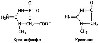
Structure of skeletal striated muscle
A Muscle consists of long, multinucleated cells known as muscle fibers (Fig. 28.1).
Fig. 28.1. Structure of a striated muscle fiber

Their cell membrane—the sarcolemma—is connected via neuromuscular junctions to nerve endings that transmit the signal triggering contraction. The membrane encloses the Cytoplasm, multiple nuclei, and numerous mitochondria. The cytoplasm is packed with numerous contractile filaments, called myofibrils, running the entire length of The Cell, each surrounded by membranous sacs of the sarcoplasmic reticulum.
Structure of a myofibril
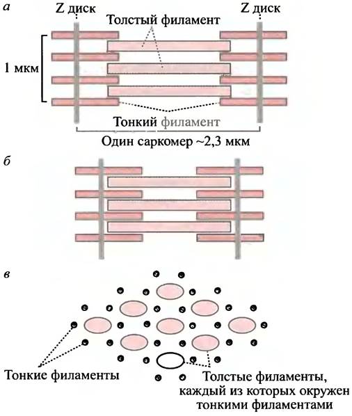
A myofibril is a contractile filament that runs the entire length of the muscle cell. It is divided into segments called sarcomeres (see Fig. 28.2), which are bounded by Z-discs (from the German Zwischen, meaning intermediate). Upon contraction, the Z-discs move closer together, and each sarcomere—and consequently the entire myofibril—shortens. This shortens the entire muscle fiber, thereby driving muscle contraction.
Fig. 28.2. Structure of thick and thin filaments in a sarcomere. a - Relaxation phase. The cross-striated appearance of a myofibril section under Electron Microscopy is due to passing protein bundles; b - contraction phase; c - cross-section. Only a few fibers are shown in the figure, although in reality each sarcomere contains A large number of myofibrils
How does a sarcomere contract?
Z-discs are robust protein discs located at each end of the sarcomere. Thin filaments are anchored to them and extend toward the center of the sarcomere. They are formed by the protein actin and firmly attached at one end to the Z-disc. Thin filaments are inert and serve as a "ratchet mechanism" against which the force pulling the Z-discs toward each other is applied. In vertebrate muscles, thin filaments have a hexagonal packing and are attached to two discs, with each sarcomere containing several such ordered structures. Inside each hexagonal "cell" formed by six thin filaments, a thick filament is located. It possesses finger-like protrusions that perform the actual work of contraction. Using the "ratchet mechanism," they slide the thin filaments toward the center, pulling the Z-discs together (see Fig. 28.2).
This is the general picture of muscle contraction. To understand how it occurs, we must examine the molecular structures involved.
Structure and Action of Thick and Thin Filaments
Thin Filaments
The monomeric globular protein actin is called G-actin ("G" stands for globular) (Fig. 28.3). Its molecular structure features two domains connected by a narrow waist that imparts polarity to the molecule. Under certain conditions within the cell, G-actin molecules readily polymerize to form a double-helical F-actin structure, in which all molecules are oriented in the same direction—from HEAD to tail (see Fig. 28.3)
Fig. 28.3. SCHEMATIC STRUCTURE OF actins. a — G-actin (globular); b - F-actin (fibrillar), formed by the polymerization of G-actin
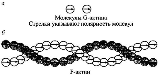
Thin filaments are formed by double-stranded structures, with their ends designated as (+) and (-).
Thick Filaments
Let us now consider the protein myosin, each molecule of which has a backbone formed by two intertwined α-helices, also referred to as myosin heavy chains (Fig. 28.4, a). The structure of each helix is such that regularly spaced hydrophobic amino acid residues engage in hydrophobic interactions with the other α-Helix, forming a rigid rod-like structure. Each chain terminates in a globular head, to which two small polypeptide chains, called myosin light chains, are attached.
A thick filament consists of several thousand myosin molecules organized in a bipolar fashion, as shown in Fig. 28.4, b. Thick filaments maintain a central position relative to the surrounding thin filaments through the action of other Proteins comprising the sarcomere. One of these is titin, named for its enormous molecular mass (derived from the word "titanic"). This central position ensures the optimal distance required for the myosin heads to contact the actin filaments.
Fig. 28.4. Organization of a thick filament. a - Structure of a myosin molecule, consisting of two heavy chains, each terminating in a myosin head. Two different light chains are attached to them; b - thick filament formed from myosin molecules connected in a bipolar manner
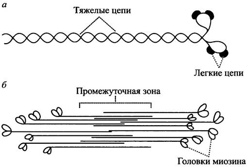
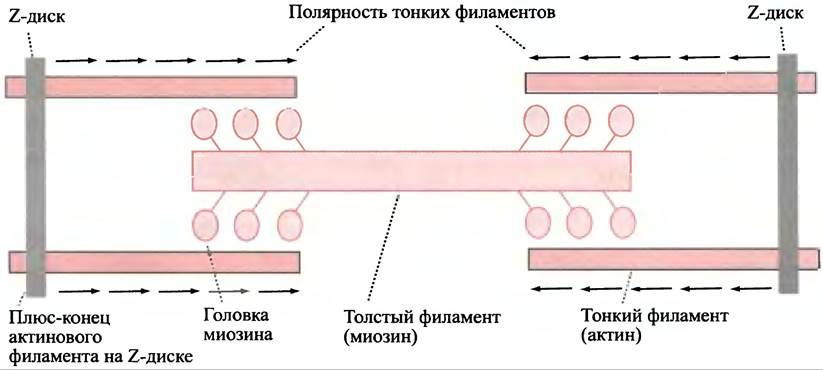
Thin filaments are anchored to the Z-discs by their (+)-ends such that the myosin heads at both ends of the thick filament are oriented identically relative to the "poles" of the thin filaments (Fig. 28.5).
Fig. 28.5. Organization of thick and thin filaments in a sarcomere. Arrows indicate the polarity of actin filaments. During contraction, the myosin heads move along the actin filaments toward their (+)-ends, thereby pulling the two Z-discs toward each other
How does the myosin head convert the energy of ATP hydrolysis into the mechanical work of actin filaments?
The myosin head possesses ATPase activity, and conformational changes occur within it during the hydrolysis of ATP to ADP and Pi. Without proper explanation, this is bound to cause confusion. After all, up to this point we have considered cases where ATP Cleavage was used to perform work, and it would be natural to assume that muscle contraction occurs via the hydrolysis of ATP to ADP and PPi. In the ATP-dependent processes discussed earlier, indirect ATP hydrolysis took place: first, a phosphoryl group or AMP was transferred to the reacting molecule, followed by the release of either PPi or AMP, i.e., coupled reactions were used. During muscle contraction, mechanical work is driven by direct ATP hydrolysis (without covalent intermediates). However, the actual "power stroke" during contraction occurs not during ATP hydrolysis, as one might expect, but after the dissociation of ADP from the protein. It is precisely when ADP leaves the protein that the primary release of free energy occurs, meaning that the "power stroke" in muscle contraction is driven by the release of ADP from the myosin head rather than by ATP hydrolysis. At the same time, during The conversion of the protein-ATP complex into the protein-ADP plus Pi complex, The change in free energy is small. In solution, however, the conversion of free ATP into free ADP and Pi
is characterized by a large negative value of ∆G°′, so the overarching energetic concept of ATP hydrolysis as the driving force remains unchanged.
The myosin head can exist in a strained conformation (primed for force generation) and a relaxed conformation (following force generation). The head attaches to the actin filament in the strained conformation and then executes a "power stroke" against the actin (Fig. 28.6), after which it adopts the relaxed conformation.
Fig. 28.6. Skeletal muscle contraction (simplified diagram)

Now let us examine the steps of this cycle in more detail, starting at the point where the myosin head has just completed its "power stroke." The myosin head is attached to actin in a relaxed conformation. The subsequent events unfold as shown in Fig. 28.7.
Fig. 28.7. Skeletal muscle contraction (simplified diagram). The numbers correspond to the stages described in the text. The myosin head has just delivered a "power stroke" along the thin actin filament. Note that the "power stroke" is coupled with the release of ADP from the myosin head.

1. ATP binds to the myosin head, causing it to detach from actin.
2. ATP is hydrolyzed to ADP and Pi; following the release of Pi, the myosin head transitions into a cocked (strained) conformation and attaches to actin.
3. The myosin head executes a "power stroke" coupled with the release of ADP. This induces a conformational change in the head, causing it to pivot at the hinge region.
4. The myosin head remains bound to actin in a relaxed conformation until it is released by an incoming ATP molecule, which resets it to its initial state.
A large number of myosin molecules participate in muscle contraction. If ATP is unavailable, all myosin heads become cross-linked to actin (since the first step of the cycle cannot occur). This leads to muscle rigidity (rigor mortis), which sets in upon the depletion of ATP reserves. The ability of a myosin head to carry out this entire process was demonstrated in vitro using latex beads coated with isolated myosin heads, which literally "walked" along immobilized actin filaments in the presence of ATP and Ca2+.
How is the contraction of voluntary striated muscles regulated?
Skeletal muscle contraction is initiated by a Nerve Impulse that triggers the release of Ca2+ ions from the sarcoplasmic reticulum into the myofibrils. Ca2+ ions are rapidly cleared from the myofibrils (see below), bringing contraction to a halt. Contraction will not occur until a sustained nerve impulse triggers the release of a large flood of Ca2+ ions.
How does Ca2+ trigger contraction?
Actin is the primary protein of thin filaments, but other protein molecules are also associated with them. One of these is Tropomyosin, which consists of two subunits. It is a relatively small, elongated molecule located in the groove formed by the two actin strands of the thin filament. There is approximately one tropomyosin molecule for every seven actin molecules. These two protein molecules overlap end-to-end to form a continuous strand. A thin filament has two grooves, with tropomyosin molecules lying in each of them (Fig. 28.8).
Fig. 28.8. Arrangement of actin and tropomyosin molecules. Each tropomyosin molecule binds seven actin monomers, forming an extended strand within the grooves of the actin filament. A troponin complex is attached to one end of each tropomyosin molecule.
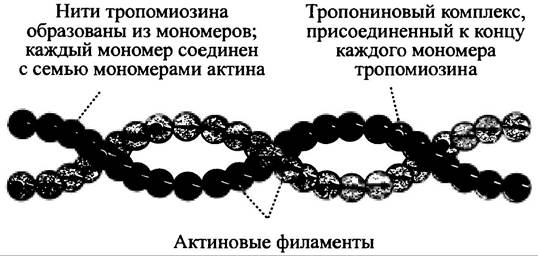
Attached to one end of the tropomyosin molecule is troponin, a complex composed of three Globular proteins. The troponin-tropomyosin complex is sensitive to Ca2+ ions, which induce a slight shift in the tropomyosin strand relative to the thin filament, thereby exposing the binding site for the myosin head on the actin fiber. While tropomyosin was traditionally thought to physically block myosin from binding to actin, other observations suggest that Ca2+ does not directly affect myosin binding to the thin filament. Nevertheless, it is well established that unless the myosin-actin power cycle is engaged, Ca2+ ions are absent.
Without going into excessive detail, it should be noted that Ca2+ ions bind to troponin, inducing a conformational change in the tropomyosin molecule that somehow activates the myosin-actin power cycle, leading to ATP hydrolysis and muscle contraction. The removal of Ca2+ ions reverses this entire sequence of events and halts contraction, bringing us to the mechanism regulating the release and uptake of Ca2+ ions.
Transport of Ca2+ Ions in Muscle
Each myofibril of a muscle cell is surrounded by membrane sacs of the sarcoplasmic reticulum, whose Ca2+-ATPase pumps Ca2+ ions from the Cytosol surrounding the myofibril into the lumen of the reticulum (in an ATP-dependent manner against a concentration gradient). This mechanism maintains a low concentration of Ca2+ within the myofibril and prevents muscle contraction. The regulation of Ca2+ levels also involves a protein in the sarcoplasmic reticulum membrane that Functions as a passive Ca2+ ion channel. Normally closed, this channel opens upon the arrival of a nerve impulse at the muscle cell, allowing Ca2+ ions to flow from the lumen of the reticulum into the myofibril and thereby triggering contraction.
To sustain muscle contraction, all sarcomeres of a myofibril and all myofibrils within a muscle must respond to the motor nerve impulse simultaneously; otherwise, the contraction would be uncoordinated and inefficient. A nerve impulse triggers the release of acetylcholine at the Neuromuscular Junction, which in turn causes a local depolarization of The Plasma Membrane that rapidly spreads across the entire membrane (see p. 358). This depolarization opens voltage-gated Ca2+ channels in the sarcoplasmic reticulum, releasing ions into the myofibril. To ensure the rapid delivery of the signal throughout the cell's sarcoplasmic reticulum, the plasma membrane forms invaginations known as transverse (T) tubules. These tubules penetrate the myofibril at the level of the Z-disc and make direct contact with the sarcoplasmic reticulum membrane, allowing the electrical signal to reach all contractile units controlled by that nerve impulse almost instantaneously (Fig. 28.9).
Fig. 28.9. Neuromuscular junction. (a) The plasma membrane (sarcolemma) of a muscle fiber forming the neuromuscular junction; (b) diagram of transverse (T) tubules that transmit the plasma membrane depolarization signal to the sarcoplasmic reticulum (SR). It is believed that plasma membrane depolarization is transmitted directly to the SR Ca2+ channels, thereby triggering a rapid release of Ca2+ from the channel.

How do smooth muscles differ in structure and regulation from striated muscles?
Smooth muscles are found in the walls of blood vessels, the intestines, the urogenital tract, and other Organ Systems. Their long, spindle-shaped cells are uninucleate and assemble to form muscles whose shape matches their specific functions (for example, circular in blood vessels or network-like in the Urinary Bladder).
The Basic principles of contraction are the same as in striated muscle, where myosin molecules exert a mechanical force on the actin filament, using ATP hydrolysis as an energy source in a similar "power cycle." However, the contractile components in smooth muscles are not nearly as highly
organized. There are no myofibrils or repeating sarcomeres, and consequently, no cross-striations are visible under a Microscope. Instead of a sarcomeric structure, actin filaments run the entire length of the cell (which is small compared to a striated muscle cell) and are anchored at one end to The cell membrane.
Regulation of smooth muscle contraction
Although Ca2+ ions are responsible for initiating contraction, its regulatory mechanism differs from that of striated muscle. The sarcoplasmic reticulum is absent. Smooth muscle contracts much more slowly (50 times) than striated muscle (taking about 5 seconds). Unlike striated muscle, smooth muscle does not require a near-instantaneous contraction of all its structures; the contraction of individual cells propagates at a more gradual pace. A nerve impulse from the autonomic nervous system opens Ca2+ channels in the smooth muscle cell membrane, causing Ca2+ ions to enter the cell from the extracellular fluid. Specialized cell-to-cell junctions allow the nerve signal to spread from Cell to Cell throughout the entire muscle.
How does Ca2+ regulate smooth muscle contraction?
As noted previously, the head of each myosin molecule contains two small Polypeptides known as myosin light chains. In smooth muscle, one of these light chains (the p-light chain) inhibits the binding of the myosin head to the actin filament, thereby preventing contraction. Ca2+ activates myosin light chain kinase, which, in the presence of ATP, phosphorylates the p-light chain and relieves this inhibition, thus triggering contraction.
Ca2+ ions activate the kinase indirectly: they bind to the protein calmodulin (see p. 353), inducing a conformational change that enables the Ca2+-calmodulin complex to attach to the inactive kinase and activate it. When Ca2+ ion levels decline, the process is reversed, and a phosphatase dephosphorylates the myosin light chain, leading to muscle relaxation. The overall regulatory pathway is illustrated in Fig. 28.10.
Fig. 28.10 Mechanism of smooth muscle contraction activation by Ca2+ ions

In addition to neural regulation, certain Hormones are involved in controlling smooth muscle contraction—for example, noradrenaline, which induces the contraction of specific blood vessel muscles.
Questions for Chapter 28
1. What are the differences between fast- and slow-twitch muscle fibers? What is their biological significance?
2. Illustrate the structure of a myofibril, sarcoplasmic reticulum, and sarcomere.
3. Explain how the myosin head converts the energy of ATP hydrolysis into mechanical work.
4. How is the contraction of a skeletal striated muscle sarcomere regulated?
5. How is smooth muscle contraction regulated?
Last update: 06/08/2026
Editorial and Educational Adaptation: This material has been compiled based on the primary/original source text. The project team performed an editorial review, corrected technical inaccuracies, structured sections, and adapted the content for an educational format.
What was processed:
- elimination of formatting defects (OCR errors, structural breaks, corrupted characters);
- editorial organization of content;
- standardization of terminology in accordance with academic sources;
- verification of factual statements against the original source text.
All mentions of the author, publication year, and origin of the primary text have been preserved in accordance with the source.