BIOLOGY Volume 2 - A Guide to General Biology - 2004
18. THE MUSCULOSKELETAL SYSTEM OF ANIMALS
18.4. The Muscular System
18.4.4. The Mechanism of Muscle Contraction; The Sliding Filament Theory
Once the ultrastructure of myofibrils was elucidated, two independent groups of researchers (H. Huxley/J. Hanson and A. Huxley/R. Niedergerke) proposed a hypothesis of Muscle contraction based on the sliding of Actin and Myosin filaments past one another (Fig. 18.20). This can be easily visualized by interleaving the fingers of one hand between those of the other: if both palms are considered equivalent to a single sarcomere, the system will shorten—that is, contract. This mechanism is supported, in particular, by the fact that During muscle contraction, the widths of the I-band and H-zone decrease, whereas the width of the A-band remains unchanged. Today, this hypothesis is definitively confirmed, universally accepted, and known as the sliding filament theory.
To understand the forces driving this sliding motion, it is necessary to examine The Structure of thin (actin) and thick (myosin) myofilaments.
Class="center">
Fig. 18.20. Diagram of sarcomere contraction. Actin filaments slide along myosin filaments. Myosin heads are shown on the thick myofilaments. (See text for explanations.)
Myosin (Thick) Myofilaments
A myosin molecule consists of two parts: a long, rod-like region (the "tail") and a globular region attached to one of its ends, represented by two identical "heads" (Fig. 18.21, A). Myosin molecules are arranged within the myosin filament such that the heads are regularly distributed along its entire length. In regions where actin and myosin filaments overlap, the myosin heads can attach to neighboring actin filaments. The Significance of this interaction will become apparent when we examine the actual mechanism of sarcomere contraction.
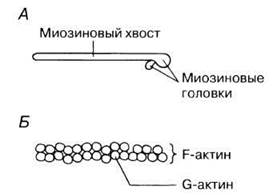
Fig. 18.21. A. Structure of myosin: showing its linear part (tail) and two heads. B. Structure of actin.
Actin (Thin) Myofilaments
Each actin myofilament is formed by two chains of globular actin molecules (G-actin) twisted around each other like a helix (Fig. 18.21, B). The entire complex of actin molecules is called F-actin (filamentous actin). It is believed that one ATP molecule is associated with each G-actin molecule.
Mechanism of Contraction
The contraction mechanism operates as follows. Where actin and myosin myofilaments overlap, the myosin heads act like hooks, "catching" onto adjacent F-actin filaments to form cross-bridges. These bridges bend like fingers in one direction, pulling the actin myofilaments along the myosin ones. The heads then detach from the actin, straighten out, attach to new sites further along, and the cycle repeats. During contraction, approximately half of the heads are "pulling" at any given moment while the rest return to their initial position, ensuring a smooth process. Energy for this process is supplied by ATP. ATP molecules are hydrolyzed to ADP and phosphate by the action of ATPase contained within the myosin heads. These events are illustrated in Fig. 18.22. A single HEAD takes 1/50 to 1/100 of a second between two successive bridge formations. Obviously, this requires a very high rate of ATP consumption. This explains the presence of a vast number of Cell/35.html">Mitochondria in muscle fibers, which replenish ATP stores via aerobic Respiration. A sarcomere is capable of shortening by 30–60% of its initial length.

Fig. 18.22. Movement of an actin filament driven by a myosin filament during muscle contraction. The dashed vertical line on the left of each stage helps to visualize the displacement.
18.2. How does the width of the A-band change during sarcomere contraction?
How is the contraction process triggered and terminated? It is activated by Calcium Ions (see "The Role of Tropomyosin and Troponin" below). They are stored in the sarcoplasmic reticulum (a specialized Endoplasmic reticulum of the muscle fiber), which forms expanded cisternae around the Z-lines (Fig. 18.23). These cisternae are in contact with transverse tubules (T-tubules, or the T-system) formed by invaginations of the sarcolemma that permeate the sarcoplasm (the Cytoplasm of the muscle fiber) (Figs. 18.23 and 18.18). When a Nerve Impulse via a motor neuron reaches the Neuromuscular Junction On the surface of the muscle fiber, an Action Potential originating from the end-plate region sweeps in a wave through the T-system, is transmitted from there to the sarcoplasmic reticulum, and triggers the release of calcium ions into the sarcoplasm.

Fig. 18.23. The sarcoplasmic reticulum and the T-system.
The Role of Tropomyosin and Troponin
Actin filaments consist of F-actin and two accessory Proteins: tropomyosin and troponin. Tropomyosin molecules form two rather elongated helical chains that wrap around F-actin, as shown in Fig. 18.24. Tropomyosin serves to switch the contraction mechanism on and off. Associated with tropomyosin is the globular protein troponin, which is capable of reversibly binding calcium ions.
When the muscle is at rest (relaxed), tropomyosin blocks the myosin head attachment sites on the thin myofilament (Fig. 18.24, A), thereby "switching off" actin. Calcium ions, released from the sarcoplasmic reticulum, bind to troponin, causing it—and simultaneously the tropomyosin with which it is associated—to shift. This unblocks the myosin head attachment sites (Fig. 18.24, B)—actin is "switched on," and filament sliding begins via the mechanism described above (Fig. 18.22). When stimulation of the muscle fiber by excitatory impulses ceases, calcium ions are pumped back from the sarcoplasm into the sarcoplasmic reticulum by a calcium pump in the membrane, that is, via Active Transport, which also requires ATP energy. Troponin and tropomyosin return to their initial configuration, the thin myofilament is "switched off," and the muscle fiber relaxes.

Fig. 18.24. Model illustrating changes in actin filament structure. A. "Off" state at low Ca2+ levels: tropomyosin blocks myosin-binding sites on actin. B. "On" state at high Ca2+ levels: tropomyosin shifts, exposing the myosin-binding sites (indicated by arrows). A — actin; T — tropomyosin. Troponin is not shown; it is located closer to the actin subunits.
Last update: 06/08/2026
Editorial and Educational Adaptation: This material has been compiled based on the primary/original source text. The project team performed an editorial review, corrected technical inaccuracies, structured sections, and adapted the content for an educational format.
What was processed:
- elimination of formatting defects (OCR errors, structural breaks, corrupted characters);
- editorial organization of content;
- standardization of terminology in accordance with academic sources;
- verification of factual statements against the original source text.
All mentions of the author, publication year, and origin of the primary text have been preserved in accordance with the source.