BIOLOGY Volume 2 - A Guide to General Biology - 2004
18. ANIMAL SUPPORT AND LOCOMOTOR SYSTEMS
18.4. The Muscular System
18.4.3. Ultrastructure of Striated Muscle
Insights into the Mechanism of Muscle contraction come from electron microscopic studies of its ultrastructure, which allows myofibrils to be examined in detail (Fig. 18.17): their diameter is approximately 1 мкм (whereas the muscle fiber is 100 times larger). They are crossed by dark bands that give the muscle fiber its striated appearance. More detailed study reveals that each myofibril is composed of Two Types of parallel filaments running side by side: thin and thick myofilaments. These consist of different Proteins: the former of Actin, and the latter of Myosin. In certain regions, the thin myofilaments extend into the gaps between the thick ones, much like the partially interleaved fingers of two hands. These overlapping regions correspond to the dark bands, which can also be easily demonstrated using one's fingers (Fig. 18.16). The dark bands are conventionally called A zones or A bands, while the lighter regions between them are called I zones or I bands. The latter contain only actin myofilaments. Within the A zone, a central region can be distinguished where no overlap occurs, meaning only myosin filaments are present; this is known as the H zone. Finally, the H zone is bisected by the M line, and the I zone by the Z line. This Structure is represented schematically in Fig. 18.17.
Class="center">
Fig. 18.17. Ultrastructure of a Skeletal Muscle fiber.
It turns out that the functional unit of a muscle is the contractile segment of a myofibril between two Z lines, known as a sarcomere. A myofibril—and consequently the muscle fiber as a whole—consists of thousands of sarcomeres. Understanding how a single sarcomere contracts makes it possible to explain The Mechanism of contraction for the entire muscle.
Figures 18.18 and 18.19 show the appearance of Actin and myosin myofilaments under an Electron microscope.
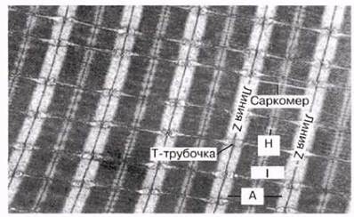
Fig. 18.18. Transmission electron micrograph of a Cytology/practical/54.html">Longitudinal section of a fish (roach) muscle fiber. Triads and detailed myofibril structure are visible, х7650.

Fig. 18.19. Electron micrograph of a transverse section of a contracted insect flight muscle (from the giant Water bug Belostoma). The Origin of the cross-bridges is explained in the text. Myosin and actin myofilaments are arranged in an orderly pattern. Thin actin filaments surround each thick myosin filament like the sides of a regular hexagon surrounding its center. In each row, thin filaments alternate with thick ones, which is also evident in longitudinal sections (Figs. 18.17 and 18.18; х137,000).
Last update: 06/08/2026
Editorial and Educational Adaptation: This material has been compiled based on the primary/original source text. The project team performed an editorial review, corrected technical inaccuracies, structured sections, and adapted the content for an educational format.
What was processed:
- elimination of formatting defects (OCR errors, structural breaks, corrupted characters);
- editorial organization of content;
- standardization of terminology in accordance with academic sources;
- verification of factual statements against the original source text.
All mentions of the author, publication year, and origin of the primary text have been preserved in accordance with the source.