BIOLOGY Volume 2 - A Guide to General Biology - 2004
18. ANIMAL SUPPORT AND LOCOMOTOR SYSTEMS
18.4. The Muscular System
18.4.2. Histology of Striated Muscle
Skeletal Muscle as viewed under a Light Microscope is illustrated in Fig. 18.16. The appearance of a Cytology/practical/54.html">Longitudinal section of muscle (or isolated muscle fibers) explains why it is referred to as striated muscle. However, the underlying causes of this characteristic banding pattern only become clear through Electron Cell/15.html">Microscopy (see the following section). Striated Muscle consists of numerous elongated Cells, known as muscle fibers due to their shape. They can reach several centimeters in length with a diameter of 0.01–0.1 mm (compared to an average cell diameter of about 0.02 mm). These fibers are cylindrical in shape and run parallel to one another. Each fiber is multinucleated, Setting it apart from Other types of muscle cells. The nuclei are located peripherally, just beneath The Cell membrane (Fig. 18.16). Bundles of muscle fibers are surrounded by Collagen fibers and Connective Tissue, with additional collagen interspersed between individual fibers. Each fiber is enclosed by a membrane called the sarcolemma, which is structurally very similar to a standard Plasma Membrane.
Under a light microscope, numerous thin myofibrils (from the Greek myo’s meaning muscle and the Latin fibrilla meaning little fiber) can be distinguished within the muscle fiber, and it is these structures that produce the characteristic cross-striations.
Class="center">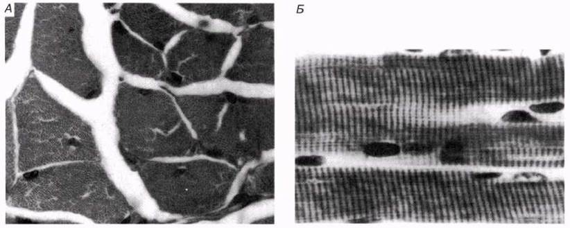
Fig. 18.16. Light micrograph of striated (skeletal, voluntary) muscle. A. Cross-section. B. Longitudinal section.
Last update: 06/08/2026
Editorial and Educational Adaptation: This material has been compiled based on the primary/original source text. The project team performed an editorial review, corrected technical inaccuracies, structured sections, and adapted the content for an educational format.
What was processed:
- elimination of formatting defects (OCR errors, structural breaks, corrupted characters);
- editorial organization of content;
- standardization of terminology in accordance with academic sources;
- verification of factual statements against the original source text.
All mentions of the author, publication year, and origin of the primary text have been preserved in accordance with the source.