BIOCHEMISTRY AND MOLECULAR BIOLOGY - W. ELLIOTT - 2002
CHAPTER 29. THE ROLE OF THE CYTOSKELETON IN MAINTAINING CELL SHAPE AND PERFORMING MECHANICAL WORK
At first glance, it might seem that non-Muscle Cells perform no mechanical work. It is also easy to picture an animal Cell as a shapeless sac filled with protoplasm. However, this is incorrect, as cells possess a Cytoskeleton (a network of internal structural elements) that is involved in positioning intracellular structures, cell motility, and maintaining cell shape.
As a first approximation, the necessity for these types of mechanical work is not immediately obvious. Without a rigid Cell wall, however, an animal cell would simply be a small, amorphous entity. The shape of most Types of Animal cells is distinct, and intracellular structures play a crucial role in maintaining it. For instance, the biconcave disc shape of a red Blood cell is maintained by specialized protein frameworks that interact with the membrane.
Many cells constantly change their shape, meaning that the cytoskeleton responsible for defining it is in a state of continuous remodeling. This dynamic cytoskeletal Organization, which differs from the structures found in erythrocytes or intestinal microvilli (see Fig. 4.1), is characteristic of animal cells. Plant and bacterial cells, possessing rigid cell walls, belong to an entirely different category.
Motility is an inherent property of animal cells; aside from the striking Examples of amoeboid movement in macrophages and other leukocytes migrating through Tissues, many cells crawl across tissue surfaces, a process vital during embryonic development and normal wound healing. Another type of active (non-muscle) contraction occurs during Cell Division, where a contractile ring forms around the equatorial zone, eventually pinching The Cell in two. Other forms of movement include the beating of microvilli, which helps transport surface layers of mucus (such as in the respiratory tract), or the lashing of sperm flagella.
A Introduction/5.html">Eukaryotic Cell is a massive object when its dimensions are considered relative to the slow rate of diffusion of molecules in solution. A bacterial cell is much smaller and therefore "relies" on the diffusion of dissolved substances, whereas many Eukaryotic cells require a sophisticated internal transport
system. Golgi vesicles must be transported to the cell surface or other specific destinations. The movement of mRNA molecules from The Nucleus to The Cytoplasm also follows a strictly regulated pathway. Another striking example is a nerve axon, which can exceed half a meter in length; Protein Synthesis AND vesicle formation occur in the cell body, yet some of these components must be delivered all the way to the axon terminal. Diffusion could never accomplish this! The giant alga cell Nitella is so large that diffusion alone could not sustain its life; consequently, the cell maintains constant cytoplasmic streaming to transport substances. Yet another example of Intracellular Transport is the segregation of Chromosomes into daughter cells during cell division.
To summarize, cells possess a complex internal organization that ensures shape maintenance, motility, and intracellular transport. This internal organization is less conspicuous than the contractile apparatus of muscle cells because: 1) the structures carrying out these Functions are only revealed under specific staining conditions; 2) many of them are constantly modified to meet the shifting demands of the cell.
Before we delve into a detailed examination of the cytoskeleton's role in determining cell shape and generating mechanical work, It is worth noting several key principles.
✵ Actin filaments are found in virtually all cells; they are involved in determining animal cell shape and driving amoeboid and crawling movements.
✵ Actin filaments also act as intracellular "railroad tracks" along which cell components are actively transported.
✵ Myosin-like molecules exist in non-muscle cells as well. One type of myosin molecule (analogous to muscle myosin) exerts force on adjacent actin filaments to drive cell motility. Other types of myosin molecules (minimiosins), positioned along actin transport tracks, carry various intracellular "cargoes."
ATP serves as the energy source.
✵ A specialized transport system is formed by microtubules, which also serve as tracks along which molecular "motors" move, dragging intracellular "cargoes" behind them. Utilizing energy from ATP, such a "motor" exerts force on neighboring microtubules, producing sliding movements and thereby performing mechanical work, much like the Actomyosin system.
✵ The third type of cytoskeletal component is represented by Intermediate filaments (so named because their diameter is intermediate between that of Actin filaments and microtubules). Some of these (lamins) form a meshwork that constitutes an integral part of the nuclear envelope. Intermediate filaments provide mechanical strength to epidermal cells, while keratin intermediate filaments do the same for Hair cells.
Following this brief introduction, let us examine the Components of the cytoskeleton in greater detail.
The Role of Actin and myosin in Non-Muscle Cells
Actin is one of the most abundant Proteins in most eukaryotic cells. Actins from various cell types are remarkably similar: they are highly conserved proteins that assemble into filaments in a manner analogous to that described for muscle cells (see Chapter 28). Myosin is also a universal component of eukaryotic cells, although it is present in much lower amounts. The predominance of actin over myosin in non-muscle cells is due to the fact that, aside from participating in contraction, actin also plays a structural role.
The Structural Significance of Actin and Its Role in Cell Motility
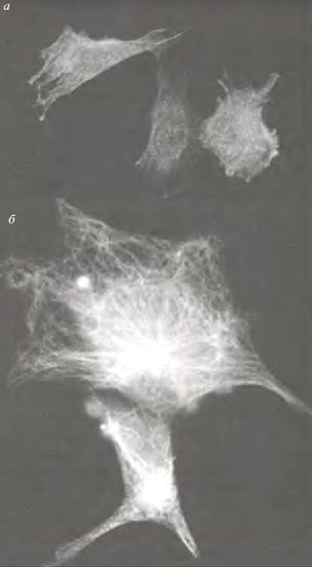
The most vivid example of the Structural Role of actin can be seen in the architecture of microvilli in brush-border cells, whose finger-like projections significantly increase the absorptive surface area of the intestine (Fig. 29.1). Actin filaments permeate most animal cells. They are particularly concentrated near The Plasma Membrane, where bundles of actin filaments are anchored, helping to maintain cell shape (Fig. 29.2, a). The organization of actin filaments into ordered arrays and networks depends on numerous actin-binding proteins. Although the actin filaments of microvilli are stable structures, they are nonetheless capable of disassembly and self-assembly. When a phagocyte migrates toward a particle to be engulfed, actin filaments can be observed extending directly into pseudopodia (paddle-like cellular extensions projecting in the direction of movement).
Class="center">Fig. 29.1. Organization of actin filaments in a microvillus. The formation of a robust cytoskeletal Structure results from the interaction of actin filaments with other proteins to form cross-links

Fig. 29.2. Micrographs of actin fibers and microtubules. a — Actin fibers in mouse myoblasts, visualized using anti-actin Antibodies. The fibers pervade the cell, and their bundles (stress fibers) are anchored at focal adhesion points on The cell membrane. These fibers rapidly assemble and disassemble. Micrograph kindly provided by R. Gunning, Children’s Medical Research Institute, Sydney; b — Microtubules in the Cell Cytoplasm, radiating from the microtubule-organizing center, or centrosome
Actin filaments are involved in amoeboid and "crawling" cell movements. This is the result of the action of myosin II (conventional muscle-type myosin molecules). Approximately 16 such molecules assemble into small bipolar filaments as needed, resembling the thick filaments of striated muscle. Their action is very similar to that of a sarcomere: bipolar filaments generate a contractile force on adjacent actin filaments and cause tension in the cell membrane to which the filaments are anchored (Fig. 29.3). The regulation of contraction (as in smooth muscle) is associated with the phosphorylation of myosin light chains (see Chapter 28).
Fig. 29.3. The pulling action of myosin II on actin filaments. The energy for the process is provided by ATP Hydrolysis. The performance of useful work by the system depends on the appropriate attachment of the actin filament to the cell membrane to alter cell shape. Each myosin molecule has a region capable of interacting with other molecules, which determines the self-assembly of the bipolar filament. The diagram shows a longitudinal section; the bipolar complexes are cylindrical in shape. Contraction is driven by the sliding of actin filaments relative to the myosin bundles.

The contractile role of the actomyosin system in cytokinesis—the division of the cytoplasm during cell division—has been defined with considerable precision. During anaphase
a ring of actin filaments assembles in the equatorial plane, where division takes place. It is believed that actin filaments are anchored in the plasma membrane and that the overlap of these fibers (with opposite polarity) enables cytoplasmic myosins to contract. As contraction progresses, this structure disassembles, and the cytoplasm is distributed between the two daughter cells. Such a system serves as an excellent example of the temporary organization of contractile complexes within non-muscle cells.
The Role of Actin and Myosin in Intracellular Transport
Actin filaments can serve as tracks along which ATP-dependent Molecular Motors transport cellular components. Chapter 28 mentioned that if individual (muscle-type) myosin heads are attached to latex beads in an experiment, they will move along immobilized actin filaments in the presence of ATP and Ca2+. The "tail" of a muscle-type myosin molecule is structured in such a way as to enable the self-assembly of myosin into a bipolar (thick) filament, which promotes the sliding of adjacent actin fibers, thereby causing contraction.
There is a family of myosin molecules with conventional myosin heads, but instead of the long rod-like tail characteristic of muscle myosin, they have a short tail, hence their name—mini-myosins. Mini-myosin molecules cannot form bipolar bundles, but utilizing ATP energy, they move along the actin filament. The tails of mini-myosins are designed to attach to other structures, such as vesicle membranes: if such attachment occurs, the mini-myosin will pull the vesicle along the actin filament. In the giant alga Nitella, mini-myosins pull The Endoplasmic reticulum across the entire cell along stationary actin filaments. The mini-myosin tails are attached to the endoplasmic reticulum, which permeates the cytoplasm. They pull it along, causing continuous Circulation of the entire cytoplasmic contents.
Many unusual (non-muscle) myosins have been identified. They have been found in both Yeasts and vertebrates. All non-muscle myosins possess globular actin-binding heads and various tails that interact with specific membrane structures.
Microtubules, Cell Movement, and Intracellular Transport
What are Microtubules?
Microtubules are formed by the polymerization of the protein tubulin, which is a heterodimer composed of α- and β-tubulin subunits. During polymerization, the α-tubulin of one dimer contacts the β-tubulin of the next to form protofilaments, 13 longitudinal rows of which make up the hollow tube. Polymerization proceeds in a HEAD-to-tail direction such that the microtubule has a defined polarity; its ends are designated as the (+) and (-) ends, respectively. Microtubules within the cell are unstable and can rapidly assemble and disassemble. This instability is due to unprotected ends, which promote rapid microtubule breakdown via depolymerization into free tubulin subunits. In the cell, the (-) ends are anchored to the microtubule-organizing center (MTOC)—a structure located near the nucleus that contains (in animal cells) a pair of small bodies called centrioles, formed from fused microtubules. The cell is permeated by microtubules radiating from the MTOC (see Figs. 29.2b and 29.4).
Fig. 29.4. Microtubules radiating from the MTOC. The (+) and (-) signs indicate microtubule polarity. Centrioles are a pair of tubular structures built from joined microtubules; microtubule outgrowth occurs in the substance surrounding the centrioles.

What Protects the (+) Ends of Microtubules?
As already noted, microtubules rapidly disintegrate unless their ends are protected. The MTOC protects the (-) end. Within the cell, microtubules grow (and disassemble) from their (+) ends. Upon reaching an appropriate target, proteins associated with the target protect the microtubule. Microtubules may grow from the MTOC in completely random directions, but only those that make contact with a suitable cellular component will be protected and stabilized by target proteins, whereas the rest will break down. It remains unclear precisely what protects a growing microtubule from disintegration before it reaches its protein target. According to the current model, a tubulin subunit binds GTP and, in this complexed form, protects the (+) end. Following its incorporation into the tubule, slow hydrolysis of GTP to GDP and Pi occurs. Thus, newly added tubulin subunits remain complexed with GTP for a short time, which temporarily protects the end of the microtubule from destruction. GTP hydrolysis acts as a familiar "molecular timer": if new tubulin molecules are added before the last incorporated subunit loses its GTP, the microtubule is protected. If the GTP at the free end is hydrolyzed sooner, microtubule disassembly is inevitable according to this model. In Cilia and flagella, where microtubules are permanent structures (see below), covalent protein modification occurs after assembly to prevent breakdown. This outlines the basic structural scheme of microtubules. We will now consider their functions.
Functions of Microtubules
Like actin filaments, microtubules participate in maintaining cell shape, cell motility, and intracellular transport.
Molecular Motors Driving Movement Along Microtubules
Two Types of ATP-dependent motors have been identified: kinesin and dynein. Kinesin moves along the microtubule from the (-) to the (+) end, while dynein moves in the opposite direction. Each has two globular heads resembling myosin heads (Fig. 29.5a), with light chains directed in opposite directions.
Fig. 29.5. Structure and function of kinesin: (a) Structure of a kinesin molecule; (b) transport of a vesicle along a microtubule by a kinesin molecule (schematic)
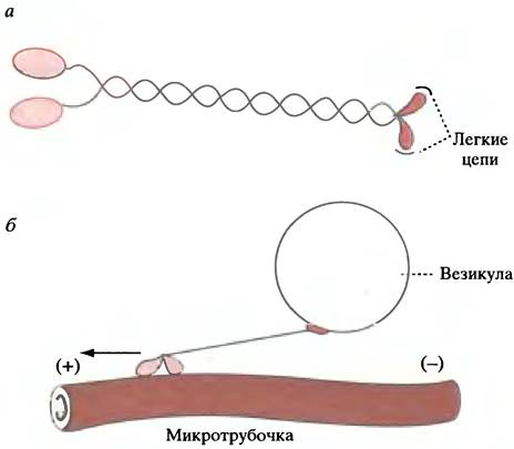
There are families of kinesin and dynein molecules specialized to perform different functions in various tissues. The motor heads that move along the microtubule probably remain unchanged, whereas the tail structure is modified to attach to various cellular structures (Fig. 29.5b). As an example of molecular motor activity, we can consider the movement of a vesicle along a microtubule (see below). In the presence of ATP, kinesin and dynein molecules (which possess ATPase activity) will move along microtubule fibers immobilized on a solid surface in the exact same way that myosin heads move along immobilized actin filaments.
The Role of Microtubules in Cell Movement
Microtubules play a vital role in the movement of cilia and flagella. Cells lining the respiratory tract possess A large number of cilia, whose beating action sweeps away mucus and removes foreign particles. Spermatozoa propel themselves by means of a rotating flagellum. Cilia and flagella
similar in structure: they originate from basal bodies resembling centrioles, with microtubules running down their entire length. Dynein molecules are attached to the microtubules and act as motors. Utilizing ATP energy, they move along the microtubules toward the (-)-end, causing the latter to slide past one another and producing the wavelike beating of cilia or flagella.
The Role of Microtubules in Intracellular Vesicle Transport
Microtubules are involved in intracellular transport. Vesicles formed by the Golgi apparatus are directed to their precise destinations within the cell. While the details of this programmed movement and vesicle translocation remain unclear, microtubules and ATP-dependent motors appear to play a key role.
As already mentioned, a nerve cell axon can be so long that its terminal may lie a considerable distance from the cell body, where vesicle formation and protein synthesis take place. To transport Materials along the axon, microtubules extend with their (+)-ends oriented toward the terminal. The molecular motor kinesin attaches by one end to the vesicle membrane and moves along the microtubule toward the (+)-end (see Fig. 29.5, b).
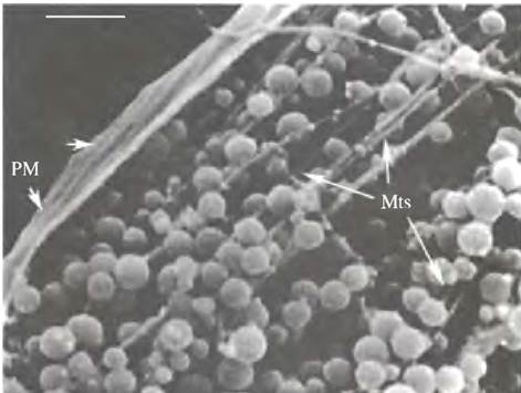
Another motor, cytoplasmic dynein, drives transport in the opposite direction, from the axon back to the cell body. A striking example of microtubule-based movement is The transport of pigment vesicles (Fig. 29.6).
Fig. 29.6 Vesicle transport along microtubules. Scanning electron micrograph showing pigment-containing vesicles being transported along microtubules in a squirrelfish chromatophore. Color Changes in the cell are driven by the movement of vesicles toward and away from the cell center. Mts — microtubules; PM — plasma membrane. (Scale bar = 0.5 µm)
The Role of Microtubules in Mitosis
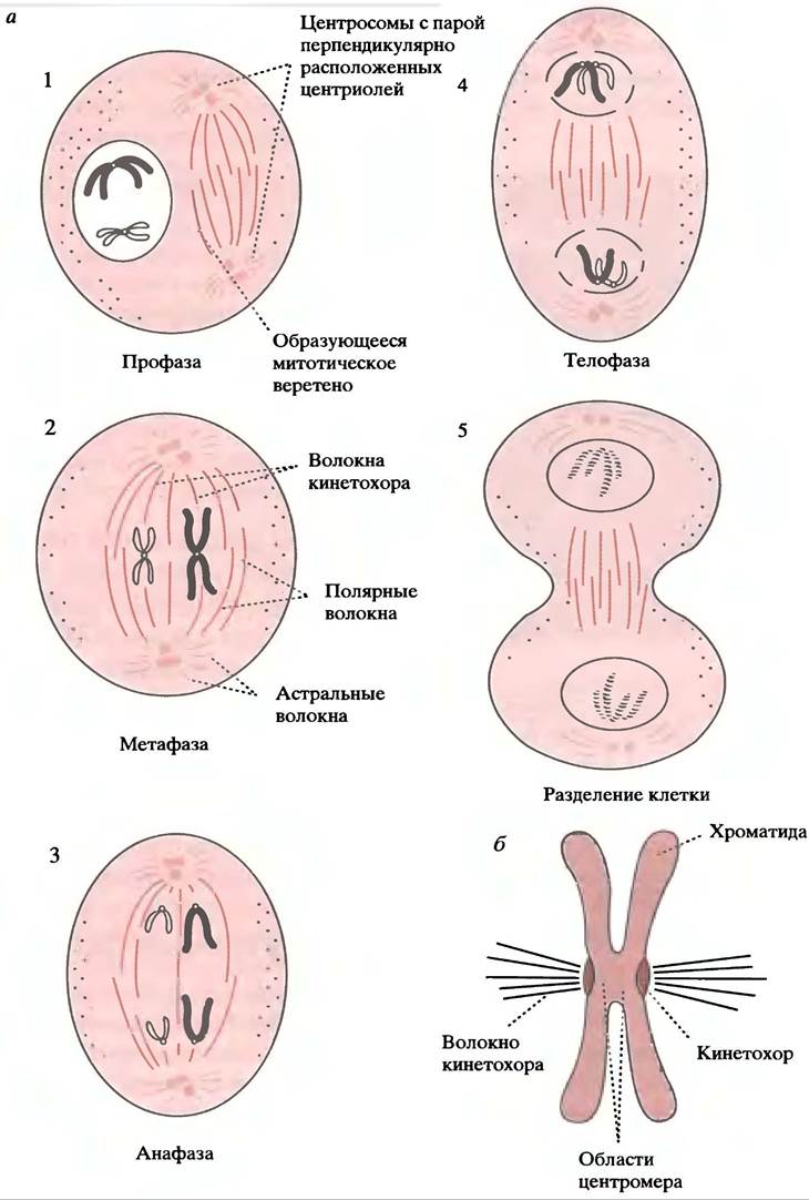
The Stages of the cell mitotic cycle are illustrated in Fig. 29.7, a.
During prophase, centrosomes (equivalent to MTOCs in non-dividing cells) duplicate and migrate to opposite poles of the cell, extending microtubules to form the mitotic spindle. DNA condenses into distinct chromosomes visible under a Light Microscope. In prometaphase, the nuclear envelope breaks down, and spindle microtubules enter the nuclear region, attaching to the kinetochores of chromatids. These represent sister chromatids formed by METABOLISM/36.html">DNA Replication during interphase, joined at their centromeres to form X-shaped structures (see Fig. 19.9). Following Separation, each chromatid will become an independent chromosome. In metaphase, chromatids align along the equatorial plane in preparation for anaphase. The chromatids then separate and migrate toward the poles (anaphase A), which subsequently move further apart from one another (anaphase B). In telophase, the nuclear envelope reforms, accompanied by
cell division via cytoplasmic Cleavage (cytokinesis), which is driven by the actomyosin system. Afterward, in each daughter cell, the centrosome becomes the organizing center for the dense array of microtubules characteristic of an interphase cell.
Mechanism of Chromosome Movement (Anaphase A)
It remains unclear what drives the movement of chromosomes toward the cell poles after their attachment to kinetochore fibers (Fig. 29.7, b), though it is evident that this work can be performed by a dynein-like molecular motor. As the chromosomes move, the kinetochore fibers shorten. Because microtubules are incapable of contraction, it is hypothesized that the chromosome crawls along the fiber while the microtubules undergo depolymerization in its wake.
Mechanism of Pole Separation (Anaphase B)
This process involves polar fibers (see Fig. 29.7, a), which do not attach to chromosomes but overlap in the equatorial region. As the poles move apart, these fibers lengthen to maintain the overlap. This suggests that pole separation is driven by molecular motors interacting with astral fibers radiating from the poles. Regardless of the exact mechanism, ATP is required for pole separation.
Fig. 29.7 Mitosis: a — Simplified diagram of mitotic stages. Polar fibers are highlighted in color and are thought to be responsible for separating the poles; black fibers represent kinetochore fibers, which drive chromosome movement toward the poles. All fibers are microtubules; b — Attachment of a kinetochore fiber (schematic)
Intermediate Filaments
The actin network described above contains microfilaments with a diameter of 6 nm, whereas microtubules have a diameter of 20 nm. A third type of cytoskeletal network in eukaryotic cells is formed by filaments averaging 10 nm in diameter, hence called intermediate filaments (IFs).
There are several types of IFs, formed by various groups of highly homologous proteins. Typically, they feature a central rod domain containing approximately 350 amino acid residues, flanked by terminal domains that vary among IF types. This protein diversity contrasts with the uniform protein subunits found in actin filaments and microtubules.
The various types of IFs found in Eukaryotic cells are expressed at different Selection/3.html">Stages of development and differentiation. However, the precise function of IF networks remains elusive. As already noted, intermediate filaments provide mechanical strength to epidermal cells.
Nuclear lamins form a meshwork underlying the inner nuclear membrane and are linked to nuclear pores. In Nerve Cells, neurofilaments provide mechanical support for long axons, while desmin filaments are localized at the Z-discs of sarcomeres. Curiously, the depletion of intermediate filaments in cultured cells—caused by Mutations or the introduction of specific antibodies—does not prevent them from growing and dividing successfully.
Chapter 29 Questions
1. What are the functions of actin in non-muscle cells?
2. What are microtubules?
3. Microtubules with unprotected ends undergo spontaneous dissociation. What protects their ends?
4. What are kinesin and dynein? Illustrate your answer with a diagram.
5. During cell division in metaphase, chromosomes move away from the equatorial plane individually. Microtubules are attached to kinetochores and shorten as the chromosomes separate. Does this mean that the microtubules themselves shorten? Explain your answer.
6. What are intermediate filaments?
7. What are the functions of intermediate filaments?
Last update: 06/08/2026
Editorial and Educational Adaptation: This material has been compiled based on the primary/original source text. The project team performed an editorial review, corrected technical inaccuracies, structured sections, and adapted the content for an educational format.
What was processed:
- elimination of formatting defects (OCR errors, structural breaks, corrupted characters);
- editorial organization of content;
- standardization of terminology in accordance with academic sources;
- verification of factual statements against the original source text.
All mentions of the author, publication year, and origin of the primary text have been preserved in accordance with the source.