BIOCHEMISTRY AND MOLECULAR BIOLOGY - W. ELLIOTT - 2002
ANSWERS TO QUESTIONS
Chapter 1
1. The amount of ATP that can be synthesized using 5000 kJ of Free energy is 5000/55, which corresponds to 90.91 mol. The amount of ATP disodium salt formed in this case would be 551 • 91 g per day, or 50,141 g (72% of human body weight). Such an amount of ATP can be synthesized because ATP in our body is continuously broken down into ADP and Pi, and then resynthesized.
2. The ∆G°′ value (-55 kJ • mol-1) refers to standard conditions where ATP, ADP, and Pi are present at a concentration of 1.0 M. In The Cell, the concentrations of these substances are significantly lower, and the actual value of ∆G for ATP synthesis will differ from ∆G°′:
Class="center">
3. ATP and ADP are high-energy phosphoric anhydride compounds, whereas AMP is a low-energy phosphoric ester. The significant exergonicity of ATP and ADP Hydrolysis is due to several factors. First, the release of phosphate relieves the strain caused by electrostatic repulsion between negatively charged phosphate groups. The released phosphate ions diffuse apart. Second, the exergonic nature of hydrolysis is promoted by the greater Resonance stabilization of the free phosphate ion compared to phosphate groups. The hydrolysis of AMP causes only a negligible increase in resonance stabilization.
4. Very negligible, because the shape of this curve depends on the time chosen for activity measurement. At higher temperatures, the probability of Enzyme inactivation increases; at any given Temperature, the amount of inactivated enzyme is proportional to the time of enzyme incubation at that temperature. When studying enzyme thermostability, it is more correct to incubate the enzyme for the same time at various temperatures and, after cooling, measure the activity in each sample at the normal incubation temperature.
5. A. Nonpolar molecules cannot form Hydrogen Bonds with Water molecules. Therefore, water molecules surrounding a benzene molecule organize into a more highly ordered Structure, forming hydrogen bonds with one another. This increase in order decreases Entropy and increases the system's energy; consequently, upon contact with water, benzene molecules are forced to minimize the benzene-water interaction surface, first by forming spherical globules and then a separate layer. This phenomenon is known as the hydrophobic effect. B. Polar glucose groups can form hydrogen bonds with water. C. Na+ and Cl- ions are hydrated, and As a result of decreased ionic attraction, the free energy decreases. Such ion Separation is characterized by a decrease in entropy.
6. The enzyme AMP kinase transfers a phosphate group from ATP to AMP in the reaction ATP + AMP —> 2ADP. Hydrolysis does not occur, so no significant changes in ∆G°′ are observed in the reaction.
7. In the cell, PPi will be hydrolyzed to 2Pi because there is no inorganic pyrophosphatase in the fully purified enzyme. In the first case, ∆G°′ of the overall reaction corresponds to -32.2 - 33.4 + 10 kJ • mol-1 = -55.6 kJ • mol-1. For the fully purified enzyme, ∆G°′ = -22.2 kJ • mol-1.
8. Ionic bonds, hydrogen bonds, and Van der Waals forces have average energies of 20, 12–29, and 4–8 kJ • mol-1, respectively. The activation energy required to form weak bonds is very small, allowing such bonds to form easily without a catalyst and break just as easily, which imparts flexibility to structures stabilized by weak bonds. It has been established that A large number of short-range weak bonds are required for molecular association. Such bonds form The basis of biological Specificity.
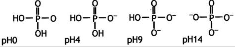
9. A.
B.

An acceptable pKa value would be 7.2.
pH = 7.2 + lg[Na2HPO4]/[NaH2PO4] = 7.2 + lg 1 = 7.2
C. Histidine with a pKa value of 6.5.
10. The ∆G°′ value of a reaction determines whether the reaction can occur, but it says nothing about the rate at which it will proceed (if at all). The reaction rate is determined by the activation energy and The rate of Transition State formation.
11. Enzymes catalyze Chemical Reactions for A number of reasons:
✵ their active sites bind the transition state of the substrate more tightly than the initial substrate, thereby lowering the activation energy;
✵ active sites establish a preferential orientation of the reacting molecules;
✵ enzymes can perform general Acid-Base Catalysis of reactions;
✵ enzymes facilitate the proper spatial orientation of metal-containing groups, thereby facilitating reaction progress.
Chapter 2
1. This is a polypeptide chain formed by a large number of Amino Acids linked by peptide (-CO-NH-) bonds.
2. The protein polypeptide chain is folded into a specific, usually compact globule, whose precise folding depends on non-covalent and covalent (disulfide) bonds between The amino acid side chains. At high temperatures, these bonds are disrupted, thereby causing the disordering of the protein polypeptide chains and turning them into a tangled, insoluble mass devoid of biological activity. This is Protein Denaturation.
3. All Examples are provided on pages 38 and 39.
4. A. About 4; B. About 10.5-12.5; C. About 6.0.
5. The net charge of this molecule depends on the content of aspartic and glutamic acids, as well as Lysine, Arginine, and histidine. The carboxyl and amino groups of the Other Amino Acids are involved in The formation of peptide bonds (excluding the two terminal ones).
6. Primary, secondary, tertiary, and quaternary (see Fig. 2.2).
7. The α-Helix, β-pleated sheet, and random coil (or connecting loop region). The α-helix and β-sheet satisfy the hydrogen-bonding potential of the main polypeptide chain (see Figs. 2.3 and 2.4). The random coil lacks a fixed structure and is typically located On the surface of the protein, where hydrogen bonding with water can occur.
8. See The structure of Collagen fibers shown in Fig. 2.8.
9. The desmosine group shown in Fig. 2.9.
Chapter 3
1. The presence of two hydrophobic tails instead of one.
2. No.
3. Only via exocytosis, endocytosis, or through specialized transport mechanisms (see Chapter 22).
4. It acts as a membrane fluidity buffer that prevents excessive association of polar Lipids.
5.
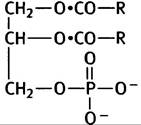
6. Lecithin (Choline), cephalin (ethanolamine), phosphatidylserine (Serine).
7. Choline is replaced by galactose and an oligosaccharide, respectively.
8. The kink formed by cis-unsaturated fatty acid tails and phospholipid membranes prevents tight packing of The Lipid Bilayer fatty acid tails, thereby increasing membrane fluidity.
9. Due to the Hydration shell surrounding the lipid membranes. Removing it is energetically unfavorable.
10. Facilitated Diffusion requires Membrane Proteins that allow solutes to cross the membrane in either direction according to their concentration gradient. This process does not require energy. An example is anion transport in red Blood Cells, which allows Cl- and HCO3- ions to pass through the membrane in either direction.
11. If the outside concentration is denoted as C1 and the inside concentration as C2, then:

Chapter 4
1. Pepsin as pepsinogen, Chymotrypsin as chymotrypsinogen, Trypsin as trypsinogen, Elastase as proelastase, and carboxypeptidase as procarboxypeptidase.
2. • Proteolytic Enzymes are potentially hazardous because they can attack the proteins lining the ducts of the digestive glands.
✵ The Small Intestine lacks the components targeted by amylase, which is therefore synthesized directly in its active form.
✵ Mucins protect cells from proteolytic attack.
3. In The Stomach, an acidic pH induces conformational changes (rearrangements) in pepsinogen, triggering self-Cleavage. This process releases a peptide that normally keeps the enzyme in an inactive state. The resulting pepsin immediately begins to activate its own synthesis from pepsinogen through an autocatalytic cascade mechanism. In the small intestine, enteropeptidase (an enzyme produced by the intestinal wall cells) converts trypsinogen into trypsin, which in turn activates the remaining zymogens.
4. Pancreatitis, or inflammation of the Pancreas, caused by proteolytic damage to pancreatic cells.
5. Because intestinal cells can only absorb monomers (and monoacylglycerols). Lipids, proteins, Polysaccharides, and Disaccharides cannot be absorbed directly.
6. Milk contains lactose, the hydrolysis of which into glucose and galactose is catalyzed by the enzyme lactase. In adulthood, many people lose The ability to synthesize this enzyme. Unabsorbed lactose undergoes Fermentation in the Large Intestine. Due to its osmotic effect, lactose draws water into the gut, causing diarrhea.
7. TAG, or triacylglycerol, is an ester of Cholesterol and Fatty acids.

8. The lipid (substrate) is emulsified by intestinal motility with the help of monoacylglycerol and free fatty acids (generated during initial Digestion, along with Bile salts), enabling lipase to act on the emulsified substrate surface. The digestion products, together with bile salts (free Fatty Acids and monoacylglycerol), are transported to the intestinal cells as disk-shaped mixed micelles, which have a high capacity for these compounds. These micelles presumably disintegrate at the cell surface.
9. Free fatty acids and monoacylglycerol are resynthesized into triacylglycerols, which are then incorporated into lipoprotein particles known as chylomicrons. TAG, cholesterol, and cholesterol esters form the Hydrophobic core of chylomicrons, which is surrounded by a shell of Phospholipids and specific proteins. Chylomicrons are absorbed into The Lymphatic system via exocytosis and are ultimately released as an emulsion into the bloodstream through the Thoracic duct.
Chapter 5
1. The Osmotic Pressure of free glucose prevents its storage in that form. The osmotic pressure of a solution depends on the number of dissolved particles. Polymerizing thousands of glucose molecules into a single Glycogen macromolecule significantly lowers this osmotic pressure.
2. Liver glycogen stores are depleted after a 24-hour fast, whereas TAG reserves typically last for several weeks. Triglycerides represent a much more potent energy source because they are more highly reduced and anhydrous. Incidentally, if the energy stored in lipids were instead stored as glycogen, body weight would be vastly greater.
3. Yes, because glucose can be converted into acetyl-CoA.
No, because acetyl-CoA cannot be converted into Pyruvate.
They do not exist, as our bodies lack specialized storage proteins dedicated to preserving amino acids (with the exception of milk).
4. • The liver maintains glucose Homeostasis by storing glucose as glycogen over extended periods. During fasting, the liver releases glucose to maintain a steady blood sugar level, which is essential for normal Brain function.
✵ During prolonged fasting, the liver synthesizes glucose to supply energy to the brain. It also converts lipids into Ketone Bodies, which the brain (and other Tissues) can use to partially meet their energy demands;
✵ The liver synthesizes fats designated for export to other tissues.
5. No.
6. Adipose cells store fat for long periods. During fasting, these fat cells release fatty acids.
7. Erythrocytes depend entirely on glucose, which they metabolize into lactate via Glycolysis. Having lost their Mitochondria during maturation, red blood cells are incapable of synthesizing ATP through any pathway other than glycolysis.
8. When blood glucose levels are high, Insulin is secreted, signaling tissues to store nutrients. As blood glucose drops, insulin levels fall while Glucagon levels rise. The rise in glucagon signals the liver to release glucose and adipocytes to release fatty acids, both of which are then utilized by tissues. Glucose uptake by brain cells is independent of insulin and can occur even during fasting when insulin levels are extremely low.
In addition, adrenaline can induce an intensive mobilization of glucose and fatty acids to cope with extreme situations.
Chapter 6
1. Glucose-1-phosphate + UTP —> UDP-glucose + PPi,
PPi + Н2O —> 2Рi,
UDPG + glycogen(n) —> UDP + glycogen(n + 1).
The hydrolysis of PPi drives the overall reaction to be strongly exergonic.
2. The reaction involving the formation of glucose-1-phosphate from inorganic pyrophosphate never occurs in the cell because its substrate (inorganic pyrophosphate) is immediately degraded. This reaction is the reverse of the one taking place in the cell; however, in accordance with the rules of systematic nomenclature, the enzyme is named after the forward reaction (see Fig. 6.3).
3. The Liver and, to a lesser extent, the Kidneys, since glucose-6-phosphatase is localized primarily in these tissues.
4. Glucose + ATP —> glucose-6-phosphate + ADP. Glucokinase has a significantly higher Km for glucose than hexokinase. During starvation, the liver releases glucose back into the blood, which is primarily essential for supplying glucose to the brain and erythrocytes. Immediately after being taken up by brain and liver cells, glucose is phosphorylated. The lower affinity of glucokinase for glucose (compared to hexokinase) prevents competition for blood sugar between the brain and the liver. The liver efficiently utilizes glucose only when its blood levels are high. Since glucokinase, unlike hexokinase, is not inhibited by glucose-6-phosphate, the liver can take up glucose and synthesize glycogen even when intracellular glucose-6-phosphate levels are high.
5. Preventing childhood galactosemia through a galactose-free diet is effective because the patients' requirement for galactose is met by the reversible epimerization of UDP-glucose to UDP-galactose. Therefore, eliminating galactose from the diet does not prevent the synthesis of Glycolipids and Glycoproteins containing galactose.
6. In the capillaries, lipoprotein lipase cleaves fatty acids from TAGs, and they are immediately taken up by surrounding cells.
7. VLDL stands for very-low-density Lipoproteins. They are similar to chylomicrons and transport cholesterol and triacylglycerols from the liver to peripheral tissues.
8. Cholesterol is transported from the liver to peripheral tissues via VLDL. As VLDL loses TAG, they are converted first into IDL and then into LDL. The latter are taken up by peripheral tissues. However, some IDL and LDL return to the liver. IDL and LDL receive cholesterol from peripheral tissues via HDL, establishing a reverse cholesterol Transport from the tissues back to the liver.
9. By conversion into bile acid salts in the liver.
10. The LCAT enzyme transfers fatty acid residues from lecithin to cholesterol.
11. TAG are not released; hormone-sensitive lipase, activated by glucagon (or epinephrine in critical situations), cleaves free fatty acids, which are transported by the blood to tissues in a complex with serum albumin.
Chapter 7
1. Glycolysis, The Citric Acid Cycle, and the Electron Transport Chain. These three stages of glucose oxidation occur in the Cytoplasm, the mitochondrial matrix, and The inner mitochondrial membrane, respectively.
2. Structures are shown on p. 100.
АН2 + NAD+ <-> А + NADH + Н+
В + NADH + Н+ <-> ВН2 + NAD+
3. FAD is a hydrogen carrier with the following structure: isoalloxazine ring system - ribitol phosphate - phosphate - ribose - adenine. FAD is a riboflavin derivative representing a prosthetic group covalently bound to the enzyme and reduced to FADH2.
4. During aerobic glycolysis, glucose is broken down into pyruvate. The resulting NADH is reoxidized by mitochondria. During anaerobic glycolysis, the rate of glycolysis exceeds the capacity of mitochondria to reoxidize NADH. This can occur, for example, during an extreme situation ("fight or flight"). If the accumulating NADH is not rapidly oxidized, glycolysis and ATP production will be inhibited due to a deficiency of NAD+. In a critical situation, NADH is reoxidized through the reduction of pyruvate to lactate.

5. The ∆G of hydrolysis is -31 kJ • mol-1 compared to 20 kJ • mol-1 for carboxylic acid esters; consequently, thiol esters are high-energy compounds.
6. Pyruvate + CoASH + NAD+ —> Acetyl-S-CoA + NADH+ + H+ + CO2
∆G°′ = -33.5 kJ • mol-1.
7. The acetyl group enters The Citric Acid cycle.
8. NAD and FADH2 are reoxidized by The electron transport chain, yielding H2O and generating ATP.
9. According to the Nernst equation, ∆G°′ and E°′ are related as follows: ∆G°′ = -nF∆E°′, where F is the Faraday constant = 96.5 kJ • mol-1, and E°′ is the difference between the redox potentials of the electron donor and acceptor. In this example, ∆E°′ = -1.035 V (-0.219 - 0.816 V). Therefore, ∆G°′ = -2 (96.5 kJ • mol-1) • (-1.035 V) = -2(-1.035) = 194.06 kJ • mol-1.
10. Breakdown of fatty acids to acetyl-CoA.
11. A. Yes. Glucose is converted to pyruvate, and pyruvate dehydrogenase converts it to acetyl-CoA, which can then be used for lipid synthesis. B. No. Fatty acids are degraded to acetyl-CoA. Pyruvate is required for glucose synthesis, but the reaction catalyzed by pyruvate dehydrogenase is irreversible. Consequently, in animals, acetyl-CoA (let alone fatty acids) cannot be converted into glucose. C. Yes, but not by reversing the pyruvate dehydrogenase reaction (see figure 7.11 legend).
Chapter 8
1. The conversion of a C6 molecule into two C3 molecules occurs via aldolytic cleavage catalyzed by the enzyme aldolase. This requires a STRUCTURE OF THE type:

The isomerization of glucose-6-phosphate to fructose-6-phosphate generates an aldol structure capable of undergoing such cleavage:

2. The process of ATP Synthesis in which a high-energy phosphoryl group, covalently bound to a substrate, is transferred to ADP. An example is The oxidation of glyceraldehyde-3-phosphate (see p. 112).
3. Pyruvate kinase catalyzes the reverse reaction, but by nomenclature, Kinases are always named after the reaction utilizing ATP. This reaction is irreversible because its product, enolpyruvate, spontaneously tautomerizes to the keto form, a process associated with a large negative value of ∆G°′.
4. 3 and 2 molecules, respectively. Phosphorolysis of glycogen using Pi yields glucose-1-phosphate, which is subsequently isomerized to glucose-6-phosphate. Conversely, the formation of glucose-6-phosphate from free glucose requires the investment of one ATP molecule.
5. Cytoplasmic NADH cannot directly cross the mitochondrial membrane; therefore, its reducing equivalents must be transported via one of two alternative shuttle mechanisms. The malate-aspartate shuttle (see Fig. 8.9) reduces mitochondrial NAD+, whereas the glycerophosphate shuttle reduces the FAD of mitochondrial inner membrane glycerophosphate dehydrogenase. Because the Redox Potential of the former is more negative, the reducing equivalents enter the electron transport chain at the level of different respiratory complexes, resulting in a lower ATP yield for the glycerophosphate shuttle.
6. The cleavage of succinyl-CoA yields a molecule of GTP from GDP and Pi. This process releases one molecule of H2O, which is consumed within the cycle (and thus does not appear as a net product), balancing the overall stoichiometry.
7. Oxidation yields a β-keto acid, which readily undergoes decarboxylation.
8. The reaction catalyzed by pyruvate carboxylase replenishes the pool of oxaloacetate:

9. Biotin, a B-group vitamin. In the presence of ATP, it forms a reactive carboxybiotin intermediate that can transfer a carboxyl group to substrates.

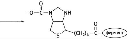
10. This is illustrated in Fig. 8.19.
11. The common feature is that both substances are mobile electron carriers. Ubiquinone links respiratory complexes I and II to complex III, whereas cytochrome c connects complexes III and IV. The former is localized within the hydrophobic lipid bilayer, while the latter resides in the aqueous phase on the outer surface of the mitochondrial inner membrane.
12. To pump protons from the mitochondrial matrix across the inner membrane and generate electrical and proton gradients that can be utilized for ATP synthesis.
13. In eukaryotes, The transfer of reducing equivalents from cytoplasmic NADH to the Mitochondrial Electron Transport chain is mediated by shuttle mechanisms. The glycerophosphate shunt results in the loss of 1 ATP molecule (from those potentially generated via NADH oxidation). Since glycolysis produces 2 NADH molecules per glucose, this leads to the loss of 2 potentially possible ATP molecules. No such problem exists in E. coli. Furthermore, unlike Eukaryotic cells, E. coli cells do not expend energy for The transport of ATP and ADP across the mitochondrial membrane.
Chapter 9
1. • Free fatty acids (derived from adipose tissue) are transported by serum albumin in the blood.
✵ Lipoprotein lipase releases free fatty acids and triacylglycerols from chylomicrons.
✵ Free fatty acids can be delivered from VLDL synthesized in the liver (their release occurs via the same pathway as in the previous case).
2. Brain cells and erythrocytes (erythrocytes lack mitochondria). See Chapter 5.
3. • Activation for the formation of acyl-CoA derivatives from fatty acids;
✵ at the outer mitochondrial membrane;
✵ in the mitochondrial matrix;
✵ as carnitine derivatives (see Fig. 9.1).
4. The reactions shown in Fig. 9.2 are analogous to the reactions: succinate -> fumarate -> malate -> oxaloacetate of the citric acid cycle (both in terms of reaction types and the electron acceptors used).
5. From 1 molecule of palmitic acid, 8 molecules of acetyl-CoA are formed, accompanied by the reduction of 7 molecules of FADH2 and NADH. The oxidation of NADH and FADH2 yields 2.5 and 1.5 molecules of ATP, respectively. Adding to this the ATP yield from the oxidation of 8 molecules of acetyl-CoA (10 ATP per molecule), the total yield is 108 molecules of ATP (counting GTP from the citric acid cycle as ATP). From this amount, 2 ATP molecules consumed in the activation reaction must be subtracted. Consequently, the net yield is 106 ATP molecules.
6. After 2 rounds of β-oxidation, cis-D3-enoyl-CoA is isomerized to trans-D2-enoyl-CoA (see the reaction on p. 134).
7. In cases of rapid fat mobilization from adipose cells, such as during starvation or diabetes, the liver converts acetyl-CoA into ketone bodies, which are released into the blood. Ketone bodies are primarily utilized by Muscles, helping to spare glucose; the brain can meet approximately half of its Energy Requirements using ketone bodies.
8. The synthesis of acetoacetate occurs in the mitochondrial matrix, whereas cholesterol is synthesized in The Endoplasmic reticulum membrane.
Chapter 10
1. Fatty acids are synthesized from two-carbon units donated by the three-carbon molecule malonyl-CoA. Acetyl-CoA is converted into malonyl-CoA via ATP-dependent carboxylation. The subsequent decarboxylation has a large negative ∆G°′ value. In other words, the carboxylation-decarboxylation cycle is essential to render The addition of two-carbon units to the growing fatty acid chain an irreversible reaction.
2. They are shown in Fig. 10.1.
3. In eukaryotes, all reactions take place on a single polypeptide chain whose enzymatic Functions are carried out by distinct domains. The functional unit of eukaryotic fatty acid synthase is a dimer uniting two molecules into a single entity. In E. coli, the various reactions are catalyzed by separate enzymes. The advantage for eukaryotes is that intermediates are directly channeled from one Active Site to another. In E. coli, intermediates must diffuse to the next enzyme, making the overall synthesis process slower.
4. See structures on p. 141. NAD+ is used in catabolic reactions—it accepts electrons during oxidation and energy-yielding processes. NADP+ participates in reductive Biosynthesis. The existence of these two compounds represents a form of metabolic compartmentalization that facilitates the independent regulation of these pathways.
5. The tissues where fatty acid synthesis is predominantly concentrated are the liver, adipose tissue, and the Mammary Glands during Lactation.
6. Acetyl-CoA is converted into citrate within the mitochondria, which is then transported into the Cytosol, where citrate lyase cleaves it into acetyl-CoA and oxaloacetate. This is an ATP-dependent reaction that drives the complete cleavage of citrate:
Citrate + ATP + CoA-SH + H2O -> acetyl-CoA + oxaloacetate + ADP + Pi.
7. Oxaloacetate is reduced to malate by mitochondrial NADH-dependent malate dehydrogenase. In the cytosol, malate is oxidized and decarboxylated to pyruvate by malic enzyme in an NADP+-dependent reaction. This pathway effectively shunts reducing equivalents from NADH to NADPH. The resulting pyruvate is returned to the mitochondria (see Fig. 10.4). In this process, only one NADPH molecule is generated per molecule of malonyl-CoA formed, whereas two are required for the reduction reactions during fatty acid synthesis. The missing molecule is provided by the glucose-6-phosphate dehydrogenase system described in Chapter 13.
8. The outline of this process is shown in Fig. 10.5.
9. There are two possible pathways for the synthesis of glycerol-based phospholipids. In the first pathway, Phosphatidic acid is attached to an alcohol such as ethanolamine (see Fig. 10.6). For this to occur, the alcohol must be activated (in phospholipid synthesis, the activated molecule is always a CDP-alcohol). For the synthesis of certain other phospholipids, diacylglycerol is activated instead (see Fig. 10.6). This takes place, once again, via the formation of a CDP-diacylglycerol complex. This situation is analogous to The Use of UDP-glucose in all cases where an activated glucose residue is required.
10. "Eicosanoids are biological regulators consisting of 20 carbon atoms (from the Greek eikosi, meaning twenty). Eicosanoids include Prostaglandins, thromboxanes, and Leukotrienes.
✵ Derived from polyunsaturated fatty acids.
✵ Prostaglandins trigger pain responses, inflammation, and fever. Thromboxanes are involved in platelet aggregation. Leukotrienes stimulate smooth Muscle contraction and, by causing airway spasms, contribute to The Development of asthma.
✵ Aspirin inhibits cyclooxygenase (an enzyme involved in prostaglandin synthesis), which allows it to suppress pain and fever as well as inhibit blood clotting.
11. Mevalonic acid is the first committed metabolite dedicated exclusively to cholesterol synthesis. Structural analogues of mevalonic acid inhibit HMG-CoA reductase, the enzyme responsible for mevalonate formation.
Chapter 11
1. The brain cannot utilize fatty acids; it must rely on a supply of glucose. The same applies to red blood cells, which lack mitochondria and can generate energy solely through glycolysis.
2. Because the substrate for pyruvate kinase is the enol form of pyruvate, and the keto-enol equilibrium strongly favors the keto form; consequently, the enzyme is left without a substrate. The solution to this problem lies in the existence of an alternative metabolic pathway that consumes two high-energy phosphate groups to drive the formation of phosphoenolpyruvate:
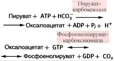
3. Fructose-1,6-bisphosphatase and glucose-6-phosphatase, which catalyze the Formation of fructose-6-phosphate and glucose-6-phosphate, respectively.
4. No. Free glucose is produced exclusively in the liver and kidneys.
5. Under normal nutritional conditions (in the absence of starvation), elevated muscle activity can lead to The production of lactate via anaerobic glycolysis. It is transported via the bloodstream to the liver, where it is converted back into glucose. The release of glucose into the Blood and Its subsequent uptake by muscles complete this cycle (see Fig. 11.4).
6. Glycerol kinase is essential for the Synthesis of glucose from glycerol (see Fig. 11.5). Glycerol is released during fasting, when maintaining blood glucose levels becomes a vital priority. Since Gluconeogenesis takes place in the liver, it makes physiological sense to transport glycerol directly there rather than metabolize it in adipose tissue, which is incapable of releasing free glucose into the bloodstream.
7. Via The Glyoxylate cycle (see Fig. 11.6).
Chapter 12
1. The first is Allosteric Regulation; the second is Covalent Modification of the enzyme, primarily achieved through phosphorylation.
2.

3. Allosteric effectors typically function by altering the enzyme's affinity for its substrate. The Substrate Concentration for such an enzyme must be subsaturating, which is normally the case. A positive allosteric effector shifts the sigmoidal curve of reaction velocity versus substrate concentration to the left, whereas a negative effector shifts it to the right (see Fig. 12.5). The sigmoidal dependence amplifies The Effect of allosteric effectors on reaction velocity, thereby increasing regulatory sensitivity (see Fig. 12.4).
4. Under these conditions, they have no effect whatsoever, because A change in enzyme affinity for the substrate at saturating substrate concentrations does not alter the reaction velocity.
5. The concerted and sequential mechanisms are illustrated in Figs. 12.7 and 12.8.
6. Because the structure of an allosteric effector differs from that of the substrate, diverse metabolic systems are able to engage in regulatory interactions via allosteric control.
7. Intracellular regulation is typically allosteric. It is this mechanism that enables every individual cell to maintain the metabolic balance of its pathways. However, allosteric regulation alone cannot dictate the overall direction of METABOLISM in The Cell as a whole in response to the demands of the entire Organism. This is The Role of the extracellular regulatory system (such as Hormones, etc.), which governs cellular activity in accordance with the physiological needs of the body.
8. The pathway is shown in Fig. 12.9. AMP activates Glycogen phosphorylase and Phosphofructokinase, whereas ATP inhibits PFK. A high ATP/ADP molar ratio inhibits glycolysis, whereas a decrease in ATP levels (leading to an increase in AMP) accelerates it. A high citrate level slows down the flux of metabolites from glycolysis into the citric acid cycle. A high acetyl-CoA level may indicate a shortage of oxaloacetate and, consequently, the need to activate pyruvate carboxylase for an anaplerotic reaction. At the same time, a high acetyl-CoA level indicates a steady supply of pyruvate from glycolysis, which at this point should appropriately be inhibited at the PEP level.
9. Direct allosteric regulation; phosphorylation of pyruvate dehydrogenase (and its inactivation) by a kinase; dephosphorylation by a protein phosphatase, which reverses the effect of phosphorylation (see Fig. 12.10).
10. As shown in Fig. 12.11.
11. High blood glucose levels stimulate insulin release, while low levels stimulate glucagon release.
12. The primary messenger is a hormone of the adrenaline type. Upon interacting with the cell receptor, it causes an increase in the intracellular concentration of a secondary messenger, which in turn affects metabolism. For the two hormones mentioned in the question, The secondary messenger is cAMP. It allosterically activates protein kinase A (PKA), which mediates various metabolic effects.
13. By mobilizing glucose transporters to The cell membrane (see Fig. 12.17).
14. cAMP triggers a cascade activation that begins with the activation of PKA (see the scheme on p. 170).
15. The differences are summarized in Fig. 12.22.
16. Different cells have receptors for different hormones. If cell A has a receptor for hormone X, then cAMP in this cell produces an effect characteristic of hormone X. Cell B lacks the receptor for hormone X but has a receptor for hormone Y; therefore, cAMP in cell B produces effects characteristic of hormone Y, but not hormone X.
17. Fructose-2,6-bisphosphate. cAMP increases its level (see Fig. 12.25).
18. Glucagon, by activating the synthesis of its secondary messenger cAMP, stimulates gluconeogenesis. Concurrently, cAMP activates a kinase that phosphorylates pyruvate kinase, causing its inhibition. In muscle, adrenaline increases the production of the secondary messenger cAMP, which stimulates glycolysis to generate energy; therefore, inhibiting pyruvate kinase here would be inappropriate.
19. cAMP activates hormone-sensitive lipase, which catalyzes the hydrolysis of triglycerides.
Chapter 13
1. The Pentose Phosphate Pathway provides ribose-5-phosphate for nucleotide synthesis, and the generated NADPH is used in lipid synthesis. In The pentose phosphate pathway, pentoses undergo complex interconversions to form molecules containing from 3 to 7 carbon atoms.
2. The oxidative phase involves the conversion of glucose-6-phosphate into ribose-5-phosphate with the release of CO2 and the reduction of NADP+.
3. Transaldolase and transketolase are the primary enzymes (see Fig. 13.2), but glycolytic enzymes may also participate.
4. Since transaldolase and transketolase use only ketoses as Donors, a portion of the ribose-5-phosphate is converted into xylulose-5-phosphate. Subsequently, the following transformations take place:
1. 2 C5 → C3 + C7 (transketolase),
2. C7 + C3 → C4 + C6 (transaldolase),
3. C5 + C4 → C3 + C6 (transketolase).
The 2 C5 in reaction 1 are ribose-5-phosphate and xylulose-5-phosphate; the final C3 component is glyceraldehyde-3-phosphate, which is converted into glucose-6-phosphate with the loss of Pi. Ultimately, 6 molecules of ribose-5-phosphate are converted into 5 molecules of glucose-6-phosphate and Pi. Thus, the cell can produce NADPH without increasing the total content of ribose-5-phosphate.
5. In the oxidative phase, glucose-6-phosphate is converted into ribose-5-phosphate with the release of CO2. Considering that 6 molecules of glucose-6-phosphate yield 6 molecules of ribose-5-phosphate, from which 5 molecules of glucose-6-phosphate can be resynthesized, the overall equation will feature 6 molecules of CO2. In other words, on paper this looks like the Complete oxidation of a glucose molecule. In reality, however, a single glucose-6-phosphate molecule is not converted into 6 molecules of CO2.
6. NADPH is required for the reduction of Glutathione, a molecule essential for protecting erythrocytes. Patients with a hereditary glucose-6-phosphate dehydrogenase deficiency are sensitive to the antimalarial drug pamaquine, which induces hemolytic anemia.
Chapter 14
1. The "light" reactions involve the splitting of water using light energy and the reduction of NADP+ to NADPH. During the "dark" reactions, NADPH is used to reduce CO2 and water into CARBOHYDRATES. The term "dark" implies that light is not required for these reactions to occur (it does not mean they take place only in the dark). In fact, dark reactions proceed most intensively in bright sunlight.
2. This is chlorophyll, which captures light photons. Upon excitation by a photon, one of the electrons in the chlorophyll molecule moves to a higher energy level. Resonance energy transfer allows this excitation to be passed from one molecule to another until it is captured by molecules of a special reaction center, which do not participate in resonance energy transfer but instead direct the electrons into the electron transport chain (see Fig. 14.4).
3. • ATP synthesis via a chemiosmotic mechanism during electron transport through the bf complex of Photosystem II (see Fig. 14.6).
✵ ATP synthesis under conditions of complete reduction of NADP+ molecules, when electrons transported by Photosystem I carriers are fed into the bf complex, promoting the formation of a larger amount of ATP (see Fig. 14.7).
4. Chlorophyll P680+, which serves as the reaction center of the photosystem II pigment: it is excited by resonance energy transfer from an antenna chlorophyll molecule and transfers an electron to pheophytin, the first component of the photosystem II electron transport chain. P680+ exhibits strong electron-accepting properties, i.e., it is a strong oxidizing agent.
5. Thylakoids are formed by the invagination of the inner chloroplast membrane (compare with the inner mitochondrial membrane), which explains the apparent opposite orientation of the proton pump.
6. The enzyme Rubisco (ribulose-1,5-bisphosphate carboxylate-oxygenase) cleaves ribulose-1,5-bisphosphate into two molecules of 3-phosphoglycerate; this reaction involves the fixation of one molecule of CO2.
7. See Fig. 14.10.
8. The sequence of these reactions is shown in Fig. 14.9.
9. If CO2 is initially fixed by the enzyme Rubisco into a 3-phosphoglycerate molecule, the plant is classified as a C3 plant. In the reaction catalyzed by this enzyme, oxygen and CO2 compete with each other. However, at high temperatures and under intense sunlight in C4 plants, CO2 is initially fixed by pyruvate-Pi dikinase and PEP carboxylase into oxaloacetate (see Fig. 14.11), which is reduced to malate. The latter then enters The Calvin Cycle. Decarboxylation of malate by malic enzyme leads to a significant increase in the CO2/O2 ratio (the concentration of CO2 in the cells can increase by a factor of 10–60). The complete pathway is shown in Fig. 14.11. Minor variations of this scheme may occur in various C4 plants, but the basic strategy of avoiding oxygenation by ribulose bisphosphate carboxylase remains unchanged.
10. In animals, pyruvate kinase cannot form phosphoenolpyruvate from pyruvate. However, plants possess the enzyme pyruvate-Pi dikinase, which utilizes two ATP phosphate groups to carry out this conversion.
Chapter 15
1. Oxidation leads to the formation of a Schiff base, which is hydrolyzed in an aqueous environment.

2. Glutamic acid.
3. Transdeamination is the most common mechanism. The amino group of Most amino acids is transferred to α-ketoglutarate to form glutamate. The latter is deaminated by Glutamate dehydrogenase:
1. Alanine + α-ketoglutarate —> pyruvate + glutamate.
2. Glutamate + NAD+ + Н2O —> α-ketoglutarate + NADH + NH4+.
Overall reaction:
Alanine + NAD+ + Н2O —> pyruvate + NADH + NH4+.
4. Pyridoxal phosphate (see Fig. 15.2). The Mechanism of Transamination is shown in Fig. 15.3.
5. By the removal of H2O and H2S, respectively (see Fig. 15.4).
6. Glucogenic Amino acids are those whose deamination ultimately yields pyruvate (or phosphoenolpyruvate). Their conversion to pyruvate may involve a number of intermediate steps leading, for example, to the formation of citric acid cycle intermediates. Acetyl-CoA is derived from ketogenic amino acids. Only leucine and lysine are exclusively ketogenic, while Some amino acids, such as phenylalanine, are mixed. Ketone bodies are formed from ketogenic amino acids only in exceptional cases, such as during starvation. In all other cases, acetyl-CoA is oxidized in the normal manner.
7. Phenylalanine normally does not undergo transamination; it is converted into Tyrosine and subsequently metabolized (see Fig. 15.5). If the conversion of phenylalanine to tyrosine is impaired, phenylalanine undergoes transamination to form phenylpyruvate, which causes irreversible brain damage in children and leads to early death.
8. Tetrahydrobiopterin serves as a donor of reducing equivalents for the formation of H2O from one atom of the oxygen molecule used in the hydroxylation reaction.
9. Activation occurs via the formation of S-adenosylmethionine (SAM). SAM has a sulfonium ion structure that significantly facilitates the Cleavage of the methyl group. The formation of SAM is illustrated in Fig. 15.6.
10. See Fig. 15.8.
11. Both high-protein diets and starvation are characterized by a high rate of AMINO ACID DEAMINATION (in muscles, proteins are degraded during starvation to sustain glucose synthesis), and the amino nitrogen must be converted into urea.
12. • Ammonium is converted into glutamine, which is transported to the liver and hydrolyzed.

✵ Amino nitrogen is transported from the muscles in the form of alanine. The alanine cycle is shown in Fig. 15.9.
Chapter 16
1. For the degradation of unwanted molecules and structures internalized via endocytosis, as well as for completing The life cycle of cellular components.
2. Primary Lysosomes are vesicles formed by the Golgi apparatus. They contain hydrolytic enzymes—acid Hydrolases with an optimum pH of 4.5–5.0, which is maintained by a proton pump.
3. The target object resides within either an endocytic vesicle or an autophagosome. The primary lysosome fuses with these structures to form a single vesicle—the secondary lysosome—where digestion takes place.
4. There is a large group of inherited lysosomal storage diseases caused by the deficiency of specific hydrolytic enzymes, which leads to the overload of lysosomes with substances that the cell normally gets rid of.
5. In Pompe disease—an inherited disorder characterized by a deficiency of lysosomal α-1,4-glucosidase—lysosomes are engorged with glycogen. However, it remains unclear why glycogen is cleared via this specific pathway; lysosomes do not readily fit into the well-established scheme of Glycogen Metabolism.
6. Peroxisomes are membrane-bound cytosolic vesicles containing oxidases. Oxidases oxidize a variety of substrates using oxygen to generate H2O2:
R′Н2 + O2 —> R′ + Н2O2.
The peroxide is then utilized to oxidize other substrates:
RН2 + Н2O2 —> R + 2Н2O.
Peroxisomes are the site of very-long-chain fatty acid shortening. It is hypothesized that the oxidation of the cholesterol side chain to form bile acid salts also occurs here. In certain severe Genetic Disorders, peroxisomes are absent in a number of tissues.
Chapter 17
1. The initial signal triggering blood clotting is very small (quantitatively), so achieving a rapid response requires a cascading response that promotes the multiple Amplification of the initial signal. At The final stage, prothrombin is activated and converted into Thrombin, an active proteolytic enzyme.
2. In its monomeric state, the fibrinogen protein is protected from spontaneous polymerization by the mutual repulsion of negatively charged fibrinopeptides (see Fig. 17.2). The removal of fibrinopeptides by thrombin promotes the association of fibrin monomers (see Fig. 17.3).
3. Cross-links are formed between the monomers making up the polymer through enzymatic transamidation between the side chains of glutamine and lysine:
СОNН2 + Н3N+ —> СОNH + NН4+.
4. Vitamin K acts as a cofactor in the carboxylase reaction. During the conversion of prothrombin to thrombin, a glutamic acid residue is carboxylated, and the resulting carboxyglutamate binds Ca2+.
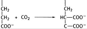
5. Cytochrome P450 is involved in converting hydrophobic xenobiotics into water-soluble ones by catalyzing the following reaction:
АН + O2 + NADPH + Н+ —> АОН + Н2O + NADP+.
6. To reduce a single oxygen atom to Н2O.
7. The glucuronide residue attaches to the OH group of a foreign molecule, making it more polar (see Fig. 17.5).
8. This is an ATP-dependent membrane transport system that pumps various substances out of cells. It transports Steroids in the secretory Cells of the adrenal cortex, but since all transported substances (including anticancer drugs used in Chemotherapy) are lipid-soluble amphipathic compounds, this indicates a broader physiological significance for the system.
9. Neutrophils secrete elastase into the mucus lining the pulmonary tissue, which degrades Elastin, the structural protein of the Lungs. This process converts small alveoli into larger structures with a significantly reduced surface area for gas exchange. Normally, blood α1-antitrypsin diffuses into the lungs and inhibits elastase, preventing alveolar damage. Cigarette smoking leads to two consequences: 1) it promotes the inactivation of α1-antitrypsin by converting a Methionine side chain into a sulfoxide (S —> S = 0); 2) lung irritation attracts neutrophils, which increases the amount of elastase released.
10. This is an oxygen molecule that has acquired an extra electron.
O2 + е- —> O2-.
Superoxide is an extremely reactive molecule capable of causing macromolecular damage.
11. Superoxide is dangerous because, by attacking molecules, it initiates the Formation of other free radicals, triggering a cascading chain reaction that disrupts vital cellular processes. Most Eukaryotic cells contain superoxide dismutase and catalase, which catalyze the following reactions:
2O2- + 2Н+ —> Н2O2 + O2 (dismutase),
2Н2O2 —> 2Н2O + O2 (catalase).
Additionally, cells contain antioxidants such as ascorbic acid and vitamin E. These antioxidants act as scavenging agents. When attacked by superoxide, they form free radicals whose reactivity is too low to sustain the chain reaction.
Chapter 18
1. 5'-phosphoribosyl-1-pyrophosphate (PRPP) is a versatile agent. It is synthesized from ribose-5-phosphate and ATP.

3. Serine hydroxymethylase transfers the -CH2OH group of serine to FH4, releasing Glycine and forming N5, N10-methylene-FH4, which is oxidized to N5, N10-methenyl-FH4 in an NADP-dependent reaction. Hydrolysis of the latter yields formyl-FH4.
4. It ribosylates guanine and hypoxanthine during purine salvage.
5. Children with Lesch-Nyhan syndrome lack HGPRT, and therefore purine salvage does not occur. However, the brain possesses a pathway that leads (due to elevated PRPP levels) to the overproduction of purine NUCLEOTIDES de novo. While patients exhibit hyperuricemia, Treatment with allopurinol (which inhibits uric acid production) does not alleviate the neurological symptoms, the causes of which remain unexplained. Patients with Gout also have elevated uric acid levels, but show no neurological symptoms.
6. Allopurinol is converted into alloxanthine, which inhibits xanthine oxidase. Because xanthine oxidase itself catalyzes this conversion, this is an example of suicide inhibition (self-inactivation).
7. See Fig. 18.8.
8. Thymidylate synthase transfers a C1 group from methylene-FH4 to dUMP and reduces it to a methyl group. FH2 serves as the hydrogen donor, becoming converted into FH2.
9. It inhibits the reduction of FH2 to FH4. The latter is required for the thymidylate synthase reaction, and its deficiency halts dTMP formation, thereby arresting DNA Synthesis AND Cell Division.
10. Vitamin B12 is required for the methylation of homocysteine to methionine, with methyl-FH4 serving as the methyl group donor. A vitamin B12 deficiency traps FH4 as a methyl derivative, rendering it unavailable for other folate-dependent reactions.
Chapter 19
1. Genetic material must possess maximum chemical stability. DNA is more stable than RNA because the 2'-OH group of ribose enables nucleophilic attack on the phosphodiester bond, thereby reducing the stability of RNA compared to DNA.
2. A linear DNA chain structure would force the hydrophobic surfaces of the bases to face the surrounding water. Driven by hydrophobic interactions, the bases stack together, bending the phosphodiester backbone (see Fig. 19.3a).
3. B-DNA: right-handed, 10 Base Pairs per turn.
4. In a DNA double helix, the two strands run in opposite directions: one goes 5' —> 3' and the other 3' —> 5'. Consequently, each end of a linear DNA molecule features the 5'-end of one strand and the 3'-end of the other.
5. The 5' —> 3' direction means you move from the terminal 5'-OH group to the 3'-OH group along the polynucleotide chain.
6. The structure shown below represents one strand of the following duplex:
5' CATAGCCG 3'
3' GTATCGGC 5'
Watson-Crick base pairing accounts for the complementary sequence. By convention, the linear sequence of a single DNA strand is written with the 5'-end on the left.
7. • Eukaryotic chromosomal DNA wrapped twice around a histone octamer core (146 base pairs). Nucleosomes are consecutively linked by linker DNA.
✵ When Chromatin is digested with DNase, the DNA fragments consisting of 146 base pairs are protected from enzymatic degradation; this observation led to the discovery of nucleosomes (see Fig. 19.7).
Chapter 20
1. A DNA fragment whose Replication initiates from a single origin of replication.
2. Positive supercoils are generated ahead of the Replication fork.
3. In E. coli, gyrase (topoisomerase II) introduces negative supercoils. In eukaryotes, topoisomerase I relaxes positive supercoils.
4. See Figures 20.6 and 20.7.
5. Wrapping DNA around a nucleosome introduces local negative supercoils, but because the strands remain unbroken, there is no net negative supercoiling. These local negative supercoils are balanced by local positive supercoils elsewhere. The latter are relaxed by the action of topoisomerase I, leaving net negative supercoils in the DNA.
6. dATP, dGTP, dCTP, and dTTP.
7. Cytosine readily deaminates to form uracil; if uracil were a normal component of DNA, it would be impossible to recognize and correct Mutations, since A-U base pairing is identical to A-T pairing.
8. • No. A primer is required. DNA polymerase cannot initiate the synthesis of de novo chains.
✵ Synthesis proceeds in the 5' —> 3' direction; consequently, the new strand is elongated in the 5' —> 3' direction, with incoming nucleotides added to the free 3'-OH end of the preceding nucleotide.
9. Hydrolysis of inorganic pyrophosphate: nucleotide base pairing.
10. High processivity means that once bound to the template, DNA polymerase III travels a long distance along it without dissociating while synthesizing the new strand. This effect is achieved by a "sliding clamp"—a ring of proteins assembled behind the polymerase through which the DNA chain glides (see Fig. 20.14).
11. Polymerase I is the enzyme that joins Okazaki fragments into a continuous lagging strand. Its activities are summarized in Fig. 20.16.
12. Accurate pairing of incoming nucleotide triphosphates with the template is of paramount importance. E. coli polymerase III also possesses proofreading ability. Its 3' —> 5' exonuclease activity removes the last incorporated nucleotide in the event of a mismatch.
13. A methyl-directed Mismatch Repair system exists in E. coli (see Fig. 20.19). Humans also possess proteins with an analogous function; deficiencies in these proteins increase the risk of carcinogenesis.
14. Exposure of DNA to ultraviolet light induces the formation of thymine dimers, where two adjacent thymine bases become covalently linked. Repair can be carried out either by a direct light-dependent system that breaks the bonds and restores individual bases, or via nucleotide Excision Repair (see Fig. 20.20).
15. As shown in Fig. 20.22, an unreplicated region remains after the removal of the 3' primer of the Okazaki fragment.
16. By means of telomeric DNA. The scheme of its synthesis is shown in Fig. 20.23.
Chapter 21
1. RNA polymerase uses ribonucleotides ATP, CTP, GTP, and UTP as substrates, whereas DNA polymerase uses dATP, dCTP, dGTP, and dTTP. RNA polymerase can initiate the synthesis of new strands, whereas DNA polymerase requires a primer for this. Unlike DNA synthesis, RNA Synthesis never involves proofreading of reading errors.
2. They are shown in Fig. 21.4. The promoter contains a Pribnow box and a -35 element.
3. RNA polymerase binds to DNA nonspecifically, but upon interaction with a sigma factor molecule, the enzyme binds precisely to the promoter. The Pribnow box and the -35 element facilitate the correct orientation of the polymerase. The enzyme synthesizes several phosphodiester bonds, after which the sigma factor dissociates, and the polymerase continues further Transcription of mRNA.
4. One of them is due to the structure of the G-C hairpin loop shown in Fig. 21.7. Within it, G=C base pairing in the mRNA prevents binding to the DNA template strand. Uridine nucleotide sequences further weaken the bond with the DNA (the U=A pair contains only two hydrogen bonds), facilitating dissociation. The second termination mechanism involves the Rho factor, a helicase that unwinds the mRNA-DNA hybrid. At the termination region, the polymerase pauses (likely due to the elevated G-C content in this DNA region); the Rho factor catches up with it and releases the mRNA.
5. The exact base sequence of the Pribnow box and the -35 element, the distance between them, as well as the bases in the region from +1 to -40.
6. See Fig. 21.10.
7. In eukaryotes, the primary transcript contains introns that must be removed; the 5' end is capped; the termination mechanism is largely unknown; mRNA (with rare exceptions) is polyadenylated at the 3' end.
8. The mechanism is shown in Fig. 21.13. The consensus sequence determines the splicing site, and the transesterification reaction is accompanied by a negligible change in free energy. Interrupted genes may facilitate evolution by promoting exon shuffling. Alternative Splicing can also result in the same Gene directing the synthesis of different proteins.
9. Eukaryotic RNA polymerase III binds not to DNA directly, but to a protein complex assembled on the DNA (Fig. 21.19). In addition, several transcription factors can be associated with this complex (see Fig. 21.20).
10. Helix-turn-helix proteins, leucine zipper proteins, zinc finger proteins, and homeodomain proteins.
Chapter 22
1. If only 20 codons were used, 44 non-coding triplets would remain. Any mutation in the coding region of a gene would have a high probability of inactivating the gene by prematurely introducing a stop codon. With 61 codons, a base substitution either causes no change (due to the degeneracy of The Genetic Code) or leads to the replacement of one amino acid with another (often structurally similar). Three triplets do not code for Amino Acids and serve as stop codons.
2. Because a wobble mechanism (ambiguous pairing) exists.
3. The base sequence of RNA is always written with the 5' end on the left. When mRNA is depicted in this way, the tRNA anticodon bases must be presented in an antiparallel orientation, which is why the tRNA molecule is drawn inverted with the 5' end on the right.
4. Proofreading of the correct attachment of the amino acid to tRNA. A pause in the EF-Tu-catalyzed GTP hydrolysis during the elongation stage gives unmatched aminoacyl-tRNAs time to leave the ribosome.
5. The hydrolysis of GTP to GDP and Pi leads to Conformational Changes in G proteins. GTP participates in the assembly of the E. coli initiation complex and in the delivery of aminoacyl-tRNA to the ribosome via the EF-Tu factor. GTP is also utilized at the translocation stage.
6. Fig. 22.11 is required for the explanation. The "rocking" mechanism illustrated in the figure (Model I) has the advantage that as the tRNA moves from one site to another, it never completely dissociates. This also means that the peptidyl group does not need to move relative to the ribosome. Fig. 22.11 presents two possible mechanisms. Model II explains why the ribosome has two subunits.
7. This incompatibility is explained by Fig. 22.13. The mechanism of locating the initiating AUG codon involves the existence of only a single start site. As shown in Fig. 22.9, initiation in prokaryotes can occur simultaneously from multiple start sites.
8. Chaperones bind to growing Polypeptides and prevent their premature misfolding. The release of chaperones at the right moment facilitates proper folding, although the exact details of this process are not yet fully understood (for details, see p. 298).
9. Prion diseases (see p. 298).
10. See Fig. 22.16.
Chapter 23
1. Through the standard receptor-mediated endocytosis pathway (see Fig. 23.1).
2. The latter must deliver RNA replicase into the host cell; the viral (+)-strand RNA functions as the mRNA that encodes RNA replicase.
3. Vaccinia virus replicates in the cytoplasm, whereas the host polymerase is located in The Nucleus.
4. Retroviral RNA is reverse-transcribed into double-stranded DNA, which then integrates into the host chromosome (see Fig. 23.5).
5. The mechanism is illustrated in Fig. 23.3.
6. The protective action of Antibodies against Influenza targets the hemagglutinin protein. It undergoes continuous mutation, but because the hemagglutinin molecule possesses a large number of epitopes, Immunity is lost only gradually. This antigenic drift leaves individuals susceptible to infection, though partial immunity mitigates the severity of the disease. The Emergence of a completely novel hemagglutinin variant via recombination between two different strains can trigger a devastating, life-threatening pandemic.
7. Viral surface glycoproteins specifically bind neuraminic (sialic) acid, which is also present at the terminal end of erythrocyte Glycophorin molecules. Consequently, when mixed with red blood cells, the Viruses bind to them, causing hemagglutination.
8. The virus attaches to the host cell via a glycoprotein terminating in neuraminic acid. The enzyme catalyzing The breakdown of neuraminic acid—essential for viral cell entry—may serve to: a) facilitate the release of new Viral Particles that would otherwise remain tightly bound to cellular glycoproteins; b) thin mucus containing neuraminic acid, thereby clearing a path for the virus to reach the cell surface. Mucus thinning may also aid viral dissemination through sneezing.
9. Bacteriophages can enter either the lytic or the lysogenic cycle (see Fig. 23.7).
10. A. A small, infectious, naked RNA molecule lacking a protein coat. B. No. Viroids lack the long base-coding sequences necessary to encode protein Amino acid sequences.
Chapter 24
1. Pancreatic DNase cleaves DNA nonspecifically. Restriction enzymes, by contrast, cleave DNA at strictly defined sites by recognizing short base sequences.
2. The adenine bases within all sequences flanking the hexamer in the E. coli R-strain are methylated, preventing the enzyme from recognizing them. Foreign DNA lacks this protective modification.
3. "Sticky ends" refer to the overhangs generated by the staggered cuts produced by the majority of restriction enzymes.

The protruding DNA ends will spontaneously "anneal" (stick together) via base pairing.
4. A gene clone is a cloned DNA segment identical to a chromosomal DNA sequence. A cDNA clone refers to complementary DNA identical to the mRNA of a gene. A genomic gene clone contains introns, whereas a cDNA clone does not.
5. The steps for preparing a genomic library are shown in Fig. 24.1.
6. The isolation steps are outlined in Fig. 24.2.
7. A nucleotide lacking a 3'-OH group. When DNA polymerase incorporates a dideoxynucleoside triphosphate into the newly synthesized DNA strand, further chain elongation is terminated.
8. A specific region of chromosomal DNA can be amplified. This process involves copying a DNA fragment and repeatedly replicating those copies. The Significance of this technique is that starting with a minimal amount of DNA (otherwise insufficient for analysis), one can generate ample quantities of the target fragment for experimental use. In addition to the enzyme and substrates, primers are required to direct DNA synthesis in both directions; therefore, the base sequences at the ends of the target fragment must be known.
9. A plasmid containing a convenient insertion site for cloning a specific protein's cDNA, alongside bacterial transcription (promoter) and Translation signals. The DNA is transcribed, and the mRNA is translated within the bacterial cell. Such a plasmid vector can be utilized to produce large quantities of the protein of interest.
10. Restriction analysis is an analytical technique used to detect genetic defects. When DNA is digested with restriction enzymes and the resulting fragments are separated by gel Electrophoresis, Hybridization Methods can be used to visualize the electrophoretic bands. Mutations will alter this banding pattern.
Chapter 25
1. See Fig. 25.3.
2. The generation of multiple immunoglobulin genes is illustrated in Fig. 25.4.
3. B lymphocytes (B cells) produce antibodies (following activation and maturation into plasma cells). This process requires B cells to interact with helper T lymphocytes (T cells). Cytotoxic T cells (or killer cells) bind to host cells presenting a foreign antigen, causing their lysis either through membrane perforation or by stimulating apoptosis.
4. An antigen-presenting cell is a type of phagocyte that engulfs a foreign antigen, processes it into fragments, and displays the antigen in a complex with class II MHC molecules. When inactive helper T cells interact with the antigen–MHC complex, the T cells become activated (see Fig. 25.4).
5. In both cases, this is a class I MHC.
6. Clonal Selection theory refers to the model explaining the selection process of cells that secrete antibodies. Each B cell produces a unique antibody. For any given antigen, there are several specific B cells; however, proliferation into a clone is triggered only when the antigen binds to an antibody anchored on the B-cell membrane. Thus, the antigen automatically selects the B cells that undergo proliferation.
7. All Antigens that bind during the primary maturation of B cells in the Bone Marrow or T cells in the Thymus are autoantigens. Because these must not elicit an Immune Response, cells reactive to autoantigens are destroyed at this developmental stage. Once they leave the bone marrow or thymus, subsequent antigen binding activates the cells.
8. Antibody secretion is achieved through alternative splicing of introns: near the 3'-end of the immunoglobulin gene, there is an exon that encodes the anchor polypeptide sequence. At the onset of secretion, splicing switching leads to the disappearance of these anchors.
9. The initiation of secretion in B cells is accompanied by somatic mutation, which modifies the variable region. Since antigen binding triggers rapid, active cell proliferation, this modification serves as a selection mechanism for cells producing superior antibodies.
10. CD4 is expressed on The surface of helper T cells. It binds to the constant protein domain of class II MHC molecules and therefore interacts exclusively with B cells, helper T cells, and antigen-presenting cells that contain proteins of this class. The surface of cytotoxic T cells bears the CD8 protein, which binds to class I MHC molecules; consequently, killer T cells interact only with cells carrying this type of molecule. The CD4 protein also serves as the receptor through which the AIDS virus infects helper T cells.
Chapter 26
1. Endocrine hormones, growth factors, and Neurotransmitters.
2. The modes of action of lipid-soluble and lipid-insoluble signaling molecules are shown in Fig. 26.1.
3. The regulation pathway of adenylate cyclase, the enzyme responsible for cAMP synthesis, is depicted in Fig. 26.11.
4. GTP hydrolysis acts as a molecular clock. In cholera, the toxin irreversibly inactivates GTPase, causing cAMP levels to remain elevated. This leads to severe loss of Na and water from the body.
4. cAMP allosterically activates protein kinase A (PKA), which phosphorylates various target proteins, including enzymes and transcription factors. Furthermore, certain genes contain cAMP response elements (CREs), indicating that cAMP likely plays a vital role in gene regulation.
5. Nitric oxide (NO) activates cytoplasmic guanylate cyclase, which synthesizes cGMP. The latter acts as a second messenger and activates PKG.
6. An example of such a system is the phosphoinositide cascade. It involves receptor-mediated activation of membrane-bound phospholipase C, which releases Inositol trisphosphate (IP3) and diacylglycerol (DAG). IP3 increases the concentration of cytoplasmic Ca2+, while DAG activates protein kinase C (PKC). Thus, IP3 and DAG function as second messengers (see Fig. 26.16).
7. The Ras pathway utilizes receptor tyrosine kinases and involves the phosphorylation of intermediary proteins (see Fig. 26.18).
8. Such a protein can bind to a phosphorylated receptor tyrosine kinase (see Fig. 26.21) and activate cellular regulatory pathways.
9. If a receptor or, for example, a component of the Ras pathway becomes aberrantly and constitutively active, the overexpression of genes encoding transcription factors can drive uncontrolled cell division (see Fig. 26.22).
10. See Fig. 26.22.
11. • Adrenergic receptor activation induces an allosteric change in the hormone-binding cytoplasmic domain.
✵ Activation of the EGF receptor in the Ras pathway triggers receptor dimerization and autophosphorylation of tyrosine residues.
✵ Interferon receptor activation involves receptor dimerization and binding to a specific tyrosine kinase, which subsequently phosphorylates the receptor (see Figs. 26.6, 26.17, and 26.22).
12. Such Ca2+ channels are present, for example, in the membranes of nerve terminals that release acetylcholine to initiate muscle contraction. The channel opens when a wave of neuronal membrane depolarization reaches it. The abrupt change in Ca2+ concentration triggers the release of acetylcholine (see Fig. 26.24).
13. A Ligand-gated channel is defined as a channel that opens upon binding to a specific ligand. During photoreception, cationic channels in rod membranes are opened by cGMP. Light causes a decrease in cGMP concentration, which leads to channel closure. The resulting hyperpolarization of rod membranes is converted into an optic Nerve Impulse.
Chapter 27
1. In The First stage, 5-aminolevulinate (ALA) synthase catalyzes the Condensation of glycine with succinyl-CoA (see Fig. 27.5), and In the second stage, ALA dehydratase forms the pyrrole porphobilinogen (see Fig. 27.6).
2. Increased hepatic ALA synthase activity has been observed in acute intermittent porphyria, although the relationship between ALA production and neurological symptoms remains unclear.
3. The mechanism of activity regulation is shown in Fig. 27.8.
4. Myoglobin has a higher affinity for oxygen. Unlike the sigmoidal curve of Hemoglobin, the oxygen saturation curve of myoglobin is hyperbolic in shape (see Fig. 27.11).
5. According to the accepted model, hemoglobin exists in low (T) and high (R) oxygen-affinity states. Oxygen binding shifts the equilibrium toward an increase in the number of molecules in the R state.
6. Upon oxygen binding to the heme iron, the iron atom moves into the plane of the porphyrin ring. For this to occur, a certain flattening of the tetrapyrrole is required. The iron attaches to the F8 histidine of the protein, inducing a rearrangement of the protein molecule that results in the flattening of the tetrapyrrole. All these displacements affect subunit interactions, triggering a conformational transition of the tetramer from the T to the R form (see Fig. 27.13).
7. Deoxygenated adult hemoglobin contains a cavity in which 2,3-diphosphoglycerate (DPG) binds to positively charged protein groups. DPG binding is possible only in the deoxygenated state, and therefore it decreases the oxygen affinity of hemoglobin. Fetal hemoglobin contains a y-subunit instead of the adult $\beta$-subunit. The y-subunit lacks one of the charged groups involved in DPG binding. Consequently, DPG binds less tightly to fetal hemoglobin, and its oxygen affinity is higher than that of maternal hemoglobin.
8. This is necessary for the transport of CO2 in the form of HCO3- (see Fig. 27.15).
Chapter 28
1. Fast fibers derive their ATP from glycolysis, whereas slow fibers rely on oxidative metabolism. Fast fibers provide immediate escape or combat responses (“fight or flight”), but fatigue develops rapidly in such fibers. Slow-twitch fibers generate ATP via oxidative metabolism and can function significantly longer without signs of fatigue, though generating additional energy takes time. Humans possess both types of muscle. Postural back muscles, which maintain posture over extended periods, belong predominantly to the slow-twitch type, whereas extraocular eye muscles are fast-twitch.
2. See Figs. 28.1 and 28.2.
3. The response is summarized in Fig. 28.7.
4. Stimulation of the muscle receptor by acetylcholine leads to the release of Ca2+ ions from the sarcoplasmic reticulum. Tropomyosin molecules are associated with Actin filaments and, in turn, contain a Ca2+-sensitive troponin complex. In the absence of Ca2+, tropomyosin blocks the interaction between the Myosin HEAD and the actin filament. When Ca2+ binds to the troponin complex, a conformational change occurs that promotes myosin binding to the thin filament and initiates contraction. Ca2+-ATPase pumps Ca2+ back into the sarcoplasmic reticulum, bringing contraction to an end.
5. The myosin heads of smooth muscle contain a p-light chain that inhibits the binding of the myosin head to the actin filament, thereby preventing contraction. Neurogenic stimulation opens channels that allow Ca2+ to enter the cell. Ca2+ binds to the regulatory protein calmodulin, which activates myosin light-chain kinase. Phosphorylation of the light chain relieves the inhibitory effect, and contraction ensues.
Chapter 29
1. In nonmuscle cells, Actin forms filaments. Actin filaments anchored to the cell membrane allow small bundles of myosin molecules to generate pulling force. In addition to participating in nonmuscle cell contraction, actin filaments form tracks along which minimyosin molecules move. These molecules have conventional myosin heads, but the long tail-like structure characteristic of muscle myosin is replaced by a short tail to which transport vesicles can attach.
2. Microtubules are hollow tubes formed by the polymerization of tubulin protein subunits. They possess a defined polarity, and their ends are designated as the (+) end and (-) end, respectively.
3. The microtubule-organizing center caps the (-) end. The (+) ends of growing microtubules are capped by a tubulin-GTP complex. GTP is slowly hydrolyzed, removing the protective cap. If new subunits add on after GTP hydrolysis, microtubule depolymerization occurs. When a microtubule reaches its “target,” its (+) end becomes capped.
4. Kinesin and dynein are Molecular Motors that move along microtubules, hauling intracellular “cargo.” Kinesin and dynein travel along the microtubule in opposite directions: from the (-) to the (+) end and from the (+) to the (-) end, respectively.
5. Microtubules cannot contract. Shortening occurs as a result of microtubule depolymerization, though what precisely drives chromosome movement remains unknown.
6. Intermediate filaments are so named because their diameter is 10 nm, placing them intermediate in size between microtubules (20 nm) and actin filaments (6 nm).
7. Intermediate filaments provide the structural framework for Hair and impart mechanical rigidity to structures such as nerve fibers and sarcomeric Z-disks.
Last update: 06/08/2026
Editorial and Educational Adaptation: This material has been compiled based on the primary/original source text. The project team performed an editorial review, corrected technical inaccuracies, structured sections, and adapted the content for an educational format.
What was processed:
- elimination of formatting defects (OCR errors, structural breaks, corrupted characters);
- editorial organization of content;
- standardization of terminology in accordance with academic sources;
- verification of factual statements against the original source text.
All mentions of the author, publication year, and origin of the primary text have been preserved in accordance with the source.