Biochemistry: The Chemical Reactions of Living Cells, Volume 2 - D. Metzler 1980
Enzymes: The Protein Catalysts of Cells
Mechanisms of Enzymatic Catalysis
Acid-Base Catalysis
Many reactions accelerated by Enzymes can also be catalyzed by acids or bases, and frequently by compounds of both types. A well-studied example of this is mutarotation—the reversible interconversion of the α- and β-anomeric forms of sugars, particularly glucose [see scheme (6-75)]. This reaction is catalyzed by the specific enzyme mutarotase, as well as by inorganic acids and bases. These findings indicate that simple acids and bases, on the one hand, and enzymes, on the other, share a common mechanism in terms of their catalytic action. Since many amino acid side chains contain acidic and basic groups, it is quite natural to conclude that these groups must participate in catalysis as acids and bases. However, to understand precisely how they function in catalysis, we need to know the numerical values of certain equilibrium and rate constants.
a. Strong and Weak Acids
Acids are proton Donors, whereas bases are proton acceptors. An acid can be characterized by its dissociation constant or by the negative logarithm of this constant, pKa (see also Chapter 4, Section B):
Class="center">
where Ka = [H+] [B-]/[HB]. Note that [H+] represents the concentration (or, more precisely, the activity) of H3O+ ions rather than free protons. Hereinafter, we will denote acids by the symbols HB or H+B, and the conjugate bases formed upon acid dissociation by the symbols B- or B.
Recall that strong acids have low pKa values, and their conjugate bases are weak. Conversely, very Weak acids have high pKa values, and their conjugate bases are strong. When examining the mechanisms of enzymatic reactions, it is useful to know the pKa values of the following groups:
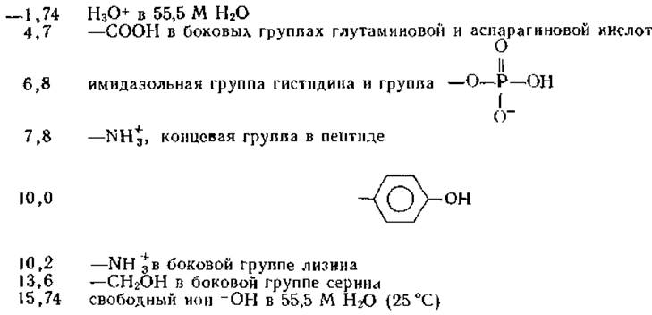
These values are not constant and may vary by up to 0.5 units higher or lower, depending on the compound's Structure and the microenvironment of the given side group within the protein. In some cases, deviations are even more pronounced.
b. Acid-Base Catalysis of Mutarotation
The mutarotation of glucose proceeds via The formation of a free aldehyde intermediate:
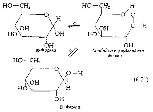
During the reaction, a hydrogen atom is abstracted from the OH group at the C-1 atom of α-glucose, and a proton (presumably a different one) is transferred to the ring oxygen, accompanied by the Cleavage of the C—O bond. The reverse process can also proceed via pathway b, leading to the Formation of the β-isomer.
Proton transfer (most commonly between oxygen, nitrogen, and sulfur atoms) occurs in numerous biochemical reactions. The bonds between a hydrogen atom, on the one hand, and oxygen, nitrogen, and sulfur atoms, on the other, are typically strongly polarized, resulting in a slight positive charge on the hydrogen atoms. Consequently, these groups acquire a weakly acidic character, allowing protons to dissociate relatively easily and transfer to other groups. It is logical to assume that the catalytic properties of acids and bases are closely related to such hydrogen atom transfers.
c. General Base and General Acid Catalysis
Base-catalyzed mutarotation proceeds as follows: a base, such as an OH- ion, attacks the proton of the hydroxyl group, converting the latter into an anion while simultaneously generating the conjugate acid BH+ (stage a in the scheme below):
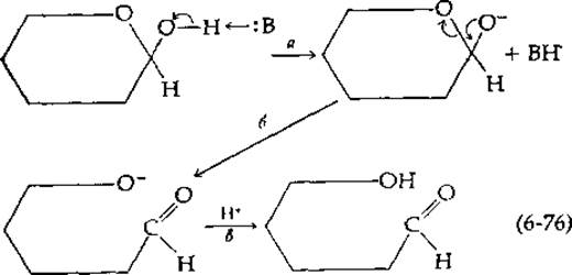
The anion then isomerizes into the open-ring form (stage b). Proton attachment (via transfer from H3O+) yields the free aldehyde form of the sugar (stage c).
The catalytic base in this scheme is not limited to the OH- ion; weaker bases, such as the ammonium ion or even Water, can also act in this capacity. As it turns out, in some cases the catalytic reaction rate is proportional solely to the concentration of OH- ions, while the presence of other, weaker bases has no effect on this value. Such catalysis is referred to as specific hydroxide-ion catalysis. In the general case, the apparent first-order rate constant (kobs) for the process under study is the sum of several terms:
kobs = kH2O + kOH-[OH-] + kB[B]. (6-77)
The term kH2O characterizes The rate of the process in pure water and corresponds to the catalytic action of water acting as either an acid or a base. The final two terms determine the respective contributions to catalysis by the OH- ion and another base. The term kB[B] characterizes general base catalysis, which is believed to play a vital role in the functioning of many enzymes. In enzymology, general base catalysis refers to the ability of a basic group within an enzyme molecule to function as a proton acceptor.
Acid-catalyzed mutarotation is observed when an acid acts as a proton donor that adds to the ring oxygen atom of the sugar molecule:
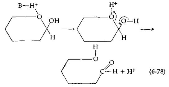
As in the previous case, either specific acid catalysis (mediated by the H3O+ ion) or general acid catalysis can take place.
It is reasonable to assume that both general base and general acid catalysis operate during enzyme function. Enzymes are incapable of locally concentrating protons or hydroxyl ions to drive specific base or specific acid catalysis. However, specific ionizable groups of enzymes, at their normal degree of protonation corresponding to the cellular pH, can act as general acid and general base catalysts.
d. Concerted Mechanisms
A third possible type of catalysis involves the concerted action of both an acid and a base, resulting in the simultaneous cleavage of an old bond and the formation of a new one. It is known, for example, that the mutarotation of tetramethylglucose in benzene containing either pyridine (a base) or phenol (an acid) proceeds very slowly. However, when both pyridine and phenol are simultaneously present in the solution, mutarotation proceeds significantly faster. Based on this observation, Swain and Brown [50] proposed a concerted mechanism in which both the acid and the base participate simultaneously:
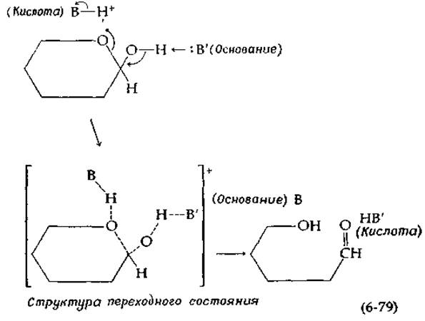
Note that during the reaction, the acid BH+ is converted into its conjugate base B, and the base B' into its conjugate HB'. It might seem that since these agents are altered during the reaction, they are not true catalysts. However, a simple proton exchange restores the original forms and completes the catalytic cycle. In aqueous solutions, water itself can act as an acid or a base, or even simultaneously as both an acid and a base in concerted catalysis.
Experimental Evidence for the existence of concerted acid-base catalysis in the mutarotation of tetramethylglucose in benzene by phenol and pyridine is currently considered insufficiently convincing [51, 52]. For non-enzymatic reactions proceeding in aqueous solutions, proving the existence of such a catalytic mechanism is quite difficult1). However, it may play an exceptionally important role in Enzymatic Catalysis, since among the side chains of Amino Acids there may be two appropriately positioned acidic and basic groups.
d. Brønsted Relations
The effectiveness of a given base as a general base catalyst can usually be related to its basicity (pKa) using the Brønsted equation [53]:
lgkB = lgGB + ß(pKa). (6-80
The constant kB in this equation is defined by expression (6-77), and GB represents a constant for a given type of reaction. A similar relation connects the rate constant kA for general acid catalysis with the pKa value:
lg kHB = lg GA — а (рКa). (6-81)
These equations are similar to the Linear Free-Energy Relationships (Chapter 3, Section D), but they deal with rates rather than equilibria. Admittedly, the Hammett equation [Equation (3-66)] is also very frequently applied not only to equilibrium constants, but to rate constants as well.
The condition for the applicability of the Brønsted equations is the existence of a direct relationship between the Free energy of activation of the reaction and the basicity (or acidity) of the catalyst.
The coefficients β and а in equations (6-80) and (6-81) characterize the sensitivity of the reaction rate to Changes in the basicity or acidity of the catalyst. It is easy to show that when β or а are close to 1, general base or general acid catalysis is usually absent, and the reaction rate is determined solely by specific hydroxide or hydrogen ion catalysis [53]. As β or а decrease toward 0, THE CONTRIBUTION OF base or acid catalysis also becomes negligibly small. Thus, general base or general acid catalysis is most significant when the coefficients β and а are close to 0.5. Under these conditions, as is easy to see, a relatively weak base such as imidazole (in the Histidine side chain) can prove to be an extraordinarily effective catalyst at pH 7.
1) Recently, however, it has been shown that the catalysis of acetone enolization by acetic acid and acetate does indeed proceed via a concerted acid-base pathway [52a].
To experimentally determine The values of β and а, it is necessary to plot lgkB or lgkHB versus pKa (a Brønsted plot) and determine the slope of the resulting straight line. In the case of ionized amino groups, where one of the three protons can dissociate from the nitrogen atom, and dicarboxylic acids, statistical corrections must be introduced (Chapter 4, Section B, 5).
Any mechanism for which general base or general acid catalysis operates is characterized by one important feature: proton abstraction or addition occurs in the rate-determining step of the reaction.
e. Tautomeric Catalysis
Swain and Brown [50] carried out a very interesting experiment showing that acidic and basic groups incorporated into the same molecule catalyze sugar mutarotation much more effectively than a simple mixture of an acid and a base. Thus, 0.001 M α-hydroxypyridine catalyzes the mutarotation of tetramethylglucose (0.1 M) in benzene 7000 times more effectively than a mixture containing 0.001 M pyridine and 0.001 M phenol. Swain and Brown proposed the following fully concerted reaction mechanism for the multifunctional catalyst α-hydroxypyridine. They postulated that the reaction is preceded by the formation of a hydrogen-bonded complex analogous to the enzyme-substrate complex:
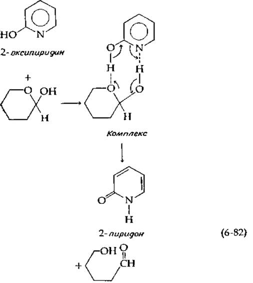
the product of the catalyst transformation is 2-pyridone, a tautomeric form of 2-hydroxypyridine, with rapid equilibrium being established between both forms.
Rony [51] suggested calling the catalysis by α-hydroxypyridine tautomeric catalysis; he believes that the high efficiency of this catalyst is due not simply to the proximity of the acidic and basic groups within the same molecule, but also to the ability of the catalyst to undergo reversible transitions from one tautomeric form to another.
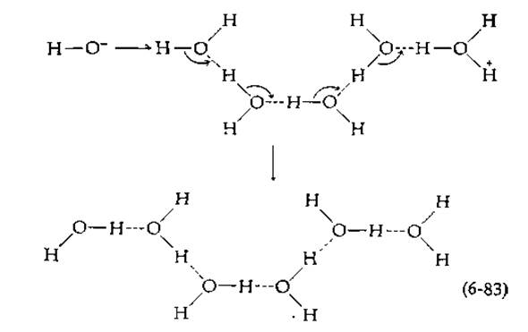
The limiting value for second-order rate constants under diffusion-controlled conditions is approximately 1010 M-1∙s-1 (Section A, 7). In 1956, Eigen [54], who developed new Methods FOR STUDYING fast reactions, made a surprising discovery, finding that protons and hydroxyl ions interact much faster when located in an ice lattice than in solution: the second-order rate constants were 1013–1014 M-1∙s-1. The corresponding reaction rates are as high as molecular vibration frequencies; for example, the frequency of the O–H bond vibration in water2) is approximately 1014 s-1. This is apparently explained as follows. The OH- ion and the proton that adds to a water molecule to form the H3O+ ion are hydrogen-bonded to neighboring water molecules. Since all water molecules in ice are connected by Hydrogen Bonds, the hydroxyl and hydrogen ions turn out to be linked by a chain of water molecules:
f. Rates of Proton Transfer1)
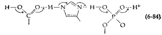
Thanks to the synchronous displacement of electrons from the OH- ion along the chain (small arrows in the scheme) during a single vibration, charge neutralization can occur. Note that the positions of the oxygen atoms remain the same by the end of the process, while the protons involved in hydrogen bonding shift slightly to the left in the scheme. A reaction of this type not only serves as Evidence of the remarkable mobility of hydrogen ions, but may also be directly relevant to tautomeric catalysis in enzymes. Thus, Wang [55] suggested that proton transfer along a rigidly fixed chain of hydrogen bonds in the ES complex is an integral part of enzymatic catalysis. It is easy to imagine that a similar concerted proton shift can take place within interconnected carboxylic acid, imidazole, and phosphate groups:
1) A detailed Discussion of the dynamics of proton transfer in solution, featuring biochemical model systems, can be found in the comprehensive review by P. Schuster, P. Wolschann, and K. Tortschanoff in Molecular Biology, Biochemistry and Biophysics, vol. 24, Chemical relaxation in molecular biology, edited by I. Pecht and R. Rigler, Berlin, Springer-Verlag, 1977, pp. 107–109. — Transl. note.
2) The frequency of the OH stretching vibration is equal to the frequency of the infrared light that excites these vibrations. The frequency v is given by the product of the wavenumber (which is 3710 cm-1 for the —OH bond) and the speed of light c (3∙1010 cm∙s-1). Consequently, the vibration frequency of the —OH bond in a water molecule is v = 3∙1010∙3710 = 1.14∙1014 s-1.
As a result, a proton is transferred from one end of the chain to the other [as shown in scheme (6-83)], facilitated by readily occurring tautomerization reactions. A similar process can also take place with the participation of protein side chains, linking two groups of the Active Site and thereby promoting concerted acid-base catalysis, much like what occurs in scheme (6-79).
Other tautomerization processes are also possible in Proteins (in the presence of less stable "minor tautomers"). For example, a shift of the following type involving peptide bonds in an a-helix or ß-Structure may take place:
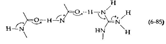
As shown in the scheme, electrons migrate toward the guanidine group of the Arginine side chain. Other variants can also be considered (and there are numerous such possibilities, especially taking into account that Coenzymes or purine and pyrimidine bases may also participate in the reaction). All such processes proceed extremely rapidly and are difficult to detect.
3. Effect of pH on Enzyme Activity
The data presented above regarding the participation of acidic and basic groups in enzymatic catalysis were obtained primarily from studies of non-enzymatic model reactions. Is there any indication that enzymes actually contain such groups? Yes, there is, and the clearest evidence is the Dependence of enzymatic activity on pH. Quite often, the Vmax versus pH curve has a bell-shaped appearance (Fig. 6-13). The optimal Vmax value is observed at a pH typically ranging between 6 and 9. Bell-shaped curves are most simply explained by assuming that the Active Site of the enzyme contains two ionizable groups, a and b. In this case, the enzyme can exist in three forms that differ in their degree of protonation: E, EH, and EH2:
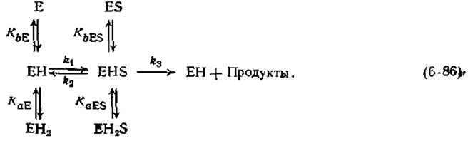
Let us denote the dissociation constants for the two groups in the free enzyme as KaE and KbE, and the corresponding values for the ES complex as KaES and KbES. The rate constants k1, k2, and k3 characterize the steps of formation and breakdown of the ES complex.
If we assume that the only reactive form of the enzyme that breaks down to form products is the EHS form, then the dependence of the maximum velocity on pH will be bell-shaped (Fig. 6-13). Such curves are frequently encountered in enzyme kinetics, which Supports the adequacy of scheme (6-86). Furthermore, if for the reaction to proceed, group a of the enzyme must be dissociated to the conjugate base and group b must be in the protonated state, it is natural to assume that these two groups participate in acid-base catalysis.
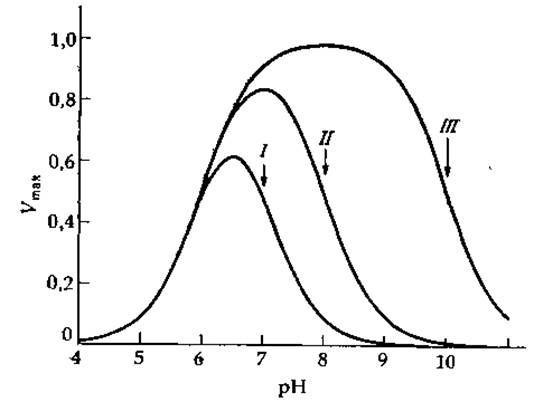
FIG. 6-13. Theoretical Vmax versus pH dependencies calculated from equation (6-87) with k3[E]t = 1, pKaES = 6, and the following pKbES values: I — 7, II — 8, and III — 10 [58]. Computer-generated plots kindly provided by C. Harris.
For the simple case described by scheme (6-86), the pH dependencies of the maximum velocity and the Michaelis constant are expressed by the following equations, respectively:
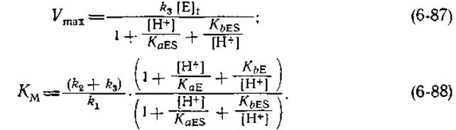
When the enzyme is fully saturated with substrate, only the EH2S, EHS, and ES forms are present. From the definition of KaES and KbES, it follows that the following relation holds under these conditions:

The term in parentheses is identical to the denominator of expressions (6-87) and (6-88); it is sometimes referred to as the Michaelis pH function [56]. A structurally analogous pH function for the free enzyme appears in the numerator of expression (6-88). The pH dependence of enzymatic activity often has a more complex appearance than the dependencies presented in Fig. 6-13 and described by the equations above. However, it is straightforward to write down the Michaelis pH Functions, as well as the corresponding equations similar to equations (6-86)–(6-89), for an enzyme with any number of ionizable groups in the E and ES forms. It should be borne in mind that if the free substrate contains groups that dissociate within the pH range where enzyme activity is being studied, the pH function for the free substrate will also appear in the numerator of expression (6-88). If changes in the conformation of the protein molecule contribute to the alteration of enzyme activity upon pH variation, cooperative binding or release of more than one proton may occur, which should be reflected in the Michaelis pH function. In some cases, this can lead to the appearance of an additional term analogous to (4-32). The Nature of the pH dependence of enzyme activity was analyzed in detail by Dixon [56], who proposed plotting lg Vmax (or the logarithm of specific activity) and —lg KM against pH.
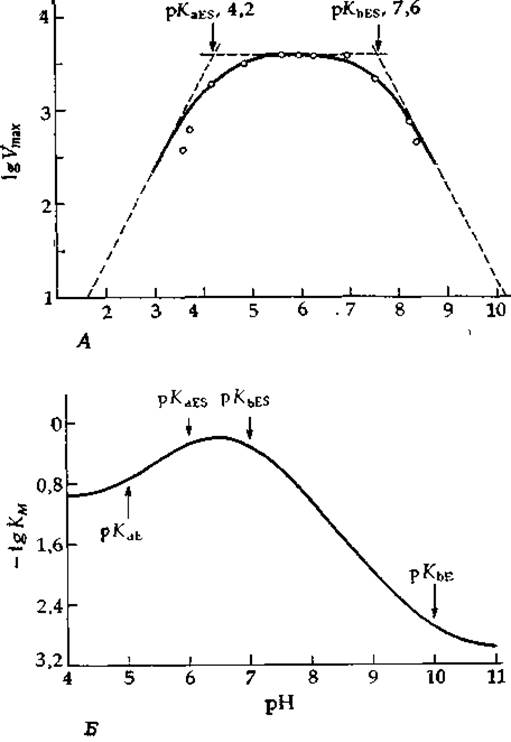
FIG. 6-14. A. Dependence of Vmax on pH for crystalline bacterial a-amylase [56a]. B. Theoretical dependence of lg KM on pH, calculated from equation (6-88) with pKaE = 5, pKbE = 10, pKaES = 6, and pKbES = 7. Computer-generated plot kindly provided by C. Harris.
Typical curves of this kind are shown in Fig. 6-14. In the region where pH = pKa ± ~ 1.5, the plot of lg Vmax versus pH is curvilinear; however, outside this interval, the Branches of the curve asymptotically approach straight lines whose slope is equal to one if a single proton participates in the dissociation process, or greater than one if cooperative dissociation of multiple protons takes place. The intersection points of the asymptotes with a line segment parallel to the abscissa axis (see Fig. 6-14) correspond to the pKa values. (It should be noted, however, that fitting all experimental points to a single theoretical curve yields more reliable results.) The curvilinear section always lies below (or above) the intersection points by an amount equal to lg 2 = 0.30 (or less, if proton dissociation occurs cooperatively).
The concave Regions of the lg KM versus pH curves yield the pKa values of the free enzyme or free substrate, whereas the convex regions yield the pKa values of the ES complex. This approach to analyzing the pH dependencies of enzyme reaction parameters is widely used by enzymologists, although it often leads to incorrect results. For example, many curves of this type exhibit a very sharp inflection, with the curvilinear region spanning a pH interval of < 3; meanwhile, the ordinate distance between the curve and the intersection point of the asymptote with the horizontal segment is much less than 0.301). This means that proton binding occurs cooperatively, and the apparent pKa in such a case resembles the constant K in equation (4-33). Readers wishing to delve deeper into these topics are encouraged to consult the theoretical paper [57], which discusses transition-state pKa values. An interesting analysis of curves with unusually sharp maxima is presented in [57a,b].
An example of an enzyme studied in detail with respect to The Effect of pH on kinetic parameters is fumarase, an enzyme that catalyzes the reversible Hydration of fumaric acid to malic acid [scheme (6-64)]. In an early and very interesting paper, Alberty et al. [58] demonstrated that a bell-shaped pH dependence occurs for both the forward and reverse reactions. Using equations (6-88) and (6-89), these researchers calculated the apparent pKa values for groups a and b of the enzyme in Buffer solutions with an Ionic strength of 0.01 (Table 6-1). It is important to realize that The kinetics of this reversible reaction are described by more complex equations than equations (6-87)–(6-89), and therefore the apparent pKa values may not coincide with the true ones. Nevertheless, it is very tempting to assume that the two pKa values for the free enzyme, equal to 6.2 and 6.8, correspond to identical groups—apparently imidazole groups—with microscopic pKa values of ~ 6.5. The properties of fumarase will be discussed further in Chapter 7, Section 3.6.
The mutarotation of glucose in E. coli is catalyzed by a specific mutarotase [59] having a turnover number of 104 s-1. The shape of the —lg KM versus pH plot indicates the presence of two ionizable groups with pKa values of 5.5 and 7.6 in the free enzyme, while the Nature of the lg Vmax versus pH dependence implies that one group with pKa = 4.75 is present in the ES complex [59]. The latter may represent a catalytic group [group B' in scheme (6-79)], possibly imidazole in the conjugate base form. Why does the group having a pKa of 7.6 in the free enzyme fail to manifest itself in the ES complex? Either this group does not participate in catalysis, or its pKa value is shifted so strongly upon substrate binding that the group cannot be detected using the lg Vmax versus pH curve. Thus, experimental data do not yet allow a definitive resolution of whether the ionizable group corresponding to the —BH+ group in scheme (6-79) participates in mutarotase action.
1) It is remarkable that for the majority of curves presented by Dixon and Webb [56] to illustrate the pH profiles of enzymatic activity, the same deviation from theoretical behavior is observed. Many of the obtained pKa values may be incorrect due to the participation of multiple protons in the dissociation process, leading to sharper pH transitions, or as a result of experimental errors (such as those associated with buffer effects).
Table 6-1 Apparent pKa values for fumarase and its complexes with fumarate and malate [58]
|
Free enzyme |
Enzyme–fumarate complex |
Enzyme–malate complex (for the reverse reaction) |
|
|
pК |
6,2 |
5,3 |
6,6 |
|
pKb |
6,8 |
7,3 |
8,5 |
Last update: 06/08/2026
Editorial and Educational Adaptation: This material has been compiled based on the primary/original source text. The project team performed an editorial review, corrected technical inaccuracies, structured sections, and adapted the content for an educational format.
What was processed:
- elimination of formatting defects (OCR errors, structural breaks, corrupted characters);
- editorial organization of content;
- standardization of terminology in accordance with academic sources;
- verification of factual statements against the original source text.
All mentions of the author, publication year, and origin of the primary text have been preserved in accordance with the source.