BIOCHEMISTRY - L. Stryer - 1984
VOLUME 3
PART V. MOLECULAR PHYSIOLOGY
CHAPTER 34. MUSCLE CONTRACTION AND CELL MOTILITY
34.5. Actin Forms Filaments That Associate with Myosin
Actin is the major component of thin filaments. In solutions of low Ionic strength, actin exists as a monomer with a mass of 42 kDa, designated as G-actin (from globular). When the ionic strength is raised to physiological levels, G-actin polymerizes into F-actin, the fibrous form that closely resembles thin filaments. In electron micrographs, F-actin fibers appear as two strands of beads wound around each other (Fig. 34.10). X-Ray Diffraction Analysis has shown that F-actin is a helix of actin monomers with a diameter of about 70 Å and a repeating structural unit of 360 Å along the helix axis (Fig. 34.11).
Class="center">Fig. 34.10. Electron micrograph of a purified F-actin filament
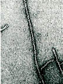
Fig. 34.11. Schematic diagram of F-actin, composed of helically arranged actin monomers

When an actin solution is added to a Myosin solution, a complex of these two Proteins, Actomyosin, is formed. The formation of this complex is accompanied by a sharp increase in solution viscosity. In the 1940s, Albert Szent-Györgyi demonstrated that this viscosity increase is reversed by The addition of ATP. This revealed that ATP causes the dissociation of actomyosin into Actin and myosin. Szent-Györgyi also obtained actomyosin threads whose molecules were oriented in a specific way by fluid flow. When these threads were placed in a solution containing ATP, K+, and Mg2+, a striking result was observed: the actomyosin threads contracted. Under the same conditions, threads formed from myosin alone did not contract. These remarkable experiments provided the basis for the concept that Muscle contraction arises from the interaction of myosin, actin, and ATP.
34.6. Actin Enhances the ATPase Activity of Myosin
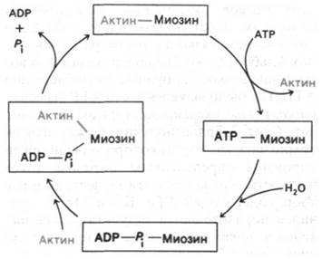
The ATPase activity of myosin increases dramatically in the presence of stoichiometric amounts of F-actin. Specifically, the turnover number increases 200-fold, from 0.05 to 10 s-1. Although the Hydrolysis of ATP by pure myosin proceeds very rapidly, the reaction products—ADP and Pi—are released slowly. Actin increases the myosin turnover number by binding to the myosin–ADP–Pi complex and accelerating the release of ADP and Pi (Fig. 34.12). Following the hydrolysis step, actomyosin binds to ATP, which
leads to its dissociation into actin and myosin. This regenerates the myosin–ATP complex, which then enters a new catalytic cycle. These reactions require Mg2+. A crucial feature of this cycle, proposed by Edwin Taylor on The basis of rapid-reaction kinetics, is that actin has a high affinity for myosin and the myosin–ADP–Pi complex, but a low affinity for the myosin–ATP complex. Consequently, actin alternately binds to and dissociates from myosin depending on ATP hydrolysis. As will be shown below, this ATP-dependent modulation of the interaction between myosin and actin underlies force generation During muscle contraction.
Fig. 34.12. ATP hydrolysis drives the cyclic formation and dissociation of the actin-myosin complex
34.7. Thick and Thin Filaments of the Muscle Fiber Have a Definite Polarity
The cyclic formation and dissociation of the myosin-actin complex produces coordinated movement because these proteins are organized into a highly structured system. By studying intact myofibres isolated from muscle, as well as synthetic filaments formed from purified myosin, Hugh Huxley demonstrated the arrangement of myosin molecules within thick filaments. A thick filament isolated from a muscle fiber has a diameter of 160 Å and a length of approximately 1.5 µm (15,000 Å). Cross-bridges project periodically in a helical pattern along the filament, except for a 1,500-Å central region that is devoid of cross-bridges (Fig. 34.13).
Fig. 34.13. Electron micrograph of a reconstituted thick filament. Projecting cross-bridges are visible on both sides of the bare zone
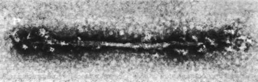
Synthetic thick filaments, formed by lowering the ionic strength of a myosin solution, exhibit a similar Structure. The shortest synthetic filament is about 3,000 Å long, with a central bare zone of 1,500 Å devoid of cross-bridges. Because this same bare-zone length is characteristic of longer synthetic filaments as well, thick filaments evidently grow by the parallel addition of molecules to an already assembled core. Myosin molecules located on one side of the bare zone are oriented in one direction, whereas those on the other side are oriented in the opposite direction. Consequently, thick filaments are bipolar by their very nature.
LMM, like myosin, aggregates in solutions of low ionic strength to form filamentous structures with an axial repeat of 430 Å—the same as the distance between cross-bridges in an intact thick filament. However, LMM filaments are smooth and lack projecting cross-bridges. This indicates that the cross-bridges reside in the HMM portion of the myosin molecule, whereas the LMM units form the backbone of the thick filaments.
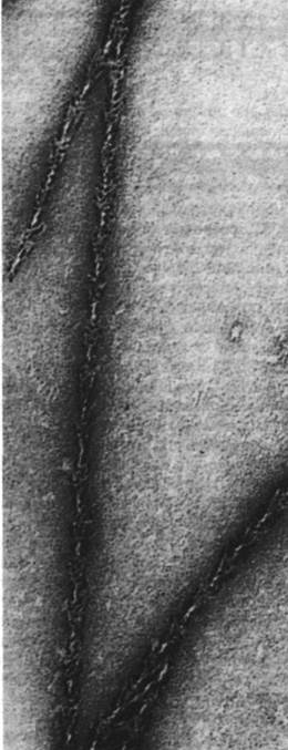
Thin filaments also possess a distinct polarity. When myosin (or HMM, or subfragment-1) is added to thin filaments or F-actin, a structure is formed that in the Electron microscope resembles a series of arrowhead patterns (Fig. 34.14). These structures have aptly been termed decorated filaments. Along the entire length of the decorated filaments, the arrowheads on both strands point in the same direction. Thus, thin filaments inherently possess a uniform polarity along both strands that make up the filament. In some preparations of thin filaments obtained from homogenized muscle, some filaments remain attached to the Z disc. When such preparations are decorated with heavy meromyosin, the arrowheads on all filaments point away from the Z disc. It follows that all thin filaments on one side of the Z disc are oriented in the same direction, whereas those on the opposite side are oriented in the reverse direction.
Fig. 34.14. Electron micrograph of an F-actin filament decorated with S1 heads of HMM. All arrowheads point in the same direction
.
34.8. Polarity of Thick and Thin Filaments Reverses in the Middle of the Sarcomere
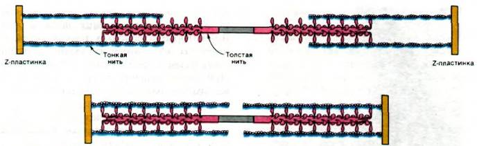
The structural polarity of thick and thin filaments plays a crucial role in coordinated movement. Because the interaction sites on actin and myosin filaments are oriented uniformly relative to each other, the forces generated at each interaction site summate during filament sliding. Furthermore, halfway between two Z-discs, the orientation of the filaments reverses (Fig. 34.15). As a result, two thin filaments interacting with a single thick filament slide toward each other, leading to a decrease in the distance between the Z-discs (Fig. 34.16).
Fig. 34.15. Reversal of polarity of thick and thin filaments midway between two Z-discs
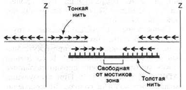
Fig. 34.16. Diagram of the interaction between thick and thin filaments during Skeletal Muscle contraction
Last update: 06/08/2026
Editorial and Educational Adaptation: This material has been compiled based on the primary/original source text. The project team performed an editorial review, corrected technical inaccuracies, structured sections, and adapted the content for an educational format.
What was processed:
- elimination of formatting defects (OCR errors, structural breaks, corrupted characters);
- editorial organization of content;
- standardization of terminology in accordance with academic sources;
- verification of factual statements against the original source text.
All mentions of the author, publication year, and origin of the primary text have been preserved in accordance with the source.