BIOCHEMISTRY - L. Stryer - 1984
VOLUME 3
PART V. MOLECULAR PHYSIOLOGY
CHAPTER 34. MUSCLE CONTRACTION AND CELL MOTILITY
34.34.9. The "Power Stroke" Involves the Rotation of the Actin-Bound Myosin S1 Head
The generation of contractile force involves the cyclic formation and dissociation of the complex between the Myosin S1 HEAD and Actin. Cyclically repeating processes of attachment, pulling, and detachment accompany even a single contraction. The Mechanism of force generation is currently being studied using biochemical, electron microscopic, and X-Ray Diffraction Methods. The obtained data show that in a resting Muscle, the S1 heads are not attached to the thin filaments (Fig. 34.17, A). In this physiological state, the S1 heads are arranged in a helix around the thick filament. Upon muscle stimulation, the S1 heads move away from the thick filaments and attach to the actin units on the thin filaments (Fig. 34.17, B). In the next step, the S1 heads change their orientation such that their long axis forms an angle of approximately 45° with the axis of the thin filaments (Fig. 34.17, C). This postulated Rotation of the myosin heads apparently constitutes the power stroke of Muscle contraction. Through the myosin S2 unit, the rotation of the S1 domain is transmitted to the thick filament. As a result, the thick filament advances relative to the thin filament by approximately 75 Å. The final step of the process is the detachment of the S1 head from the thin filaments (Fig. 34.17, I).
Class="center">Fig. 34.17. Proposed mechanism of force generation during the interaction of myosin S1 head filaments with actin filaments. The rotation of the actin-bound S1 head causes the movement of thick filaments relative to thin filaments (in the diagram, the transition from B to C)

Let us compare these hypothesized structural transitions, which provide the generation of contractile force, with the intermediate steps of ATP Hydrolysis. In the resting state, myosin contains tightly bound ADP and Pi (Fig. 34.17, A). Upon stimulation of the muscle fiber, the S1 head attaches to the thin filament in a perpendicular orientation to it (Fig. 34.17, B). Next, the ADP and Pi bound to S1 are released, and the S1 head undergoes a rotation, assuming an inclined position relative to the thin filament (Fig. 34.17, C). Thus, the "power stroke" is driven by the release of tightly bound ADP and Pi. In the next step, the S1 head dissociates from the thin filament as a result of ATP binding (Fig. 34.17, I). Subsequently, the S1 head, detached from actin, positions itself perpendicularly near the thin filament once again. The final step of the cycle is the hydrolysis of ATP by the S1 head not bound to actin.
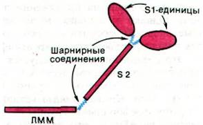
The myosin molecule contains Two Types of hinge joints, thanks to which the S1 head reversibly attaches to or detaches from actin, as well as changes its orientation while in the actin-bound state. One type of hinge is located between each S1 head and the S2 rod, and the second type of hinge is located between S2 and the LMM unit of myosin (Fig. 34.18). The hinges represent flexible Regions of the polypeptide chain that are easily cleaved by hydrolytic Enzymes. Indeed, the very fact of the Enzymatic Cleavage of myosin into LMM, S1, and S2 fragments indicates that myosin is composed of domains connected by hinge regions. The function of the S2 domain is to transmit tension from the thin filament-bound S1 head to the LMM domain, which forms part of the thick filament. Due to the hinge region between S1 and S2, the S1 head can interact with actin in various ways depending on whether tightly bound ADP and Pi are present on it or not. Another hinge region connecting S2 and LMM allows for rather large changes in THE POSITION OF S1 relative to the thick filament and thereby ensures the precision of S1 interaction with actin. As a result, tension can be generated over a large extent of the lateral surfaces of the thick and thin filaments. In general, hinge-type segmental mobility (flexibility) plays a critical role in muscle contraction, much like in the mechanism of antibody action (Section 33.7).
Fig. 34.18. Two types of hinge joints in myosin—one between S1 and S2, and the second between S2 and LMM—allow changing the position of the S1 head relative to actin during the "power stroke"
34.10. Troponin and Tropomyosin Mediate the Regulatory Effect of Calcium Ions on Muscle Contraction
The physiological regulator of muscle contraction is Ca2+. Setsuro Ebashi discovered that The Effect of Ca2+ on the interaction of Actin and myosin is mediated by Tropomyosin and the troponin complex, which are localized in the thin filaments and constitute about a third of their mass. Tropomyosin is a two-chain α-helical strand. This highly elongated protein with a mass of 70 kDa lies almost parallel to the long axis of the thin filament (Fig. 34.19). Troponin is a complex of three polypeptide chains: TnC (18 kDa), TnI (24 kDa), and TnT (37 kDa). TnC binds Calcium Ions, TnI binds to actin, and TnT binds to tropomyosin. Troponin complexes are located on the thin filaments at a distance of 385 Å from each other, this interval being dictated by the length of tropomyosin. The troponin complex associated with one tropomyosin molecule regulates The activity of approximately 7 actin monomers.
Fig. 34.19. Proposed Structure OF THE thin filament in the resting state. The tropomyosin double helix (shown in red) blocks the actin sites (blue) where myosin S1 heads bind during contraction. The attachment of Ca2+ to the TnC component of the troponin complex (shown in yellow) causes a Displacement of the tropomyosin helix, thereby exposing the S1 head binding sites on actin, which ultimately results in contraction

In the absence of Ca2+, troponin and tropomyosin inhibit the interaction of actin and myosin. In this state, tropomyosin sterically blocks the S1 binding sites on actin. A Nerve Impulse, as will be shown below, triggers the release of Ca2+ from the sarcoplasmic reticulum.
The released Ca2+ binds to the TnC component of troponin, which induces conformational shifts that are transmitted to tropomyosin and then to actin. Specifically, tropomyosin moves toward the center of the long groove running helically along the thin filament. This permits the interaction of myosin S1 heads with the actin units of the thin filaments. A contractile force is generated and simultaneously ATP is hydrolyzed; subsequently, Ca2+ is removed, and tropomyosin once again blocks the access of actin to the myosin S1 heads. Thus, Ca2+ regulates muscle contraction via an allosteric mechanism with the following sequence of signal transmission:
Ca2+ → Troponin → Tropomyosin → Actin → Myosin.
34.11. The Ca2+ Ion Flux is Regulated by the Sarcoplasmic Reticulum
A nerve impulse reaching the end plate, i.e., the region of the Neuromuscular Junction, causes depolarization of the outer membrane of the muscle fiber. Via T-tubules (T standing for transverse orientation), the depolarization of the outer membrane spreads deep into the muscle fiber. T-tubules are in close contact with a network of ultrafine channels called the sarcoplasmic reticulum, which serves as a storage site for Ca2+ (Fig. 34.20).
Fig. 34.20. Schematic representation of the sarcoplasmic reticulum
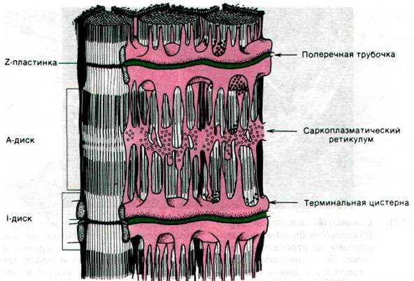
In the resting state, The Active Transport system for Ca2+ accumulates it in the sarcoplasmic reticulum (Section 36.9). The calcium pump, driven by ATP, lowers the concentration of Ca2+ in the Cytoplasm of resting Muscles to below 10-6 M, while raising it to more than 10-3 M in the sarcoplasmic reticulum. A second protein, called calsequestrin, binds Ca2+ inside the reticulum. Calsequestrin, characterized by high acidity and a mass of 44 kDa, contains more than 40 Ca2+ binding sites. Depolarization of the T-tubule membranes triggers the release of Ca2+ from the cisternae of the sarcoplasmic reticulum. The released Ca2+ binds to the TnC component of the troponin complex and stimulates muscle contraction, as described above.
34.12. Phosphocreatine as a Storage Form of ~P
The amount of ATP present in a muscle can sustain contractile activity for only a fraction of a second. However, in vertebrate muscles, energy-rich phosphate bonds are stored as phosphocreatine (creatine phosphate). This compound features a higher phosphoryl group transfer potential than ATP (see Section 11.6). Creatine kinase catalyzes The transfer of a phosphoryl group from phosphocreatine to ADP, yielding ATP:
Phosphocreatine + ADP ⇄ ATP + Creatine.
Certain invertebrates utilize phosphoarginine to store high-energy phosphoryl groups. Both phosphocreatine and phosphoarginine are referred to as phosphagens.

In working muscle, phosphocreatine reserves are rapidly depleted, leading to a corresponding decrease in ATP levels. Concurrently, the concentrations of ADP and Pi rise, alongside an increase in AMP content mediated by adenylate kinase (myokinase):
2ADP ⇄ ATP + AMP.
A decline in The energy charge stimulates Glycolysis, The Tricarboxylic Acid Cycle, and Oxidative Phosphorylation (see Section 14.13) in active muscle. The relative contribution of each pathway to ATP generation depends on the muscle type. For instance, in red muscles—whose color stems from high concentrations of Myoglobin and Respiratory Chain Cytochromes—The rate of aerobic METABOLISM is significantly higher than in white muscles.
Last update: 06/08/2026
Editorial and Educational Adaptation: This material has been compiled based on the primary/original source text. The project team performed an editorial review, corrected technical inaccuracies, structured sections, and adapted the content for an educational format.
What was processed:
- elimination of formatting defects (OCR errors, structural breaks, corrupted characters);
- editorial organization of content;
- standardization of terminology in accordance with academic sources;
- verification of factual statements against the original source text.
All mentions of the author, publication year, and origin of the primary text have been preserved in accordance with the source.