BIOCHEMISTRY - L. Stryer - 1984
VOLUME 1
PART I. CONFORMATION AND DYNAMICS
CHAPTER 10. AN INTRODUCTION TO BIOLOGICAL MEMBRANES
10.8. Reconstitution of Functioning Membrane Systems from Purified Components
A large number of Membrane Proteins have been solubilized and purified (see Chapters 34-37). Some of these retain their activity in detergent solutions. For example, the photoreceptor protein rhodopsin exhibits the same absorption band at 500 nm whether it is in a detergent solution or in the retinal membrane. Furthermore, in both cases, the prosthetic group of this protein undergoes the same structural changes upon illumination. Calsequestrin, a calcium-binding protein of the Muscle sarcoplasmic reticulum, retains its ability to bind calcium even outside the membrane. The calcium pump (the enzyme Ca2 + -ATPase) has also been isolated from the sarcoplasmic reticulum. It was found that functionally active vesicles capable of accumulating Ca2+ in the presence of ATP as an energy source can be formed from a mixture of phospholipid and the calcium pump protein. The reconstitution of functionally active membrane systems from purified components is a powerful approach to studying membrane processes.
10.9. Some Membrane Proteins Are Deeply Embedded in the Lipid Bilayer
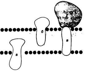
Some membrane Proteins can be isolated by mild Treatment, such as extraction with a high Ionic strength solution (e.g., 1 M NaCl). Other proteins are more tightly bound to the membrane and can be separated only with the aid of a detergent (Fig. 10.17) or an organic solvent. Based on differences in the strength of their association with the membrane, these proteins are classified as peripheral or integral (Fig. 10.18). Integral proteins form extensive interactions with the hydrocarbon chains of Membrane Lipids and can therefore be isolated only by using agents that compete in these nonpolar interactions. Peripheral proteins, in contrast, are bound to membranes by electrostatic forces and Hydrogen Bonds. These polar interactions can be disrupted by adding salts or altering the pH. According to recent data, peripheral membrane proteins in most cases bind to The surface of integral proteins.
Class="center">Fig. 10.17. Structures of detergents used for the solubilization and purification of membrane proteins
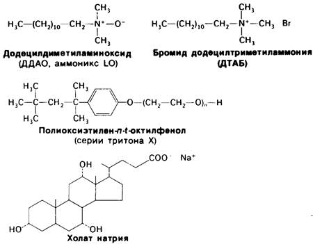
Fig. 10.18. Integral membrane proteins (a, b, c) form numerous interactions with the hydrocarbon chains of the bilayer. Peripheral membrane proteins (d) bind to the surface of integral proteins
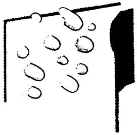
To determine whether a given protein is localized in the interior of the membrane, the freeze-fracture electron Cell/15.html">Microscopy Technique is primarily used. Cells or membrane fragments are rapidly frozen to liquid nitrogen Temperature. The frozen membrane is then fractured by the blow of a microtome knife. The fracture plane usually runs longitudinally through the middle of the bilayer (Fig. 10.19), exposing a large area of The Lipid Bilayer interior. This exposed surface is then shadowed by spraying it with a layer of carbon or platinum, creating a replica of the bilayer interior. To study the outer surface of the membrane, freeze-fracture is used in combination with deep etching. In this process, the interior of the frozen membrane is first exposed by fracturing. Then, the ice on the adjacent surface of the membrane is sublimed (dried), which is the deep-etching technique. The combination of these two Methods, known as freeze-etching Electron microscopy, provides a view of the membrane interior and both of its surfaces. The advantage of this approach is that it does not require fixation or dehydration of the biological material.
Fig. 10.19. Freeze-fracture electron microscopy. The fracture plane runs through the middle of the membrane bilayer
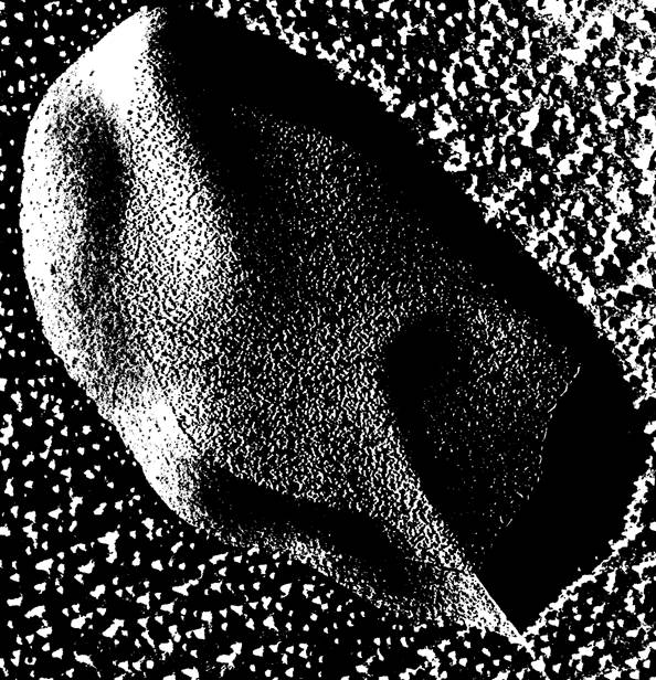
Freeze-etching studies have directly demonstrated the presence of integral proteins in many Introduction/36.html">Biological Membranes. For example, The erythrocyte membrane contains high-density globular particles about 75 Å in diameter (Fig. 10.20). A large number of globular particles are also present within the sarcoplasmic reticulum membrane. In contrast, an artificial phosphatidylcholine bilayer has a completely smooth fracture face. The fracture faces of the myelin membrane also appeared mostly smooth, as would be expected for this relatively inert membrane that Functions primarily as an insulating sheath.
Fig. 10.20. Freeze-etch electron micrograph of an erythrocyte Plasma Membrane. The interior of the membrane exposed by fracturing is rich in globular particles about 75 Å in diameter. These particles are integral membrane proteins
10.10. The Erythrocyte Membrane Contains Diverse Peripheral and Integral Proteins
Erythrocytes have proven to be the ideal system for membrane studies because they are readily available and relatively simple in Structure. Lacking internal Organelles, red Blood Cells possess only a single membrane—The Plasma Membrane. Osmotic lysis can be used to obtain erythrocyte ghosts, which are pure Plasma Membranes free of Cytoplasm. Fig. 10.21 shows the results of electrophoretic Separation of proteins from such a membrane preparation on an SDS-polyacrylamide gel. Coomassie blue staining reveals more than ten bands. The major bands are numbered and designated as bands 1, 2, 3, 4.1, 4.2, 5, and 6 (Fig. 10.21). Staining with periodic acid-Schiff reagent (PAS reaction)1 reveals several carbohydrate-rich protein fractions. These bands are designated PAS-1, PAS-2, PAS-3, and PAS-4, respectively.
1 Translator's Note: In Russian literature, the abbreviation ШИК (SHIK) is often used instead of ИКШ (IKSh) for euphonic reasons, although it does not reflect the actual sequence, as periodic acid treatment precedes the Schiff reagent. Therefore, we use the widely accepted abbreviation PAS (periodic acid-Schiff reagent).
Fig. 10.21. Electrophoretic separation of erythrocyte membrane proteins on an SDS-polyacrylamide gel. Stained with Coomassie blue
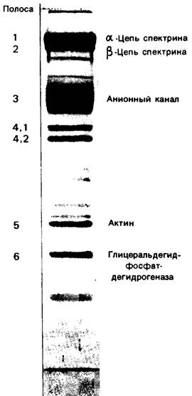
Where are these proteins located in the erythrocyte membrane? The proteins corresponding to bands 1, 2, 4.1, 4.2, 5, and 6 can be extracted from membranes by increasing the ionic strength of the solution or by altering the pH; thus, they are peripherally located. Furthermore, they are unaffected by the incubation of intact cells or sealed ghosts with various Proteolytic Enzymes. In contrast, these proteins are completely degraded when ruptured ghosts are treated with proteases. This indicates that these peripheral proteins reside on the cytoplasmic side of the erythrocyte membrane. Band 6 is the glycolytic enzyme glyceraldehyde 3-phosphate dehydrogenase (Section 12.5), and band 5 is Actin, which is essential for Muscle contraction and cell motility (Section 34.1). Bands 1 and 2 represent spectrin, whose chains associate to form a branched fibrous network. Together with other proteins, spectrin appears to stabilize and regulate the shape of the erythrocyte membrane, which undergoes deformation as the cells pass through narrow Blood Vessels (Fig. 10.22). In addition, erythrocytes are subjected to severe mechanical stress as blood is pumped by The Heart.
Fig. 10.22. Erythrocytes undergo reversible deformation as they pass through narrow blood vessels (capillaries)

As for bands 3 and 7 and all four PAS-positive bands, they can be separated from the erythrocyte membrane only by detergents or organic Solvents. Consequently, these bands represent integral membrane proteins. This Conclusion is supported by electron microscopy: freeze-fracture studies (Fig. 10.20) showed that some of the erythrocyte proteins are embedded deep within the hydrocarbon region of the membrane.
Last update: 06/08/2026
Editorial and Educational Adaptation: This material has been compiled based on the primary/original source text. The project team performed an editorial review, corrected technical inaccuracies, structured sections, and adapted the content for an educational format.
What was processed:
- elimination of formatting defects (OCR errors, structural breaks, corrupted characters);
- editorial organization of content;
- standardization of terminology in accordance with academic sources;
- verification of factual statements against the original source text.
All mentions of the author, publication year, and origin of the primary text have been preserved in accordance with the source.