BIOCHEMISTRY - L. Stryer - 1984
VOLUME 1
PART I. CONFORMATION AND DYNAMICS
CHAPTER 10. AN INTRODUCTION TO BIOLOGICAL MEMBRANES
10.6. Lipid bilayers are impermeable to ions and most polar molecules
The permeability of lipid bilayers has been measured in two well-characterized synthetic systems: lipid vesicles and planar bilayer membranes. These model systems have helped define The primary function of Introduction/36.html">Biological Membranes, namely, their role as an impermeable barrier. Crucially, lipid bilayers are inherently impermeable to ions and most polar compounds.
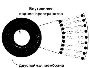
Lipid vesicles (also called Liposomes) are aqueous compartments enclosed by a lipid bilayer (Fig. 10.12). To prepare liposomes, a suspension of a suitable lipid, such as phosphatidylcholine, is made in an aqueous medium. The mixture is then sonicated to form closed lipid vesicles of nearly uniform size. Lipid vesicles can also be prepared by rapidly mixing an ethanol solution of Lipids with Water. This can be achieved by injecting the lipid solution through a fine needle, yielding nearly spherical vesicles about 500 Å in diameter. Larger vesicles (on the order of 104 Å, or 1 µm in diameter) can be prepared by slowly evaporating the organic solvent from a phospholipid suspension in a mixed solvent system.
Class="center">Fig. 10.12. Schematic representation of a lipid vesicle
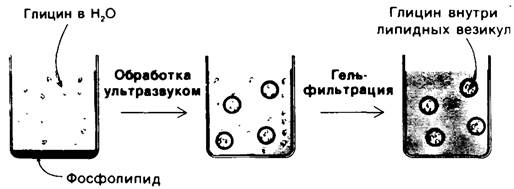
If liposomes are formed in the Presence of water-soluble ions or molecules, these substances will be trapped in the aqueous compartment of the liposomes (Fig. 10.13). For example, if liposomes 500 Å in diameter are formed in a 0.1 M Glycine solution, each vesicle will contain approximately 2,000 molecules of glycine. These glycine-containing liposomes can be separated from the surrounding glycine solution by dialysis or Gel filtration. To determine the permeability of the membrane bilayer to glycine, one measures The rate of efflux of glycine from the inner compartment of the liposomes into the external medium. Liposomes are of interest not only for permeability studies. They can also fuse with Cell/30.html">The Plasma Membrane of various Cells, which opens up the possibility of introducing A wide variety of membrane-impermeable substances into cells. The selective fusion of liposomes with specific cell types can be successfully used for targeted drug delivery to target cells.
Fig. 10.13. Preparation of a suspension of lipid vesicles containing glycine molecules
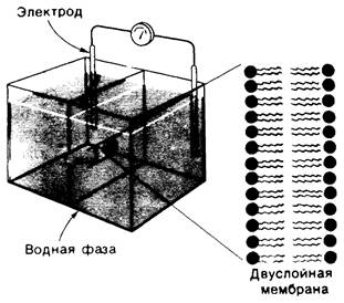
A second type of synthetic membrane is the planar bilayer membrane (or black lipid membrane). This Structure is formed across a 1-mm aperture in a partition separating two aqueous compartments. Such a membrane is highly suitable for studying electrical phenomena due to its macroscopic size and simple geometry. A method for preparing large bilayer membranes was developed by Paul Mueller and Donald Rudin. A fine paintbrush is dipped into a solution of the lipid from which the membrane is to be formed, such as phosphatidylcholine in decane. The tip of the brush is then swiped across the aperture (1 mm in diameter) in the partition between the two aqueous solutions. As a result, a thin lipid film spontaneously forms across the aperture, with excess lipid accumulating at the margins. The formation of a planar phosphatidylcholine bilayer takes a few minutes. The electrical conductivity of this macroscopic bilayer can be easily determined by placing electrodes in the aqueous phase on both sides of the membrane (Fig. 10.14). For example, the ionic permeability of the membrane can be determined by measuring the current flowing through it as a function of the applied voltage.
Fig. 10.14. Experimental setup for studying planar bilayer membranes. The bilayer membrane forms across a 1-mm aperture in a partition separating two aqueous phases
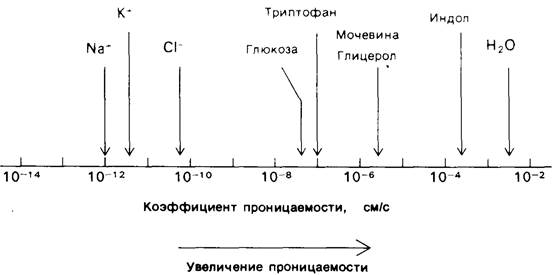
Permeability studies of membrane vesicles and conductivity measurements of planar bilayers have shown that lipid bilayer Membranes have a very low permeability for ions and most polar molecules. An exception to this rule is water, which readily penetrates such membranes. Experimentally determined permeability coefficients vary over a wide range (Fig. 10.15). For instance, Na+ and K+ traverse membranes 109 times more slowly than water. Tryptophan, which is a zwitterion at pH 7, traverses membranes 103 times more slowly than indole, which is structurally similar to tryptophan but lacks ionized groups. The permeability coefficients of small molecules correlate with their partition coefficients between nonpolar Solvents and water. This relationship suggests that small molecules traverse a bilayer membrane as follows: first, they shed their solvation shell of water; then, they dissolve in the hydrocarbon core of the membrane; and finally, they diffuse through this core to the other side of the membrane, where they become rehydrated.
Fig. 10.15. Permeability coefficients of lipid bilayer membranes for various ions and molecules
10.7. Most membrane processes are mediated by proteins
We turn now to Membrane Proteins, which are responsible for most of the dynamic processes carried out by membranes. While Membrane Lipids establish permeability barriers and thus define compartments, specific proteins mediate distinct Membrane Functions, such as transport, information transfer, and energy Transduction. In doing so, membrane lipids provide the environment necessary for the action of these proteins.
Membranes differ significantly in their protein content. For example, myelin, which serves as an electrical insulator around many nerve fibers, has a low protein content (18%). The primary component of myelin is lipid, which has excellent insulating properties. In contrast, the Plasma Membranes of most cells are much more active and contain various pumps, channels, receptors, and Enzymes. The protein content of these plasma membranes is typically about 50%. The highest protein content, up to 75%, is found in membranes that function as energy transducers, such as the inner membranes of Mitochondria and METABOLISM/14.html">Chloroplasts.
The major membrane Proteins can be visualized by sodium dodecyl sulfate (SDS) Polyacrylamide gel Electrophoresis.
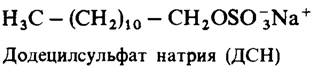
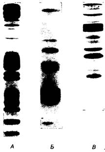
In this method, membranes are first solubilized with a 1% SDS solution. This detergent disrupts most protein-protein and protein-lipid interactions. The resulting solution is then layered onto a polyacrylamide gel, which also contains SDS, and an electric field is applied for several hours. In this system, unlike in SDS-free systems, the electrophoretic mobility of most proteins depends on their mass rather than their charge. This is because the negative charge of the SDS molecules bound to the protein far exceeds the intrinsic charge of the protein itself. The bands of separated protein fractions are visualized by staining the gel with a dye such as Coomassie blue. This technique can detect extremely small amounts of protein, on the order of a few micrograms. Fig. 10.16 shows the gel electrophoresis patterns of three membranes: the erythrocyte plasma membrane, the photoreceptor disc membrane of retinal rods, and the sarcoplasmic reticulum membrane of Muscle cells. It is clearly evident that the protein compositions of these three membranes are completely different. Furthermore, the membranes differ in their polypeptide chain composition and mass distribution. Thus, membranes performing different functions also differ in their protein composition.
Fig. 10.16. Electrophoretic patterns of various membranes in SDS-polyacrylamide gel. A - erythrocyte plasma membrane. B - retinal rod photoreceptor disc membranes. C - sarcoplasmic reticulum membranes of muscle cells
Last update: 06/08/2026
Editorial and Educational Adaptation: This material has been compiled based on the primary/original source text. The project team performed an editorial review, corrected technical inaccuracies, structured sections, and adapted the content for an educational format.
What was processed:
- elimination of formatting defects (OCR errors, structural breaks, corrupted characters);
- editorial organization of content;
- standardization of terminology in accordance with academic sources;
- verification of factual statements against the original source text.
All mentions of the author, publication year, and origin of the primary text have been preserved in accordance with the source.