Biological Membranes - A. N. Ogurtsov 2012
Electrogenesis of Biomembranes
Mechanisms of Action Potential Generation
Voltage-Gated Sodium Channels
Voltage-gated sodium channels are closed when the membrane is at rest, and the Membrane Potential in this state is referred to as the resting membrane potential.
A slight initial point depolarization of the membrane triggers a conformational change in adjacent channel Proteins, causing the sodium channels to open. Through these open channels, Na+ ions flow into The Cell, thereby further increasing the depolarization (positively charging the cytosolic side of the membrane).
The greater the depolarization, the more sodium channels open, and the more Na+ ions enter the cell. As an increasing number of Na+ ions move into the Cytosol, the zone of excess positive charge on the cytosolic side of the membrane and the corresponding zone of negative charge on the exoplasmic side (the zone of depolarization) spread outward from the initial site of depolarization.
This Propagation of the depolarization zone encompasses a progressively larger area of the membrane, within which sodium channels open, steadily amplifying the inward flux of Na+ ions.
The process develops in an avalanche-like fashion, and within a fraction of a millisecond, so many sodium channels open that the total inward flux of Na+ ions vastly exceeds the outward counter-flow of K+ ions through potassium channels.
When the membrane potential reaches the equilibrium potential for sodium ions, the movement of Na+ ions into the cell driven by the concentration gradient is balanced by their outward movement driven by the repulsive force of the excess positive charge already accumulated in the cytosol, halting any further increase in the amplitude of the membrane potential. The value of the membrane potential in this state is called the reversal potential.
Figure 128 illustrates the main structural Conformations of the sodium channel.
Class="center">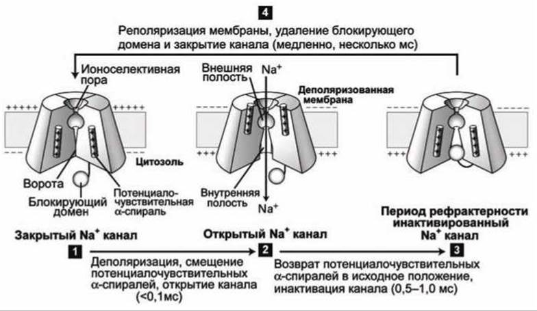
Figure 128 — Gating cycle of a voltage-gated sodium channel
In the resting state, the channel "gate" is closed (Figure 128(1)). Membrane depolarization causes the voltage-sensing α-helices (2) to shift upward toward the exoplasmic side of the membrane, inducing a conformational change in the Protein Structure that opens the gate and allows ions to move into the cell. After 1 ms, the channel is mechanically blocked by a cytosolic domain that simply acts as a plug, obstructing the channel pore (3). As long as the membrane remains depolarized, the channel stays blocked. Several milliseconds after repolarization—once the membrane potential returns to the resting level—the charged α-helices shift downward back to their initial position, "squeezing out" the blocking domain plug from the channel, and the channel returns to its resting state (Figure 128(4)).
Last update: 13/08/2026
Editorial and Educational Adaptation: This material has been compiled based on the primary/original source text. The project team performed an editorial review, corrected technical inaccuracies, structured sections, and adapted the content for an educational format.
What was processed:
- elimination of formatting defects (OCR errors, structural breaks, corrupted characters);
- editorial organization of content;
- standardization of terminology in accordance with academic sources;
- verification of factual statements against the original source text.
All mentions of the author, publication year, and origin of the primary text have been preserved in accordance with the source.