MEDICAL BIOLOGY, HUMAN ANATOMY, PHYSIOLOGY AND PATHOLOGY - Ya.I. Fedoniuk 2010
ANATOMY, PHYSIOLOGY, PATHOLOGY
SECTION 3. ANATOMICAL AND PHYSIOLOGICAL ASPECTS OF BODY FUNCTION SELF-REGULATION
BRAIN
The Brain (encephalon) is located within the cranial cavity. It consists of two hemispheres (the most evolutionarily recent part) and the Brainstem (Medulla Oblongata, Pons, cerebral peduncles) along with the Cerebellum. The Superolateral surface of the brain is convex, whereas the inferior surface (base) is irregular and gives rise to 12 pairs of Cranial Nerves (Fig. 3.10).
Class="center">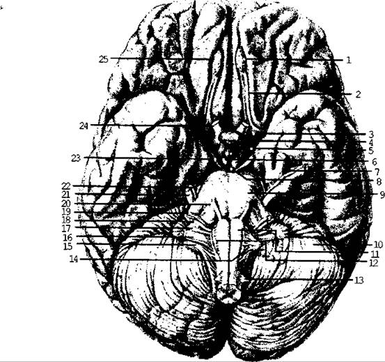
Fig. 3.10. The brain
The brain mass of an adult human ranges from 1245 to 1375 g, and in a newborn, it is 330-340 g. During the Embryonic period and the first years of life, the brain grows quite intensively, reaching its permanent size only by the age of 20.
The BRAIN AND SPINAL cord develop from the outer germ layer (ectoderm). On the DORSAL SIDE OF the embryo, a neural tube forms with an expansion at the cranial end. Initially, this expansion is represented by three primary brain vesicles: the prosencephalon (Forebrain), mesencephalon (Midbrain), and rhombencephalon (Hindbrain) (Fig. 3.11 a). Subsequently, the forebrain and hindbrain vesicles divide to form five secondary brain vesicles: the myelencephalon, metencephalon, mesencephalon, Diencephalon, and Telencephalon (Fig. 3.11 b, c). The walls of the brain vesicles grow unevenly: they either thicken or remain quite thin, protruding into the cavities of the brain vesicles to form the choroid plexuses of the ventricles. The remnants of the cavities of the brain vesicles and the neural tube are known as the brain ventricles and the central canal of the Spinal Cord, which are filled with CEREBROSPINAL FLUID. Specific Regions of the brain develop from each brain vesicle.
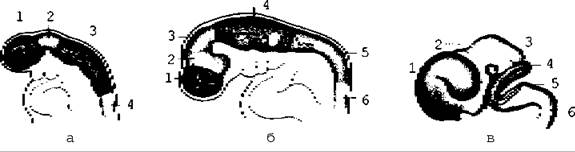
Fig. 3.11. The brain at the three-vesicle (a - 3.5 weeks) and five-vesicle (b - 6 weeks, c - 8 weeks) stages
Last update: 08/08/2026
Editorial and Educational Adaptation: This material has been compiled based on the primary/original source text. The project team performed an editorial review, corrected technical inaccuracies, structured sections, and adapted the content for an educational format.
What was processed:
- elimination of formatting defects (OCR errors, structural breaks, corrupted characters);
- editorial organization of content;
- standardization of terminology in accordance with academic sources;
- verification of factual statements against the original source text.
All mentions of the author, publication year, and origin of the primary text have been preserved in accordance with the source.