MEDICAL BIOLOGY, HUMAN ANATOMY, PHYSIOLOGY AND PATHOLOGY - Ya.I. Fedoniuk 2010
ANATOMY, PHYSIOLOGY, PATHOLOGY
CHAPTER 2. LOCOMOTOR SYSTEM (THE MECHANISM OF MOVEMENT)
SKELETON OF THE LOWER LIMB
JOINTS OF THE LOWER LIMB
2. THE PELVIS AS A WHOLE
The pelvis (pelvis) is formed by the hip bones, the sacrum, the coccyx, and their joints.
The pelvis is divided into the greater (false) pelvis and lesser (true) pelvis. The boundary between them is the linea terminalis (pelvic brim), which extends from the sacral promontory along the arcuate lines of the iliac bones, then along the superior rami of the pubic bones and the upper edge of the Pubic Symphysis.
The greater pelvis is formed by the flared wings of the iliac bones and serves as a support for the Internal Organs of the Abdominal cavity.
The lesser pelvis is formed by the pelvic surfaces of the sacrum and coccyx, the ischium, and the pubic bones (Fig. 2.60). It comprises the superior and inferior apertures (inlet and outlet) and the pelvic cavity. The lesser pelvis contains the Urinary Bladder, rectum, and internal reproductive organs (Uterus, uterine tubes, and Ovaries in females; Prostate Gland, Seminal Vesicles, and deferent ducts in males).
Class="center">

Fig. 2.60. Male pelvis (a) and female pelvis (b)
The female pelvis is wider and shorter than the male pelvis, with more widely flared iliac wings. In addition, the subpubic angle is obtuse (acute in males), and the sacrum is short, wide, and flat. These structural features are due to the function of the female pelvis as the birth canal.
In obstetric practice, measurements of both the greater and lesser pelvis are routinely performed.
The following dimensions of the female pelvis are distinguished (average values) (Fig. 2.60 a, 2.61).
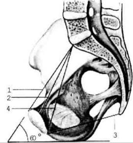
Fig. 2.61. Dimensions of the female pelvis (sagittal section)
Greater pelvis:
1) interspinous distance – the distance
between the anterior superior iliac spines (26 cm); 2) intercristal distance – the distance between the furthest1 points of the iliac crests (29 cm); 3) intertrochanteric distance – the distance between the greater trochanters of the femurs (30 cm).
Lesser pelvis:
1. External conjugate – the distance between the pubic symphysis and the sacral promontory (20–21 cm);
2. Diagonal conjugate – the distance between the lower edge of the pubic symphysis and the sacral promontory (12.5–13 cm);
3. True conjugate (obstetric conjugate) – the distance between the most prominent part of the pubic symphysis and the sacral promontory (10.5–11 cm).
4. Anteroposterior diameter of the pelvic outlet – the distance between the lower edge of the pubic symphysis and the apex of the coccyx (10 cm).
During childbirth, this dimension increases by 1–1.5 cm due to the backward deflection of the coccyx.
5. The pelvic inclination angle is the angle formed by the horizontal plane and the plane of the pelvic inlet.
Last update: 08/08/2026
Editorial and Educational Adaptation: This material has been compiled based on the primary/original source text. The project team performed an editorial review, corrected technical inaccuracies, structured sections, and adapted the content for an educational format.
What was processed:
- elimination of formatting defects (OCR errors, structural breaks, corrupted characters);
- editorial organization of content;
- standardization of terminology in accordance with academic sources;
- verification of factual statements against the original source text.
All mentions of the author, publication year, and origin of the primary text have been preserved in accordance with the source.