MEDICAL BIOLOGY, HUMAN ANATOMY, PHYSIOLOGY AND PATHOLOGY - Y.I. Fedoniuk 2010
ANATOMY, PHYSIOLOGY, PATHOLOGY
CHAPTER 2. THE MUSCULOSKELETAL SYSTEM (THE MECHANICS OF MOVEMENT)
SKELETON OF THE LOWER LIMB
JOINTS OF THE LOWER LIMB BONES
1. BONES OF THE PELVIC GIRDLE AND THEIR JOINTS
Up to the age of 16, the hip bone (os coxae) consists of separate bones: the ilium, pubis, and ischium. After 16 years of age, their bodies fuse together, forming at this site the acetabulum, which accommodates the HEAD of the Femur (Fig. 2.58).
Class="center">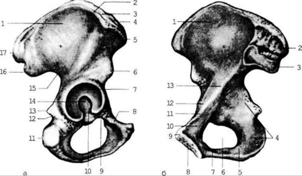
Fig. 2.58. Hip bone; right
The ilium (os ilium) consists of a body and a ala (wing). The ala is expanded, directed upward, and terminates in the iliac crest. The crest features the anterior superior and inferior iliac spines anteriorly, and the posterior superior and inferior iliac spines posteriorly. The pelvic surface of the ala presents a depression known as the iliac fossa. Posterior to it lies the auricular surface for articulation with the corresponding surface of the sacrum. The gluteal lines, which serve as attachment sites for the gluteal Muscles, are located on the external surface of the ala. The ala is separated from the body by the arcuate line.
The ischium (os ischii) comprises a body and a ramus that thickens to form the ischial tuberosity. Superior to the tuberosity lies the ischial spine. Above the spine is the greater sciatic notch, and below the spine is the lesser sciatic notch.
The pubis (os pubis) is divided into a body, a superior ramus, and an inferior ramus. The articular surface of the Pubic Symphysis is formed where these rami meet. The bodies of all three bones, along with the rami of the ischium and pubis, bound the obturator foramen.
Posteriorly, the BONES OF THE Pelvic Girdle articulate with the sacrum via the paired, flat sacroiliac joint, which has limited mobility (Fig. 2.59). It is formed by the auricular surfaces of the sacrum and ilium. Anteriorly, an unpaired amphiarthrosis (hemijoint) is formed—the pubic symphysis. Here, the pubic bones are connected by Cartilage containing a small fluid-filled cleft within its substance.
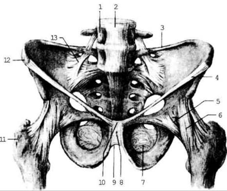
Fig. 2.59. Ligaments of the pelvis and hip joint; superior view
The proper ligaments of the pelvis include the sacrotuberous and sacrospinous ligaments. They bridge the sciatic notches, thereby converting them into the greater and lesser sciatic foramina (Fig. 2.65). Muscles, Blood Vessels, and nerves pass through these foramina.
Last update: 08/08/2026
Editorial and Educational Adaptation: This material has been compiled based on the primary/original source text. The project team performed an editorial review, corrected technical inaccuracies, structured sections, and adapted the content for an educational format.
What was processed:
- elimination of formatting defects (OCR errors, structural breaks, corrupted characters);
- editorial organization of content;
- standardization of terminology in accordance with academic sources;
- verification of factual statements against the original source text.
All mentions of the author, publication year, and origin of the primary text have been preserved in accordance with the source.