General and Sports Anatomy - L.V. Kapilevich, K.V. Davletyarova 2008
Anatomy of the Musculoskeletal System
Osteology
Osteology is The Study of bones.
Functions of bones: support, acting as levers, protection of Internal Organs, and participation in hematopoiesis.
Classification of Bones
Tubular bones (humerus, Femur, Metacarpal bones), spongy bones (carpals, tarsals), flat bones (BONES OF THE cranial vault, Sternum, Ribs), irregular bones (vertebrae), pneumatic bones (sphenoid, temporal, frontal).
The Skeleton is divided into:
The Axial Skeleton comprises (Figure 4): the Skull (29 bones), THE Vertebral Column (26), and the RIBS AND STERNUM (25).
The Appendicular Skeleton: upper extremities (64), lower extremities (62).
Class="center">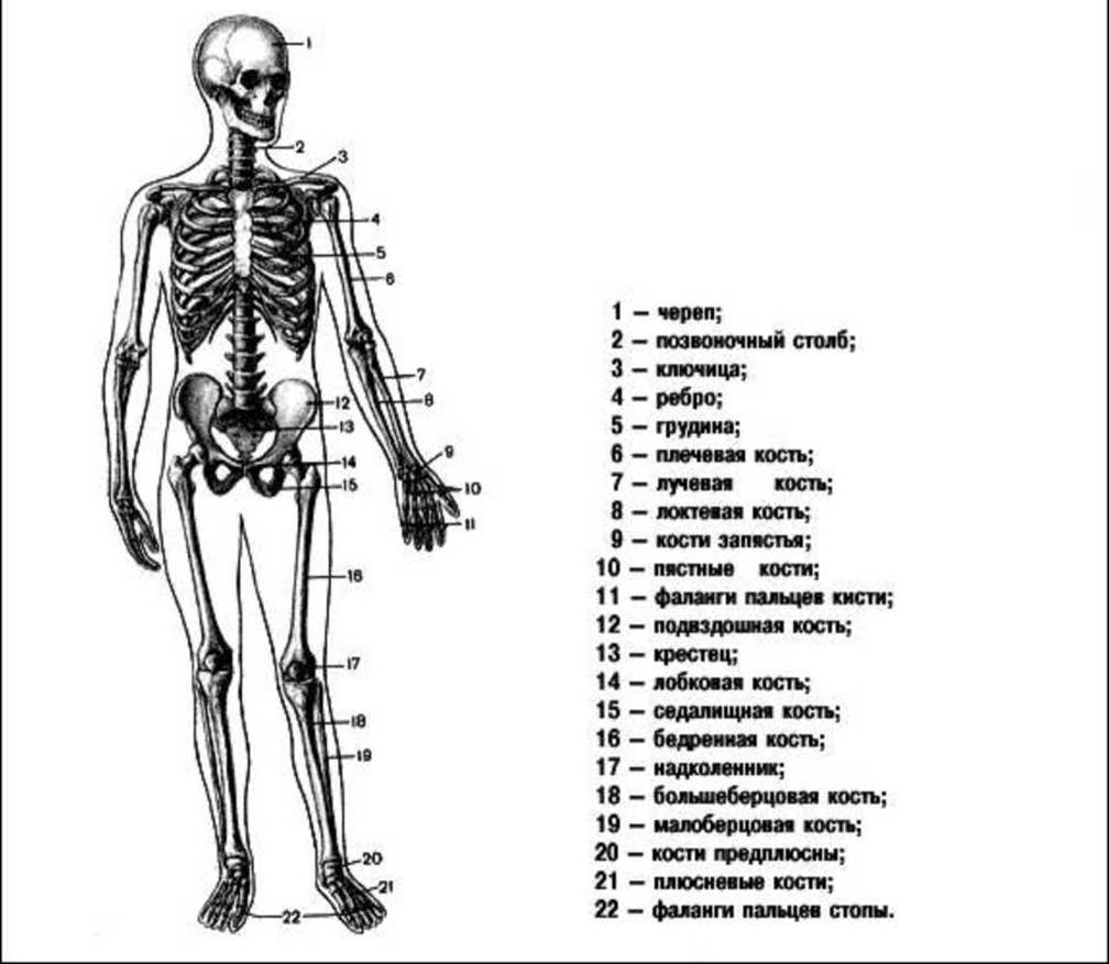
Figure 4. Human skeleton.
The outer layer of the bone is a plate of compact Bone tissue (providing support), spongy bone tissue (performing Shock-absorbing functions), with the medullary cavity located in the center of the bone (Figure 5).
Periosteum is a Connective Tissue membrane covering the outer surface of the bone. It consists of an outer fibrous layer (protective function) and an inner osteogenic (cambial) layer (responsible for bone growth).
Bone Marrow: located within the medullary cavity, Cytology/practical/86.html">Red bone marrow (hematopoiesis), yellow bone marrow (adipose Cells).
Surface irregularities of bones:
Prominences: tubercle, tuberosity, crest, process.
Depressions: fossa, fovea, sulcus.
Articular surfaces: convex, concave, flat.
Bone Development
Stages of bone development: membranous, cartilaginous, bony.
Growth of bones in thickness:
- perichondrial ossification - from the periosteum.
- endochondrial ossification - bone tissue forms inside the Cartilage.
Longitudinal bone growth occurs through epiphyseal cartilages, which form a cartilaginous layer between the diaphysis and epiphysis.
Short bones - on one side (Phalanges of the fingers).
Long bones - on both sides.
Bone growth in thickness occurs due to the inner layer of the periosteum.
The epiphyseal cartilage is replaced by bone tissue by the age of 18-20, and bone growth practically ceases.
Some bones develop and grow from several cartilages and later fuse together (the hip bone consists of 3 parts fused by the age of 14-16).
Ossification proceeds faster where mechanical stress is greater (earlier in the lower extremities than in the upper ones).
This must be taken into account when engaging children in physical education and sports.
P.F. Lesgaft formulated the law of bone growth driven by The activity of surrounding Muscles.
Laws of bone growth:
1. mechanical loads stimulating bone growth must be rhythmic.
2. bone growth activation occurs at an optimum of loads; lack or excess of loads inhibits it.
3. bones "adapt" to habitual loads (the orientation of trabeculae and the size of cellular spaces change), whereas unusual loads can lead to pathological alterations.

Figure 5. Bone structure.
1. What types of bones exist?
2. What layers does the periosteum consist of?
3. State the main laws of bone growth (according to P.F. Lesgaft).
Vertebral column
Formed by 33-34 vertebrae.
Regions: cervical (7 vertebrae), thoracic (12), lumbar (5), sacral (5), coccygeal (4-5).
The spine has curves: anterior curves are lordoses, posterior curves are kyphoses, and lateral curves are scolioses (Figure 6).
A vertebra consists of a body (facing anteriorly) and an arch, which together enclose the vertebral foramen. The vertebral body is covered with a layer of compact bone, while the interior consists of spongy bone with mutually perpendicular trabeculae. The arch features notches that, when articulated with adjacent vertebrae, form intervertebral foramina for nerves and Blood Vessels. The vertebral foramina align to form the vertebral canal, which houses the Spinal Cord.

Figure 6. The human vertebral column.
Processes: the spinous process projects posteriorly, serving as an attachment site for Ligaments and Muscles. The transverse processes (two of them) serve for Muscle and ligament attachment, and the superior and inferior articular processes (four in total) are designed for articulation with adjacent upper and lower vertebrae.
The size of the vertebrae in various regions depends on the mechanical load on that region: the greater the load, the larger the vertebrae.
Cervical vertebrae
They feature a small body, a large vertebral foramen, a bifid spinous process, and transverse foramina for cerebral vessels.
The first vertebra is the atlas.
It lacks a vertebral body and a spinous process, forming a ring-like structure. It features lateral masses on the sides for articulation with The Skull and the second cervical vertebra.
The second vertebra is the axis.
It features a dens (odontoid process) for articulation with the atlas, allowing for HEAD rotation.
3rd to 7th vertebrae: the transverse processes bear tubercles. The 7th vertebra has a long, prominent process that is easily palpable.
Thoracic vertebrae
Thoracic vertebrae are larger than cervical ones. They feature demifacets on the lateral surfaces of the body for articulation with the heads of the ribs.
The 1st vertebra has a full facet for the 1st rib and a demifacet.
The 2nd to 10th vertebrae have two demifacets each.
The 11th and 12th vertebrae have 1 facet each.
The transverse processes also bear articular facets for articulation with the ribs (except for the 11th and 12th vertebrae). The spinous processes are directed downward and overlap each other like roof tiles.
Lumbar vertebrae
They feature a massive body and a short, thick spinous process directed horizontally backward.
Sacrum
It is formed by the fusion of 5 vertebrae at approximately 20 years of age. It is a massive triangular bone with its base directed upward and its apex directed downward.
The anterior surface is the pelvic surface (concave) and smooth.
The posterior surface is the dorsal surface (rough, featuring crests: the median sacral crest and lateral crests). There are sacral foramina for nerves and vessels. In the center lies the sacral canal (which forms a part of the vertebral canal).
Coccyx
It typically consists of 4 vertebrae represented by small, spherical bones.
Thoracic cage
It is formed by the thoracic spine, 12 pairs of ribs, and the sternum.
The thoracic cage can have various shapes: cylindrical, conical (broad and short), or flat (long and narrow). The subcostal angle is formed by the costal margins. The shape of the thoracic cage can be assessed by its dimensions: the vertical dimension (from the upper margin of the sternum to the anterior margin of the 10th rib), as well as the transverse and anteroposterior dimensions, which are measured using a specialized spreading caliper.
Swimming, skiing, and wrestling increase the size and mobility of the thoracic cage, whereas cycling and speed skating decrease them.
Ribs
The upper 7 pairs are true ribs, attached to the sternum by cartilage. The lower 5 pairs are false ribs: the 8th, 9th, and 10th attach to the ribs above them to form the costal arch. The 11th and 12th are floating ribs (not connected to the ribs above).
A rib is a narrow, curved plate and a long spongy bone. It consists of a body and two ends, comprising both osseous and cartilaginous parts. The posterior end features the head, neck, and tubercle for articulation with the vertebra. A costal groove for nerves and vessels runs along the rib. Rib length increases from the 1st to the 7th and decreases from the 8th to the 12th.
Sternum
A long, flat, spongy bone divided into three parts: the manubrium, the body, and the xiphoid process. The manubrium features notches: the jugular, clavicular, and costal notches. The body has articular facets for articulation with the cartilages of the true ribs.
Skull
Functions of the skull:
✵ Protection of the Brain (bones of the cranial vault);
✵ structural support (for the brain);
✵ protection for the upper sections of the gastrointestinal and respiratory systems;
✵ protection of the Sensory Organs.
It is divided into two parts: the neurocranium (braincase) and the viscerocranium (facial skeleton). It features bony buttresses (system of buttresses).
Buttresses are structural thickenings that mitigate shocks and mechanical impacts.
1. frontonasal buttress – extending from the Frontal bone along the Maxilla to the alveolar sockets of the canine
2. zygomaticosphenoid buttress (the most prominent) – extending from the molar Teeth along the auditory bone to the zygomatic process of the Temporal bone and to the frontal bone;
3. pterygopalatine buttress – formed by the pterygoid process of the Sphenoid bone and the perpendicular plate of the Palatine bone;
4. mandibular buttress – extending from the body of the Mandible along the ramus to the head and abutting against the temporal bone.
Neurocranium
Two parts are distinguished: the upper part, or cranial roof (calvaria), and the lower part, or cranial base.
The cranial base contains numerous foramina, the largest being the foramen magnum for the Medulla Oblongata, while the others transmit nerves and vessels.
It consists of 8 bones: 4 unpaired and 2 paired. Flat bones include the parietal and occipital bones. Pneumatic (air-containing) bones include the frontal, sphenoid, ethmoid, and temporal bones.
The inner surface of the bones features depressions and elevations that correspond to the cerebral gyri.
1. Occipital bone. Forms the base and the posterior part of the cranial roof. It features the foramen magnum; anterior to it lies the basilar part (which fuses with the body of the sphenoid bone by age 18–20), laterally lie the paired lateral parts, and posteriorly lies the occipital squama.
2. Frontal bone. Forms the roof and Base of the skull. It consists of four parts: the frontal squama (directed superiorly), 2 orbital parts (positioned horizontally), and the nasal part (located between the orbital parts). The orbital parts form the superciliary arches, superior to which are the frontal eminences, and between the superciliary arches lies the glabella. On the internal side of the orbital parts is the fossa for the lacrimal gland.
3. The Parietal bone is a paired bone that forms the central part of the cranial vault. It is rectangular in shape and features the parietal eminence on its upper surface.
4. The temporal bone. A paired, complexly shaped bone that contributes to The formation of the cranial vault and roof. It consists of the following parts:
- the petrous part (pyramid), shaped like a three-sided pyramid with the mastoid process posteriorly, located in the middle portion of the bone;
- the squamous part, a semicircular, vertically oriented plate forming the roof of the cranium, from which the zygomatic process (zigomaticus) extends;
- the tympanic part, a curved plate forming the inferior portion of the bone and bounding the external acoustic meatus.
On the Inferior surface of the lateral parts lie the occipital condyles, which articulate with the atlas. The hypoglossal canal passes through the condyles, and they feature the jugular notch, which forms the jugular foramen together with the temporal bone. Internally, the occipital bone is adjacent to the cerebellar hemispheres (inferiorly) and the occipital lobes of the cerebral hemispheres (anteriorly). Externally, it presents the external occipital protuberance and nuchal lines for the attachment of Neck Muscles.
5. The Ethmoid bone. A lightweight and fragile bone composed of plates: the cribriform plate (from which the crista galli projects superiorly), the perpendicular plate, and the ethmoidal labyrinth. It forms the medial wall of the Orbit.
6. The sphenoid bone. It features a body and three pairs of processes: the lesser wings directed superiorly, the greater wings laterally, and the pterygoid processes inferiorly.
The body is cuboidal in shape, contains the sphenoid sinus, and bears a depression on its superior surface known as the sella turcica. The optic canal is located at the base of the lesser wings. There are several openings for Cranial Nerves and the superior orbital fissure; the greater wings inferiorly form the floor of the orbital cavity.
Facial Skeleton
The facial bones form several cavities: the orbital, nasal, and oral cavities.
1. The Zygomatic bone is a paired, quadrilateral, and exceptionally strong bone. It articulates with the frontal, sphenoid, and temporal bones, as well as the maxilla. Its shape largely determines facial contours.
2. The maxilla is a paired bone comprising the greater part of the facial skeleton. It consists of a body and four processes: the frontal process extending superiorly, the zygomatic process laterally, the alveolar process bearing eight tooth sockets, and the palatine process extending horizontally.
3. The Nasal bone is a small, paired, trough-shaped bone situated between the processes of the maxilla, forming the bridge of the Nose.
4. The palatine bone is a paired bone located between the maxilla and the sphenoid bone. It forms the posterior part of the facial skeleton and includes a horizontal plate (forming the hard palate) and a perpendicular plate (forming the wall of the Nasal cavity).
5. The Lacrimal bone is a paired, thin, quadrilateral plate located in the anterior part of the medial orbital wall.
6. The Inferior nasal concha is a paired bone forming the lateral wall of the nasal cavity.
7. The Vomer is an unpaired, quadrilateral plate that forms the nasal septum together with the perpendicular plate of the ethmoid bone.
8. The mandible consists of a body, and right and left rami. The body forms the alveolar arch (housing the tooth sockets), the mental protuberance, the masseteric tuberosity (externally), and the pterygoid tuberosity (internally). The rami form the angle of the mandible with the body and bear two processes: the anterior coronoid process and the posterior condylar process (which articulates with the temporal bone).
9. The Hyoid bone does not articulate directly with other bones. It consists of a body (easily palpable beneath the Skin) from which two lesser horns project superiorly and two greater horns project posteriorly (serving as attachment sites for muscles and ligaments).
Age-Related Changes of the Skull
All bones feature cartilaginous interlayers; the frontal bone consists of two halves, and supraciliary arches and frontal sinuses are absent. The mandible is also divided into two halves. There are six fontanelles: the anterior fontanelle (which closes during the 2nd year), the posterior fontanelle (closing in the 2nd month), and two lateral fontanelles on each side—the sphenoid and mastoid fontanelles (closing in the 2nd to 3rd month). Serrated sutures between the bones begin to form by the 3rd year.
By age 7, the frontal and ethmoid bones fuse.
By age 13, the individual bone parts fuse completely.
By ages 20–23, the bones fully fuse with one another.
Individual characteristics.

less than 74.9 — dolichocrania
75–79.9 — mesocrania
greater than 80 — brachycrania.
Skeleton of the Upper Limb
The upper limb consists of the Pectoral Girdle (scapula and clavicle) and the free limb (arm, forearm, hand).
1. Scapula — a flat, triangular bone. It features three borders: superior, medial (facing the spine), and lateral (facing the axillary fossa); three angles: lateral, superior, and inferior; and two surfaces: costal and dorsal. The lateral angle bears the glenoid cavity for articulation with the humerus. The acromion, which is the most prominent lateral part, determines shoulder width. The coracoid process (located below the acromion) serves for the attachment of muscles and ligaments.
2. Clavicle — an S-shaped, mixed curved bone featuring a sternal end and an acromial end. The clavicle holds the scapula and shoulder joint away from the thoracic cage.
3. Humerus — a long tubular bone comprising a body (shaft), a distal end, and a proximal end. The proximal part presents the head and the anatomical neck, below which are the greater and lesser tubercles (for muscle attachment). The narrowed region is termed the surgical neck. The distal end forms the condyle, which includes the trochlea of the humerus (medially, articulating with the ulna) and the capitulum of the condyle (laterally, articulating with the radius).
4. Ulna — a three-sided tubular bone. Proximally, it features the trochlear notch (articulating with the humerus); distally, it has the head (articulating with the radius) and the styloid process.
5. Radius — a long tubular bone with a thickened distal end. Proximally, it bears a head with an articular facet (fovea). The distal end features an articular surface for the Carpal Bones, a styloid process, and the ulnar notch (for articulation with the ulna).
Hand.
1. Carpus (wrist) — 8 short spongy bones arranged in 2 rows. Proximal row (from the thumb side): scaphoid, lunate, triquetrum, pisiform. Distal row: trapezium, trapezoid, capitate, hamate.
2. Metacarpus — 5 short tubular bones, each consisting of a base, a body, and a head.
3. Phalanges (digits) — short tubular bones, with 3 phalanges per digit (the thumb has 2). Each phalanx consists of a base (proximally), a body, and a head (distally).
Sesamoid bones are embedded within the tendon thickness of the thumb and index finger muscles, increasing the leverage of muscle force.
The lower limb comprises the Pelvic Girdle (hip bone) and the free limb: thigh, leg, and FOOT.
Bones of the pelvic girdle
Hip bone (coxal bone). It consists of 3 parts: the ilium, ischium, and pubis (at birth, these are separate bones that fuse by 14–16 years of age). The 2 hip bones and the sacrum form the pelvis: posteriorly, the hip bones articulate with the sacrum, while anteriorly they form the Pubic Symphysis.
1. Ilium — located in the upper part of the bone; its upper margin forms a thickening known as the iliac crest. It consists of a body and a ala (wing).
2. Ischium — located inferiorly, featuring a body, a ramus, and a prominent ischial tuberosity.
3. Pubis — comprises a body, a superior ramus, and an inferior ramus. Together with the ischium, it forms the obturator foramen, while all three parts combine to form the acetabulum for articulation with the femoral head.
Male and female pelvis
The female pelvis is wider and shorter than the male pelvis. The iliac wings of the female pelvis are more flared and positioned more vertically. The pelvic inlet is larger in women.
Bones of the free lower limb
1. Femur. This is a long tubular bone, the largest and strongest (compressive strength of 1,500 kg). Proximally, it features the head (with a fovea for the ligament in the center) and the neck (forming a 1300 angle with the bone shaft). There are 2 prominences: the greater and lesser trochanters. Distally, there are 2 condyles—medial and lateral (bearing articular surfaces for articulation with the Tibia and Patella).
2. Patella — the largest sesamoid bone, located within the tendon of the quadriceps femoris muscle. It protects the joint from trauma.
3. Tibia — located on the medial side of the leg, tubular. Proximally, it has 2 condyles; distally, it features the medial malleolus (a prominence).
4. Fibula — a long tubular bone on the lateral side of the leg, featuring a head (proximally) and a lateral malleolus (distally).
1. Tarsus: talus, calcaneus, navicular, cuboid, and three cuneiform bones (medial, intermediate, lateral); these are short spongy bones;
2. Metatarsus — 5 short tubular bones, each having a base, body, and head;
3. Phalanges of the toes. The 1st toe has 2 phalanges, while the others have 3 phalanges each. These are tubular bones.
Sesamoid bones are located in the region of the joints between the first and fifth Metatarsal Bones, and are sometimes found between the proximal and distal phalanges of the great toe.
Self-assessment questions:
1. List the bones of the neurocranium.
2. Which bones of the upper limb are classified as tubular bones?
3. Which bones form the pelvic girdle (girdle of the lower limb)?
4. Which bones of the lower limb are classified as spongy bones?
Last update: 08/08/2026
Editorial and Educational Adaptation: This material has been compiled based on the primary/original source text. The project team performed an editorial review, corrected technical inaccuracies, structured sections, and adapted the content for an educational format.
What was processed:
- elimination of formatting defects (OCR errors, structural breaks, corrupted characters);
- editorial organization of content;
- standardization of terminology in accordance with academic sources;
- verification of factual statements against the original source text.
All mentions of the author, publication year, and origin of the primary text have been preserved in accordance with the source.