General and Sports Anatomy - L.V. Kapilevich, K.V. Davletyarova 2008
Introduction to Anatomy
Fundamentals of Anatomical Terminology
Anatomy (from the Greek for "dissection") is the science that studies the external form and internal Structure OF THE human body. Modern anatomy is referred to as Functional Anatomy because it examines body structure in close connection with its Functions.
Branches of anatomy: normal (human) and pathological.
Levels of anatomical study: MACROSCOPIC AND MICROSCOPIC (Histology, The Study of tissues; Cytology, the Study of Cells).
Types of anatomy: systematic, topographical, comparative, developmental (age-related), plastic, and dynamic.
Terminology.
In 1955, the current International Anatomical Nomenclature (in Latin) was adopted in Paris. The corresponding Russian nomenclature was approved in 1974.
To describe THE POSITION OF The Human Body in space and the relative locations of its parts and Organs, specific planes, axes, and lines are used.
Standard anatomical position: standing upright, arms at the sides, palms facing forward (Fig. 1).
Anatomical Planes.
Horizontal (transverse) plane: divides the body into upper (superior) and lower (inferior) halves.
Frontal (coronal) plane: divides the body into anterior and posterior halves.
Sagittal plane: divides the body into right (dexter) and left (sinister) halves.
Axes of the Body.
Vertical axis (runs lengthwise through the body of a standing person, regardless of posture).
Frontal axis (runs horizontally from left to right).
Sagittal axis (runs horizontally from front to back).
Position of Organs.
1. Relative to the midline: medial (closer to the midline) and lateral (further from the midline/closer to the side);
2. Relative to other structures: internal and external;
3. Relative to the body surface: deep and superficial;
4. Relative to the limbs: distal (further from the trunk) and proximal (closer to the trunk);
5. Surfaces of the hand: PALMAR AND DORSAL;
6. Surfaces of the FOOT: plantar and dorsal;
Class="center">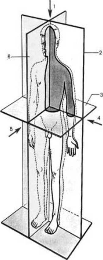
Figure 1. Axes and Planes of the human body.
Lines.
1. anterior median line (along the anterior median surface);
2. posterior median line (runs along THE Vertebral Column over the tips of the vertebral spinous processes);
3. sternal line - along the edge of the Sternum;
4. midclavicular line - through the midpoint of the clavicle and the nipple;
5. anterior axillary line - the anterior point of the axillary fold;
6. midaxillary line - the deepest point of the axillary fossa;
7. posterior axillary line - the posterior point of the axillary fold;
8. scapular line - through the inferior angles of the scapulae;
9. paraspinal line - along the vertebral column through the costovertebral joints.
1. What are the known branches of anatomy?
2. What language is used for the international anatomical nomenclature?
3. What axes are used in anatomy to describe joint movements?
4. What planes are used in anatomy to describe the position of organs and their parts?
5. What lines are used in anatomy to describe the Location of organs?
The Cell is the fundamental and ordered structural, developmental, and functional unit of all living systems. Sizes range from 2-3 µm to 200-300 µm. Cell processes can reach up to 1.5 m in length (for example, in Nerve Cells). In the human body, cells vary in shape, size, structure, and functional significance.
Cell Structure (Figure 2).
1. membrane - Lipids, Proteins;
2. Nucleus - hereditary apparatus;
3. Cytoplasm;
4. Golgi apparatus;
5. Mitochondria;
7. Ribosomes;
8. Lysosomes.
Methods of cellular research.
Light and Electron Microscopy.
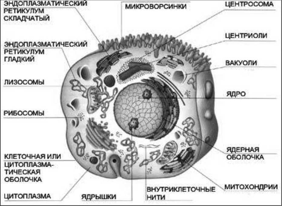
Figure 2. Cell Organelles.
Tissues
A tissue is an established system of cells united by a common origin, structure, and function.
Types of tissues (Figure 3).
I. epithelial;
II. connective;
III. muscular;
IV. nervous.
I. Epithelial Tissues form a sheet of cells resting on a basement membrane. Skin, glands, and all mucous membranes.
Epithelium:
1. simple: columnar, cuboidal, squamous;
2. stratified: keratinized, non-keratinized, transitional.
Functions: protective, secretory.

Figure 3. Types of tissues.
II. Connective Tissue.
Consists of cells and an Extracellular matrix composed of Collagen, elastic, and reticular fibers, as well as ground substance.
Functions: supporting, protective, trophic.
Types of connective tissue.
✵ loose connective tissue (accompanying Blood Vessels, contains more cells and fewer fibers);
✵ Dense connective tissue (contains more fibers and fewer cells):
- irregular (contains more elastic fibers, forms the dermis, periosteum, and Heart Valves);
- regular (ligaments, tendons).
2. Cartilage tissue consists of chondrocytes and a gel-like extracellular matrix.
Types of cartilage:
✵ hyaline (transparent, gel-rich, coats joint surfaces and epiphyses);
✵ elastic (rich in elastic fibers, yellowish, resilient; found in the laryngeal cartilages, auricle, and epiglottis);
✵ fibrocartilage (fiber-rich, forms the annulus fibrosus of intervertebral discs).
3. Bone tissue. It is composed of bone cells (osteocytes, osteoblasts, osteoclasts) and an intercellular matrix consisting of organic and inorganic substances.
Function: structural support.
Blood consists of plasma and formed elements (Blood Cells).
Erythrocytes: 4.5 million/mm3 (4.5–5×10¹²/L). Primary function: Oxygen transport and gas exchange.
Leukocytes: 4,000–9,000/mm3 (4–9×10⁹/L). Primary function: immune defense.
Leukocytes:
- granulocytes: neutrophils, basophils, eosinophils
- agranulocytes: monocytes, lymphocytes
Platelets (thrombocytes): 150,000/mm3 (150–300×109/L). Primary function: blood clotting.
Lymph is a whitish, turbid fluid containing lymphocytes and monocytes.
III. Muscular tissue. The basis of movement.
1. Smooth Muscle (found in hollow organs such as Bronchi, Esophagus, Stomach, intestines, Urinary Bladder, blood vessels, and Ureters). Contracts slowly and rhythmically.
2. Striated cardiac muscle (myocardium of The Heart walls).
3. Striated Skeletal Muscle (skeletal Muscles).
IV. Nervous tissue.
It consists of nerve cells (Neurons) and neuroglia.
Neurons consist of a cell body and processes. The longest process is called the axon, while the shorter ones are called dendrites.
Organ Systems and apparatuses.
An organ is a part of the body with a specific shape, structure, location in the body, and function. It consists of various tissues.
An organ system consists of organs that perform a common function and share a common embryonic origin. Examples include the digestive, respiratory, cardiovascular, urogenital, lymphatic, and nervous systems.
Organ apparatuses (which have a heterogeneous origin) include: the musculoskeletal apparatus (Skeleton, muscles) and the endocrine apparatus.
In sports anatomy, organs and systems are divided into three groups:
I. Organs of movement execution
II. Organs and systems supporting motor activity
III. Organs and systems regulating and controlling motor activity
1. The Musculoskeletal System. It consists of the Skeleton and Muscles. The skeleton includes Bones and Their Joints.
Bones (Osteology). Over 200 bones in total. About 30 are unpaired, and the rest are paired. 29 in the Skull, 26 in the vertebral column, 25 in the Ribs AND STERNUM, 64 in the upper extremities, and 62 in the lower extremities. The Axial Skeleton comprises The Skull and the trunk skeleton (Vertebral Column and rib cage). The Appendicular Skeleton consists of the limbs.
II. 1. Digestive System — a long muscular tube, with The Liver and Pancreas as its Major Digestive Glands. It ensures the intake of nutrients into the body and their Processing.
2. Respiratory system. It ensures the intake of oxygen into the body and the elimination of carbon dioxide.
3. Cardiovascular system (heart and blood vessels: Arteries, Veins, capillaries; 2 circuits). It delivers oxygen and nutrients to tissues and organs while removing Metabolic waste products from them.
4. Urogenital System. It ensures The excretion of metabolic waste products from the body as well as reproductive functions.
III. 1. Nervous system: central (Brain, Spinal Cord) and peripheral.
It is also subdivided into two divisions:
✵ somatic
✵ autonomic:
- sympathetic
- parasympathetic
2. Endocrine System: Pineal Gland, Pituitary Gland, Hypothalamus, Thymus, Adrenal Glands, Thyroid Gland, Parathyroid glands, pancreas, and Gonads.
3. Sense Organs: Vision, Hearing, smell, Touch, taste.
1. What are the main organelles present in the cell?
2. Characterize the MAIN TYPES OF tissues.
3. Into which groups are all organ systems classified according to their relation to locomotor activity?
Last update: 08/08/2026
Editorial and Educational Adaptation: This material has been compiled based on the primary/original source text. The project team performed an editorial review, corrected technical inaccuracies, structured sections, and adapted the content for an educational format.
What was processed:
- elimination of formatting defects (OCR errors, structural breaks, corrupted characters);
- editorial organization of content;
- standardization of terminology in accordance with academic sources;
- verification of factual statements against the original source text.
All mentions of the author, publication year, and origin of the primary text have been preserved in accordance with the source.