MEDICAL BIOLOGY, HUMAN ANATOMY, PHYSIOLOGY AND PATHOLOGY - Ya.I. Fedonyuk 2010
ANATOMY, PHYSIOLOGY, PATHOLOGY
CHAPTER 8. URINATION PROCESS AND ITS PATHOLOGY
ANATOMY AND PHYSIOLOGY OF THE URINARY ORGANS
MACROSCOPIC AND MICROSCOPIC STRUCTURE OF THE KIDNEY
The Kidney (lat. *ren*) is a paired organ located in the lumbar region on the posterior wall of the Abdominal cavity, in a retroperitoneal position. The right and left Kidneys lie at the level of the XII thoracic and I-II lumbar vertebrae. The right kidney is situated slightly lower than the left one (Fig. 8.1).
Class="center">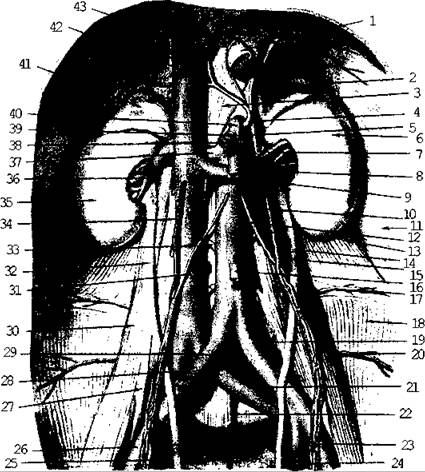
Fig. 8.1. Topography of the kidneys:
1 - Esophagus; 2 - left Adrenal gland; 3 - lumbar part of the Diaphragm; 4 - inferior phrenic artery; 5 - splenic artery; 6 - left kidney; 7 - left renal artery; 8 - superior mesenteric artery; 9 - left renal vein; 10 - left testicular artery; 11 - transverse abdominal Muscle; 12 - Abdominal Aorta; 13 - quadratus lumborum muscle; 14 - left Ureter; 15 - left testicular vein; 16 - inferior mesenteric artery; 17 - IV lumbar artery; 18 - iliac muscle; 19 - left common iliac artery; 20 - iliolumbar artery; 21 - left common iliac vein; 22 - median sacral artery; 23 - left external iliac artery; 24 - left internal iliac artery; 25 - right internal iliac artery; 26 - right external iliac artery; 27 - psoas minor muscle; 28 - right common iliac vein; 29 - right common iliac artery; 30 - psoas major muscle; 31 - lumbar Arteries; 32 - right ureter; 33 - right testicular artery; 34 - right testicular vein; 35 - right kidney; 36 - right renal vein; 37 - right renal artery; 38 - common hepatic artery; 39 - left gastric artery; 40 - celiac trunk; 41 - right adrenal gland; 42 - INFERIOR VENA CAVA; 43 - hepatic Veins.
External Structure. The kidney is bean-shaped. It has a convex anterior surface (Fig. 8.2) and a flat posterior surface (Fig. 8.3), a rounded superior pole and a pointed inferior pole, a concave medial margin and a convex lateral margin. The renal hilum is located on the medial margin, through which the renal artery and nerves enter the organ, while the ureter, renal vein, and Lymphatic vessels exit it. All these anatomical structures form the renal pedicle, which fixes the organ in a specific anatomical position. In addition to this, the fixation apparatus includes: the fibrous capsule closely adhering to the renal parenchyma, followed by the adipose capsule, and externally the renal fascia, which has anterior and posterior layers. The kidney lies within a muscular bed formed by the psoas major and quadratus lumborum Muscles.
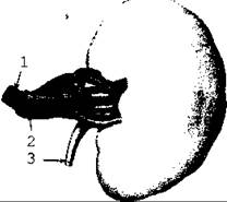
Fig. 8.2. Anterior surface of the kidney: 1 - renal artery; 2 - renal vein
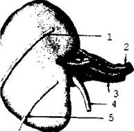
Fig. 8.3. Posterior surface of the kidney: 1 - subcostal nerve; 2 - renal artery; 3 - renal vein; 4 - ureter; 5 - iliohypogastric nerve (diagram)
A frontal section of the kidney reveals that the renal sinus is located in the region of its medial margin. It is filled with Blood Vessels, nerves, and adipose tissue; furthermore, it contains minor and major calyces that open into the renal pelvis.
The renal parenchyma consists of the cortex (Fig. 8.4) located peripherally, and the medulla, which has the appearance of renal pyramids. The bases of the pyramids face The surface of the kidney, while their apices face the sinus. The apices of the pyramids join in pairs or groups to form elevations known as renal papillae. There are about 12 papillae in total in the kidney, each containing orifices through which urine is discharged into the minor calyces. Renal tissue extends between the pyramids to form renal columns. The medullary tissue projects into the cortex at the Base of the pyramids, forming medullary rays.
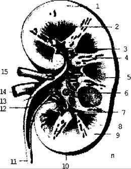
Fig. 8.4. Frontal section of the kidney
Microscopically, the kidney is a complex tubular gland, the tubules of which are called renal (uriniferous) tubules. The blind end of each renal tubule forms a double-walled cup—the glomerular capsule (Bowman's capsule)—which encloses a tuft of blood capillaries and closely adheres to them (Fig. 8.5). This allows Water and dissolved substances to easily pass into the capsular space. The glomerulus together with the capsule constitutes the renal corpuscle, which is located in the renal cortex. The structural morphofunctional unit of the kidney is the nephron (Fig. 8.6 - see Appendix). It consists of a renal corpuscle, from which extend the proximal convoluted tubule, the nephron loop (Loop of Henle), and the distal tubule. The nephron is the site of Urine Formation. The distal segments of the nephrons empty into collecting ducts. These tubules lie within the pyramids and transport urine into the minor calyces, opening at the apices of the pyramids. From there, urine flows into the major calyces, and then into the renal pelvis. The pelvis continues as the ureter, which empties into the Urinary Bladder.
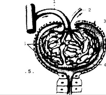
Fig. 8.5. Microscopic STRUCTURE OF THE kidney
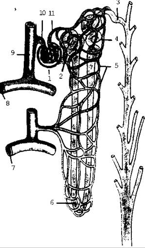
Fig. 8.6. Structure of the nephron and its connections with blood vessels (diagram)
Blood Circulation in the kidney. Arterial blood is brought to the kidney by the renal artery (Fig. 8.7), which divides into 4-5 branches at the hilum. Each branch supplies a specific area known as a renal segment. Interlobar arteries branch off from these vessels and form arches at the corticomedullary junction. The latter give rise to short afferent arterioles that form capillary glomeruli (glomerular capillaries). Efferent arterioles emerge from each glomerulus to form a capillary network around the tubules. Veins originate from this network, running alongside the corresponding arteries and subsequently merging into the renal veins, which empty into the inferior vena cava. Thus, There are two capillary networks in the kidney: blood passes from an artery into capillaries and then into another arteriole. This specific pattern of arterial branching is known as the "miraculous network" (rete mirabile) or portal System of the kidney.
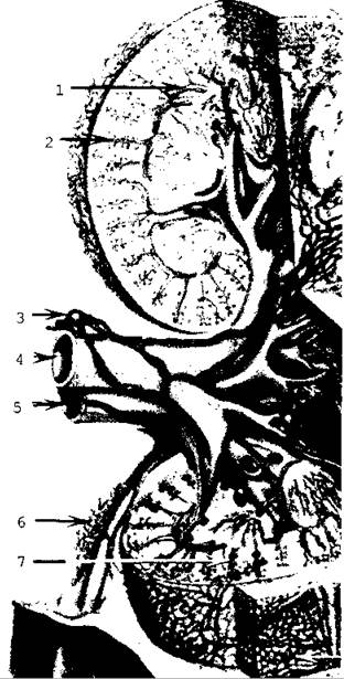
Fig. 8.7. Blood vessels of the kidney (diagram):
1 - arcuate artery and vein; 2 - interlobular vein; 3 - regional Lymph node; 4 - renal vein; 5 - renal artery; 6 - Blood and Lymphatic Vessels of the ureter; 7 - renal corpuscles.
Last update: 08/08/2026
Editorial and Educational Adaptation: This material has been compiled based on the primary/original source text. The project team performed an editorial review, corrected technical inaccuracies, structured sections, and adapted the content for an educational format.
What was processed:
- elimination of formatting defects (OCR errors, structural breaks, corrupted characters);
- editorial organization of content;
- standardization of terminology in accordance with academic sources;
- verification of factual statements against the original source text.
All mentions of the author, publication year, and origin of the primary text have been preserved in accordance with the source.