Human Anatomy - Kotsan I. Y. 2009
Splanchnology
Major Digestive Glands
Liver
The Liver (hepar) is the largest gland in The Human Body, with its mass in adults reaching 1.5–2 kg. The liver performs A wide variety of Functions. First and foremost, it carries out a detoxifying function, neutralizing toxic substances formed in the intestine that enter the liver via the bloodstream. As a digestive gland, the liver produces Bile, which enters the intestine and takes part in fat Digestion. In addition, the liver synthesizes urea and Proteins, and converts glucose into Glycogen, which is stored within it. The liver is also involved in METABOLISM and in the phagocytosis of dead erythrocytes coming from the Spleen, as well as microorganisms and other Cells. A significant volume of Blood is pooled in the liver, and during the Embryonic period, it also acts as a site for The formation of Blood Cells (erythrocytes).
The liver has a reddish-brown color and a soft consistency. In shape, it resembles a large mushroom cap or a wedge. It is located primarily in the upper right region of the Abdominal cavity. The Diaphragm lies superior to it, The Stomach is on the left, and the right Kidney, right Adrenal gland, duodenum, and INFERIOR VENA CAVA are located inferiorly and to the right. Almost the entire surface of the liver is covered by the Peritoneum, with the exception of the fossa for the Gallbladder, the porta hepatis, and its posterior border, where it fuses directly with the diaphragm. Beneath the serous membrane (peritoneum) lies the fibrous capsule. It covers the entire liver as well and is particularly well-developed in areas where the serous membrane is absent. THE POSITION OF the liver is secured by ligaments (falciform, coronary, and triangular) and Blood Vessels. Furthermore, intra-abdominal pressure and the partial attachment of the liver to the diaphragm play a major role in stabilizing its position.
The liver features two surfaces: the diaphragmatic (superior) surface and the visceral (inferior) surface (Fig. 156).
The diaphragmatic surface (facies diaphragmatica) of the liver is convex, directed upward and forward, and lies against the Inferior surface of the diaphragm. Extending from the diaphragm and the anterior abdominal wall to the diaphragmatic surface of the liver is the falciform ligament (lig. falciforme hepatis), which is a peritoneal duplication (double fold). Situated in the sagittal plane, this ligament divides the diaphragmatic surface of the liver into two lobes: the right and left lobes (lobus hepatis dexter and lobus hepatis sinister). The right lobe of the liver is significantly larger than the left. Posteriorly, the falciform ligament connects to the coronary ligament (lig. coronarium), which is also a peritoneal fold transitioning from the diaphragm to the posterior border of the liver. The coronary ligament lies in the frontal plane. The right and left ends of the coronary ligament expand into a triangular shape, forming the right and left triangular ligaments (lig. triangulare dextrum and lig. triangulare sinistrum). On the diaphragmatic surface of the left lobe of the liver, There is a small cardiac impression (impressio cardiaca), formed As a result of The Heart pressing against the diaphragm and, consequently, against the liver.
Class="center">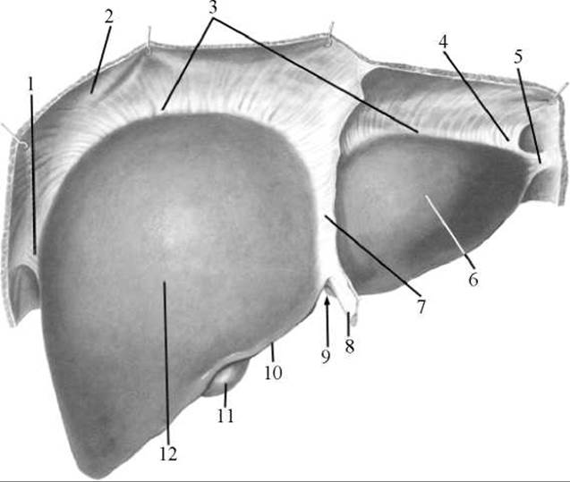
Fig. 156. The liver. Diaphragmatic surface
1 — right triangular ligament; 2 — diaphragm; 3 — coronary ligament; 4 — left triangular ligament; 5 — fibrous appendage of the liver; 6 — left lobe; 7 — falciform ligament; 8 — round ligament; 9 — notch of the round ligament; 10 — inferior border; 11 — Cytology/practical/108.html">Fundus of the gallbladder; 12 — right lobe
The visceral surface (facies visceralis) of the liver is somewhat concave, directed downward and posteriorly, and comes into contact with Internal Organs. Three sulci (grooves) are distinguished on it: two sagittal and one transverse. The sagittal grooves run anteroposteriorly, while the transverse groove connects their midpoints like a crossbar. The right sagittal groove is wider than the left; in its anterior part, it forms the fossa of the gallbladder (fossa vesicae biliaris), and in its posterior part, the sulcus for the inferior vena cava (sulcus venae cavae). The gallbladder is lodged within the gallbladder fossa, and the inferior vena cava passes through the sulcus of the inferior vena cava. In its anterior part, the left sagittal groove forms the fissure for the round ligament (fissura ligamenti teretis), and in its posterior part, the fissure for the ligamentum venosum (fissura ligamenti venosi). The first fissure contains the round ligament of the liver (lig. teres hepatis), which extends anteriorly and inferiorly from the umbilicus and contains the obliterated (obliterated) umbilical vein. The fissure of the ligamentum venosum contains the ligamentum venosum (lig. venosum), which is the fibrous remnant of the ductus venosus that connected the umbilical vein to the inferior vena cava in the fetus. The transverse groove is the region through which the portal vein, proper hepatic artery, and nerves enter the liver, while the common bile duct and Lymphatic vessels exit. This area is called the porta hepatis (Fig. 157).
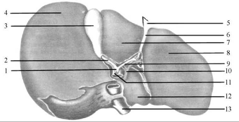
Fig. 157. The liver. Visceral surface
1 — common bile duct; 2 — cystic duct; 3 — gallbladder; 4 — right lobe; 5 — peritoneal fold; 6 — round ligament of the liver; 7 — quadrate lobe; 8 — left lobe; 9 — common hepatic duct; 10 — hepatic artery; 11 — portal vein; 12 — caudate lobe; 13 — inferior vena cava
The three grooves described divide the visceral surface of the liver into four lobes: the right lobe (lobus hepatis dexter), located to the right of the right sagittal groove; the left lobe (lobus hepatis sinister), located to the left of the left sagittal groove; the quadrate lobe (lobus quadratus), bounded posteriorly by the porta hepatis and laterally by the right and left sagittal grooves; and the caudate lobe (lobus caudatus), which is also situated between the right and left sagittal grooves, but posterior to the porta hepatis.
The visceral surface of the liver is in contact with several organs, resulting in the formation of impressions upon it. The left lobe of the liver bears the gastric impression (impressio gastrica), marking the contact site of the anterior surface of the stomach. The esophageal impression (impressio oesophagea) is noticeable on the posterior part of the left lobe. Transverse across the quadrate lobe and part of the right lobe adjacent to the gallbladder fossa lies the duodenal impression (impressio duodenalis). To the right of it on the right lobe is the renal impression (impressio renalis), and more to the left, near the sulcus for the inferior vena cava, is the suprarenal impression (impressio suprarenalis). Near the inferior border of the right lobe is the colic impression (impressio colica), formed by the contact of the right colic flexure.
At the junction of the diaphragmatic and visceral surfaces of the liver anteriorly, a sharp inferior border of the liver (margo inferior hepatis) is formed, whereas posteriorly, a rounded posterior border of the liver (margo posterior hepatis) is found. The posterior border is so obtuse that it can be regarded as the posterior surface of the liver (facies posterior hepatis).
Structure OF THE liver. Externally, the liver (except for a small area in the posterior part known as the bare area [area nuda]) is covered by a serous membrane (tunica serosa), which is represented by the visceral peritoneum. It can be considered that the liver is located intraperitoneally with respect to the peritoneum, although some authors consider its position to be mesoperitoneal. Beneath the serous membrane lies a thin, dense fibrous capsule (tunica fibrosa) (Glisson's capsule), which consists of elastic and collagenous fibers. At the porta hepatis, the fibrous capsule extends deep into the substance of the organ as a perivascular fibrous capsule (capsula fibrosa perivascularis), accompanying the hepatic blood vessels along all their branches down to their terminal endings.
In terms of internal structure, the liver is a complex branched tubular gland whose excretory ducts are the bile ducts. The structural unit of the liver is the prismatic hepatic lobule (lobus hepatis), through the center of which runs the central vein (Fig. 158). Each lobule consists of hepatic trabeculae (plates or cords) formed by two rows of epithelial secretory cells (hepatocytes), between which runs a narrow channel—the intralobular bile ductule (ductus biliferi), representing the initial segment of the biliary tract.
The trabeculae radiate from the center of the lobule toward its periphery. Bile produced by hepatocytes enters the intralobular ductule, from which it flows into interlobular ductules (ductuli interlobulares) located at the periphery of the lobules.
By fusing together, the interlobular ductules form larger ducts that ultimately give rise to the right and left hepatic ducts (ductus hepaticus dexter and ductus hepaticus sinister). At the porta hepatis, these two ducts merge to form the common hepatic duct (ductus hepaticus communis). The common hepatic duct unites with the cystic duct (ductus cysticus) to form the common bile duct (ductus choledochus), which empties into the duodenum either independently or after previously joining the pancreatic duct.
Lately, it is believed that the primary morphofunctional unit of the liver is not the classical hepatic lobule, but rather the hepatic acinus, which includes adjacent portions of two classical lobules. It has a rhombus-like shape, with its acute angles located near the central Veins and one of its obtuse angles—the portal zone—situated near the vessels from which the perilobular branches originate at a more or less right angle. From these vessels, sinusoidal vessels extend in both directions toward the central veins.
Numerous blood vessels run between the hepatic trabeculae, as well as externally and internally within the hepatic lobule. Blood enters the liver via the hepatic artery and portal vein. Venous blood arriving via the portal vein from the stomach, intestine, and spleen undergoes purification in the liver to remove harmful chemical impurities. As it flows through the sinusoidal capillaries located between the hepatic trabeculae, the blood interacts with hepatocytes, which absorb certain substances from the blood and secrete others into it. In this manner, the blood is purified and alters its composition. The purified blood enters the central vein running through the center of the hepatic lobule. From the central veins, blood flows into the hepatic veins, which direct it into the inferior vena cava. The liver is the only organ in the entire body where a vein enters and divides into capillaries, only to reunite into veins once again. Such capillary networks occupying an unusual position between two veins (in this case, the portal vein and hepatic veins) or two Arteries (as occurs in the Kidneys) are called portal systems or marvelous networks (rete mirabile).
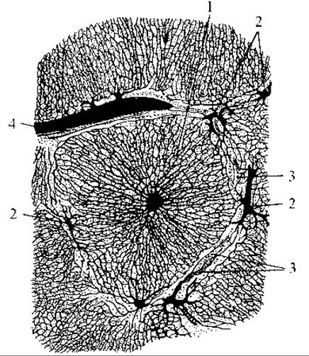
Fig. 158. Hepatic lobules
1 — hepatic lobule shown entirely in cross-section (the central vein is in the middle of the lobule); 2, 4 — interlobular veins; 3 — interlobular bile ductule
Due to advancements in surgery and hepatology, a theory of the segmental structure of the liver has been developed in modern times, superseding the earlier concept of division solely into lobes and lobules.
Five tubular systems are distinguished in the liver: 1) the biliary tract; 2) the arteries; 3) the Branches of the portal vein (portal system); 4) the hepatic veins (caval system); and 5) the lymphatic vessels. The portal and caval venous systems do not coincide with each other, whereas the remaining tubular systems accompany the branches of the portal vein, running parallel to one another and forming neurovascular bundles accompanied by nerves. Some of the lymphatic vessels exit together with the hepatic veins.
A hepatic segment is defined as the area of hepatic parenchyma surrounding a third-order branch of the portal vein, along with its adjacent branch of the hepatic artery and bile duct.
The liver is traditionally divided into eight segments (moving counterclockwise, starting from the sulcus of the inferior vena cava):
I — caudate segment of the left lobe (corresponds to the caudate lobe of the liver);
II — posterior segment of the left lobe (located in the posterior region of the left lobe);
III — anterior segment of the left lobe (located in its anterior region);
IV — quadrate segment of the left lobe (corresponds to the quadrate lobe of the liver);
V — medial superior-anterior segment of the right lobe;
VI — lateral inferior-anterior segment of the right lobe;
VII — lateral inferior-posterior segment of the right lobe;
VIII — medial superior-posterior segment of the right lobe.
Grouping radially around the porta hepatis, these segments form larger independent Regions of the liver known as zones, or sectors. Each sector represents a liver region supplied by a second-order branch of the portal vein, its corresponding branch of the hepatic artery, and nerves, and drained by a sectorial bile duct. There are five such sectors:
1. Left dorsal sector (corresponds to segment I).
2. Left lateral sector (corresponds to segment II).
3. Left medial sector (formed by segments III and IV).
4. Right medial sector (formed by segments V and VIII).
5. Right lateral sector (includes segments VI and VII).
Blood supply to the liver is provided by branches of the proper hepatic artery (a. hepatica propria), which originates from the celiac trunk. Additionally, the liver receives venous blood from the portal vein (v. portae). The proper hepatic artery delivers oxygenated arterial blood, whereas the portal vein carries venous blood from the stomach, small and large intestines, Pancreas, and spleen. Within the liver, the proper hepatic artery and portal vein branch into interlobular arteries (aa. interlobulares) and interlobular veins (vv. interlobulares). These vessels lie between the hepatic lobules alongside interlobular bile ductules. Wide interlobular sinusoidal capillaries branch off from the interlobular veins into the lobules, running between the hepatic trabeculae (plates) and emptying into the central vein. Arterial capillaries originating from the interlobular arteries empty into the initial segments of these sinusoidal capillaries. The central VEINS OF THE hepatic lobules unite to form sublobular (collecting) veins (vv. sublobulares), which ultimately converge into 2–3 large and several smaller hepatic veins that exit the liver near the groove for the inferior vena cava and drain into the inferior vena cava.
There are no lymphatic capillaries within the hepatic lobules; they are concentrated exclusively in the perivascular fibrous capsule. Efferent lymphatic vessels drain into the hepatic, celiac, right lumbar, diaphragmatic, and posterior mediastinal Lymph Nodes.
Innervation of the liver is supplied by branches of the vagus nerves and the hepatic (sympathetic) plexus.
The gallbladder (vesica biliaris) is a pear-shaped organ, 8–12 cm long, 4–5 cm wide, with a capacity of 40–60 ml (Fig. 159). It acts as a reservoir where bile is accumulated and stored. The gallbladder is situated in the fossa for the gallbladder on the visceral surface of the liver. Anatomically, it is divided into the fundus (fundus vesicae biliaris), body (corpus vesicae biliaris), and neck (collum vesicae biliaris).
The neck of the gallbladder gradually transitions into the cystic duct (ductus cysticus). Upon joining the common hepatic duct, the cystic duct forms the common bile duct (ductus choledochus (biliaris)). The common bile duct runs within the hepatoduodenal ligament alongside the proper hepatic artery and portal vein, passes between the descending part of the duodenum and the HEAD of the pancreas, pierces the medial wall of the descending duodenum, and opens at the apex of the major duodenal papilla after uniting with the pancreatic duct. Upon the merging of these ducts within the duodenal wall, an expansion is formed—the hepatopancreatic ampulla (ampulla hepato-pancreatica). Its orifice is guarded by a sphincter—the sphincter of the hepatopancreatic ampulla (m. sphincter ampullae hepatopancreaticae)—which regulates the flow of bile from The Liver and gallbladder into the duodenal lumen and prevents duodenal contents from refluxing into the common BILE AND PANCREATIC ducts. Just proximal to the junction of the common bile duct and the pancreatic duct, the circular Muscle layer of the common bile duct wall thickens to form the sphincter of the common bile duct (m. sphincter ductus choledochi), which controls the influx of bile into the duodenum. During fasting, these sphincter Muscles remain contracted, directing the continuously produced hepatic bile into the gallbladder. Inside the gallbladder, bile is not only stored and concentrated (becoming denser and more viscous) due to Water absorption, but its composition is also modified. Food intake triggers reflex contraction of the gallbladder and relaxation of the sphincters, the strength of these responses depending on the nutrient COMPOSITION OF THE meal (such as its protein or fat content).
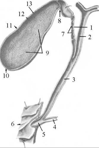
Fig. 159. Gallbladder and biliary ducts (longitudinal section)
1 — cystic duct; 2 — common hepatic duct; 3 — common bile duct; 4 — pancreatic duct; 5 — hepatopancreatic ampulla; 6 — duodenum; 7 — spiral fold; 8 — neck of the gallbladder; 9 — folds of the mucous membrane; 10 — fundus of the gallbladder; 11 — body of the gallbladder; 12 — muscular coat; 13 — mucous membrane
The wall of the gallbladder consists of three layers: 1) an outer serous coat (tunica serosa); in areas where the serosa is absent, the outer layer is represented by the adventitia;
2) the muscular layer (tunica muscularis), which consists of Smooth Muscle tissue (predominantly with a circular orientation of fibers);
3) the mucous membrane (tunica mucosa), which is lined with a simple columnar epithelium featuring a microvillar brush border capable of intensive water absorption. As a result, gallbladder bile is 3 to 5 times more concentrated than bile from the common hepatic duct. The mucosa of the gallbladder forms numerous folds that give the inner surface of its wall a reticular appearance. In the neck of the bladder and in the cystic duct, the mucosa forms a spiral fold (plica spiralis), The structure of which facilitates the flow of bile into the gallbladder (when the sphincters of the bile duct and ampulla are contracted and prevent bile from entering the duodenum).
The gallbladder receives its blood supply from the cystic artery (a branch of the proper hepatic artery). Venous blood drains through the corresponding vein.
The gallbladder is innervated by branches of the vagus nerves and the hepatic plexus.
Last update: 08/08/2026
Editorial and Educational Adaptation: This material has been compiled based on the primary/original source text. The project team performed an editorial review, corrected technical inaccuracies, structured sections, and adapted the content for an educational format.
What was processed:
- elimination of formatting defects (OCR errors, structural breaks, corrupted characters);
- editorial organization of content;
- standardization of terminology in accordance with academic sources;
- verification of factual statements against the original source text.
All mentions of the author, publication year, and origin of the primary text have been preserved in accordance with the source.