Harper's Illustrated Biochemistry, Volume 1 - Murray R. 1993
Structure and Functions of Proteins and Enzymes
Amino Acids
Properties of Individual Amino Acids
Glycine, the smallest of the Amino Acids, can fit into Regions of the Three-Dimensional Cell/13.html">Protein Structure that are inaccessible to Other Amino Acids.
The aliphatic R-groups of Alanine, valine, leucine, and isoleucine, as well as the aromatic R-groups of phenylalanine, Tyrosine, and Tryptophan, are hydrophobic. This property has a critical consequence: The formation of an ordered Hydration shell of Water molecules around the protein surface where nonpolar R-groups are exposed. The charged R-groups of basic and acidic amino acids play a vital role in stabilizing specific protein Conformations through salt bridge formation. In addition, amino acids with positively and negatively charged R-groups, along with Histidine, can participate in charge-transfer systems that facilitate long-range charge migration during Enzymatic Catalysis. Finally, histidine plays a unique and essential role in enzymatic catalysis—the pK of its imidazole group is such that at pH = 7, this amino acid can act alternately as a general acid or general base catalyst.
The primary alcoholic group of Serine and the primary thiol (—SH) group of Cysteine serve as potent nucleophiles under certain conditions and participate in enzymatic catalysis. Although the secondary alcoholic group of Threonine is also a nucleophile, there is no evidence for its potential catalytic role. Besides its catalytic function, the —OH group of serine is involved in regulating The activity of several key metabolic Enzymes whose activity depends on the phosphorylation of specific serine residues.
Amino acids do not absorb light in the visible spectrum (in other words, they are colorless). With the exception of the aromatic amino acids tryptophan, tyrosine, phenylalanine, and histidine, they do not absorb in the ultraviolet region at wavelengths above 240 nm either. As seen in Fig. 3.8, protein absorption in this region is primarily due to tryptophan.
Class="center">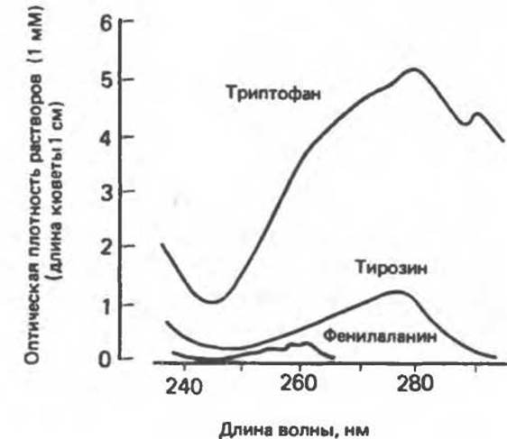
Fig. 3.8. UV absorption spectra of tryptophan, tyrosine, and phenylalanine.
Last update: 06/08/2026
Editorial and Educational Adaptation: This material has been compiled based on the primary/original source text. The project team performed an editorial review, corrected technical inaccuracies, structured sections, and adapted the content for an educational format.
What was processed:
- elimination of formatting defects (OCR errors, structural breaks, corrupted characters);
- editorial organization of content;
- standardization of terminology in accordance with academic sources;
- verification of factual statements against the original source text.
All mentions of the author, publication year, and origin of the primary text have been preserved in accordance with the source.