Biochemistry of Amino Acids - A. Meister 1961
Naturally Occurring Amino Acids
Amino Acids Commonly Found in Proteins
Introduction
"Upon closer investigation of plant-derived products, modern chemists have discovered numerous compounds unknown to earlier researchers; yet it has been a long time, I believe, since a compound as exceptional and interesting as the one we are now examining was found in plants... In a quantity of asparagus juice concentrated by evaporation, I discovered a quite large number of crystals, Two Types of which, it seems to me, belong to new substances; since these crystals differed in shape, transparency, and taste, I had no difficulty in separating them." Vauquelin (1806).
There are 22 Amino Acids regularly or frequently found in protein hydrolysates. The discovery of these amino acids was a major milestone in The Development of modern biochemistry. More detailed information on The history of the fascinating foundational works that led to the discovery of these amino acids is presented in the review by Vickery and Schmidt [1]. According to these authors, the presence of a given amino acid in protein hydrolysates can be considered established only if it has been isolated by at least two independent researchers and if its Structure has been confirmed by synthesis. These criteria remain valid today when evaluating reports on newly discovered amino acids. At the same time, they cannot be considered infallible, as evidenced by the mere fact that Vickery and Schmidt themselves made a mistake by classifying β-hydroxyglutamic acid among the "recognized" products of Protein Hydrolysis (p. 88). Norleucine was also classified among the "recognized" amino acids for a certain period.
The amino acids discussed below have been repeatedly isolated from protein hydrolysates, and their structures have been definitively established. This list includes all amino acids enumerated in 1931 by Vickery and Schmidt, with the exception of β-hydroxyglutamic acid and iodine-containing amino acids, as well as asparagine, glutamine, Cysteine, and Threonine. It should be noted that the expression "commonly detected in hydrolysates amino acids" used in the text is somewhat arbitrary and allows for some divergence in interpretation. For instance, it is known that 3,5-diiodotyrosine and thyroxine are present in thyroglobulin; the presence of 8-hydroxylysine in certain Proteins has been precisely established. These Amino Acids and several others less commonly found in protein hydrolysates will be discussed in the relevant section (p. 62).
The amino acids discussed in this section are presented in alphabetical order. They can be classified as follows:
Aliphatic amino acids
Monoaminomonocarboxylic
Isoleucine
Leucine
Valine
Hydroxymonaminomonocarboxylic
Threonine
Monoaminodicarboxylic
Aspartic acid
Glutamic acid
Amides of Monoaminodicarboxylic Acids
Asparagine
Glutamine
Diaminomonocarboxylic
Sulfur-containing
Cysteine and cystine
Aromatic amino acids
Phenylalanine
Heterocyclic amino acids
Class="center">L-Alanine (α-aminopropionic acid)
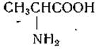
Alanine is among those amino acids that were first obtained synthetically and only later recognized as natural products. In 1850, Strecker [2], attempting to synthesize lactic acid, treated a Condensation product of acetaldehyde and ammonia with hydrogen cyanide and Hydrochloric acid; the resulting crystalline alanine was converted into lactic acid by Treatment with nitrous acid. The Strecker reaction leads to The formation of an aminonitrile, which upon hydrolysis yields the corresponding amino acid; this reaction proved to be applicable for the synthesis of A number of Other Amino Acids from their respective aldehydes. Thirty-eight years after Strecker synthesized alanine, Weyl [3] isolated this amino acid from the acid hydrolysate of silk, a protein exceptionally rich in alanine. Later, Fischer and Skita [4] obtained L-alanine from silk and established its structure and configuration by converting it into lactic acid.
L-Arginine (α-amino-δ-guanidinovaleric acid)
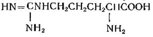
Schulze and Steiger [5] isolated arginine from etiolated lupine seedlings in 1866. In 1895, Hedin [6] reported the Isolation of the silver-nitrogen salt of arginine from horn hydrolysates. Subsequently, Kossel and Gross [7] found that arginine is one of the primary constituents of basic fish sperm proteins. The structure of arginine was established by its alkaline hydrolysis to Ornithine and urea [8] and by synthesis from benzoylornithine [9].
During the acid Hydrolysis of Proteins, arginine can be cleaved to ornithine, which is consequently sometimes detected in protein hydrolysates. The conversion of arginine to citrulline during alkaline hydrolysis has also been described [10]. Upon treatment with 1-naphthol and sodium hypochlorite or hypobromite, arginine yields a red color (a reaction first described by Sakaguchi [11]). Arginine can be precipitated from solutions as the mono- or diflavianate [7]; this reaction is used for the isolation of arginine from protein hydrolysates.
Arginine is not only a constituent of proteins but also occurs in the free state and as phosphoarginine

in invertebrate Muscles, where this compound performs a function analogous to that of phosphocreatine in higher animals [12, 13]. In addition, arginine is a component of octopine (p. 57) and argininosuccinic acid (p. 339). A chemically related compound, canavanine, was isolated from Canavalia beans (p. 49)
L-Asparagine (β-amide of α-aminosuccinic acid)

Asparagine was the first amino acid to be isolated from natural products. In 1806, Vauquelin and Robiquet [14] obtained it from asparagus juice. Acid hydrolysis of proteins leads to the deamidation of asparagine into aspartic acid. The presence of ammonia in acidic protein hydrolysates led Hlasiwetz and Habermann in 1873 [15] to suggest that the amide groups of glutamine and asparagine might serve as the source of this ammonia. However, the presence of asparagine in proteins was proven only in 1932, when Damodaran [16] described the isolation of asparagine from enzymatic hydrolysates of edestin.
Asparagine is a widely distributed compound; it accumulates in significant concentrations in certain species of Higher Plants and is also found in the free state in animal Tissues. During the acid Cleavage of proteins, asparagine undergoes hydrolysis; however, its amide group is relatively more stable than that of glutamine.
Unlike Most amino acids, which react with ninhydrin to form a purple-colored product, asparagine and certain other β-aspartyl derivatives yield a brown color upon reaction with ninhydrin. It has been noted that the appearance of the brown color in the reaction of ninhydrin with asparagine is not accompanied by the release of СО2 [17]. Data obtained from Infrared Spectroscopy [18] and X-Ray Diffraction studies [19, 20] indicate the existence of intramolecular interaction between the amide and carboxyl groups of the asparagine molecule; it is quite possible that this interaction is responsible for the anomalous behavior of asparagine in the ninhydrin reaction.
The hypothesis put forward by Steward and Thompson [17] that asparagine might exist in the form of a cyclic imide hydrate was disproved when it was established that synthetic α-aminosuccinic acid imide is not identical to asparagine [21].
L-Aspartic acid (a-aminosuccinic acid)

Aspartic acid was first described as a product of asparagine hydrolysis. Later, Ritthausen [22] isolated aspartic acid from a protein hydrolysate. Piria [23] obtained malic acid from aspartic acid by treating the latter with nitrous acid, after which the synthesis of aspartic acid was also accomplished [24, 25].
N-Acetyl-L-aspartic acid has been found in cat Brain extracts at concentrations around 100 mg per 100 g of tissue; it is also present in rat brain and, in smaller amounts (1 to 3 mg per 100 g), in the Liver, Kidneys, muscles, and urine of the cat [26].
L-Valine (a-aminoisovaleric acid)
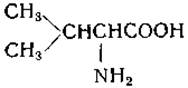
Valine was discovered in pancreatic extracts by Gorup-Besanez in 1856 [95]; however, Schutzenberger [96] was the first to demonstrate that valine is a product of protein (albumin) hydrolysis. The structure of valine was finally elucidated in 1906 by Fischer [97], who identified natural valine with one of the stereoisomers obtained upon the resolution of the synthetic amino acid. Valine is present in many proteins, but usually in relatively small amounts.
L-Histidine (a-amino-β-imidazolepropionic acid)

Histidine was isolated by Kossel [50] in 1896 from sulfuric acid hydrolysates of sturine (a sturgeon sperm protamine). In the same year, Hedin [51], independently of Kossel, isolated histidine from protein hydrolysates. Pauli proved the presence of the imidazole ring in the histidine molecule and found that the reaction of histidine with diazotized sulfanilic acid in an alkaline solution produces a red coloration (Pauli reaction).
As a result of the work of Pauli [52] and other authors [53, 54], the STRUCTURE OF THE histidine molecule was clarified, which was finally proven by the synthesis of histidine carried out by Pyman [55] in 1911. Histidine is present in relatively large amounts in Hemoglobin; in addition, it is a component of ergothioneine (in the form of thiolhistidine), carnosine, and anserine (pp. 55 and 70).
Glycine (aminoacetic acid)
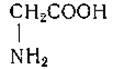
Glycine was the first amino acid isolated from a protein hydrolysate. In 1820, Braconnot [46] obtained glycine from a sulfuric acid hydrolysate of gelatin and noted the sweet taste of this amino acid. Subsequently, the "sugar of gelatin" described by Braconnot was named glycocolle and later glycine. Braconnot was unaware of the presence of nitrogen in the glycine molecule; later work, culminating in the studies of Curtius [47, 48], led to the ESTABLISHMENT OF THE structure of glycine and its synthesis from monochloroacetic acid and ammonia.
Glycine is present in large quantities in gelatin and is a component of many other proteins. In the form of an amide, it occurs in oxytocin and vasopressin (p. 72). Glycine is a constituent of a number of natural substances, such as Glutathione, as well as hippuric and glycocholic acids. In addition, the N-methyl derivative of glycine, Sarcosine, occurs in nature; this substance has been shown to be a product of tissue METABOLISM in mammals (p. 329). Sarcosine has also been detected in peanut protein [49] and in the hydrolysates of certain Antibiotics (p. 77).
L-Glutamine (y-amide of a-aminoglutaric acid)

It is interesting that, whereas the isolation of asparagine preceded that of aspartic acid, glutamine was first obtained only 17 years after the discovery of glutamic acid in protein hydrolysates.
Schulze and Bosshard [39] obtained it from beet juice in 1883; Hlasiwetz and Habermann [15] were the first to suggest the presence of glutamine in protein—a prediction confirmed relatively recently by Damodaran et al. [40], who isolated glutamine from enzymatic hydrolysates of edestin. The first Synthesis of Glutamine was accomplished by Bergmann et al. [41]. Glutamine accumulates in significant amounts in certain higher plant species and serves as one of the major amino acid components of mammalian Blood.
Upon treatment with a mixture of nitrous and acetic acids, 1 mole of glutamine, unlike asparagine and certain other amides, yields nearly 2 moles of nitrogen. A distinctive property of glutamine is the relatively high lability of its amide group, due to which it easily undergoes cyclization to form the ammonium salt of pyrrolidonecarboxylic acid. In glutamine peptides where the a-Amino groups are substituted, the amide groups are relatively stable [42–44]. Other y-glutamyl derivatives (y-glutamyl peptides, y-ethyl ester of glutamic acid) and homoglutamine also exhibit a tendency toward cyclization [45]. The process of cyclization of glutamine and related compounds is catalyzed by phosphates and certain other anions (p. 315).
L-Glutamic acid (a-aminoglutaric acid)

Glutamic acid was isolated by Ritthausen [35] in 1866 from hydrolysates of wheat endosperm gluten. It was subsequently shown that glutaric acid is formed from glutamic acid upon treatment with nitrous acid followed by reduction [36, 37]. In 1890, Wohl [38] accomplished the first Chemical synthesis of glutamic acid. This acid is among the most widely distributed amino acids and is of great importance in metabolism.
Glutamic acid crystallizes from aqueous solutions in the presence of hydrochloric acid as a sparingly soluble hydrochloride. Upon boiling an aqueous solution of this acid, it is converted into pyrrolidone-a-carboxylic acid (pyroglutamic acid, 5-oxo-2-pyrrolidinecarboxylic acid):

L-Glutamic acid in the form of its monosodium salt is widely used as a flavoring seasoning.
L-Isoleucine (a-amino-β-methylvaleric acid)

Isoleucine was isolated by Ehrlich from beet sugar molasses in 1904. Later, Ehrlich isolated this amino acid from an incomplete fibrin hydrolysate obtained by treatment with pancreatic juice, as well as from wheat gluten, egg albumin, and beef.
According to Ehrlich's observations, the product he isolated had the same chemical composition as leucine, but differed from it in a number of properties (solubility, melting point, solubility of the copper salt). Ehrlich [60–62] succeeded in cleaving L-isoleucine into d-amylamine and synthesizing the isoleucine epimer from d-isovaleraldehyde.
It is interesting to note that several years prior to Ehrlich's work, Fischer [63] had obtained fractions possessing different optical activities and varying solubilities from "leucine" preparations.
L-Leucine (a-aminoisocaproic acid)
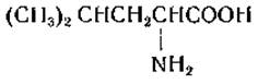
Proust [64] obtained leucine in an unpurified form from cheese in 1819. In 1820, Braconnot [65] isolated a crystalline amino acid from acid hydrolysates of Muscle and wool and named it leucine. Leucine was synthesized via the Strecker reaction from isovaleraldehyde, and the synthesis product proved to be identical to the racemized natural compound [66].
L-Lysine (a, ε-diaminocaproic acid)
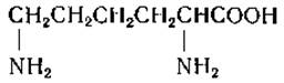
Lysine was first isolated from a casein hydrolysate by Drechsel [67] in 1889. Drechsel assumed that lysine was a diamine; the correct structure was established in 1902 by Fischer and Weigert [68], who synthesized lysine and showed that the synthesized product was identical to the racemized natural material. The lysine content in proteins varies over a wide range; it frequently occurs in animal proteins, but may be absent or present in very small amounts in Plant-derived proteins (e.g., in zein and gliadin). Upon treatment of proteins with nitrous acid, the free s-amino groups of lysine are converted into hydroxyl groups; apparently, in protein-bound lysine, most, if not all, ε-amino groups are in the free state (see, however, p. 277). Lysine bound via the ε-NH2 group is contained in biocytin.
L-Methionine (a-amino-γ-methylthiobutyric acid)

Mueller [69] discovered this amino acid in 1922 while attempting to determine The Nature of the growth factors for hemolytic streptococcus. Methionine was isolated from an acid hydrolysate of casein and its elemental composition was determined. In 1928, Barger and Coyne [70] synthesized methionine using the Strecker reaction and established the structure of this compound. Subsequently, Windus and Marvel [71] resolved methionine into its optical antipodes. Interestingly, Osborne [72] had long before noted the presence of two types of sulfur in proteins—alkali-labile and alkali-stable; sulfur of the first type is part of cysteine and cystine, whereas sulfur of the second type, as is now known, belongs to methionine.
The presence of methionine sulfoxide in natural sources is probably due to the non-enzymatic oxidation of methionine; the process of the enzymatic reduction of methionine sulfoxide back to methionine is described on p. 373.
4-Hydroxy-L-proline (4-hydroxypyrrolidine-2-carboxylic acid)

Hydroxyproline was isolated in 1902 from acid hydrolysates of gelatin by Fischer [56], who converted this amino acid into proline by reducing the hydroxyl group. Leuchs and his coworkers [57–59] succeeded in synthesizing hydroxyproline and obtaining its four stereoisomers.
Hydroxy-L-proline is found exclusively in the hydrolysates of Elastin and Collagen, where it accounts for up to 13% of all amino acid residues. Upon treatment with ninhydrin, hydroxyproline yields a yellow color on chromatograms, whereas treatment with isatin allows its detection as a blue spot. The interaction of hydroxyproline with hydrogen peroxide followed by acidification of the solution leads to the formation of pyrrole-2-carboxylic acid (p. 352), which gives an intense red-violet coloration with p-dimethylaminobenzaldehyde (Ehrlich's reagent).
L-Proline (pyrrolidine-2-carboxylic acid)

Proline was synthesized in 1900 by Willstätter [75] from a, δ-dibromopropylmalonic ester. In 1901, Fischer [76] obtained L-proline and DL-proline from casein hydrolysates and demonstrated that the latter product was identical to synthetic proline prepared from phthalimidopropylmalonic ester. Proline is present in collagen, gelatin, and other proteins. An interesting property of proline is its solubility in alcohol. On paper chromatograms, proline yields a yellow color when treated with ninhydrin, and a blue color upon reaction with isatin.
L-Serine (a-amino-β-hydroxypropionic acid)

Serine was first isolated by Cramer in 1865 from silk protein [77]. Cramer noted that in its structure serine is closely related to alanine and cystine, and concluded that serine is an oxyamino acid. The structure of serine was established in 1902 by Fischer and Leuchs, who accomplished its synthesis [78]. Serine is widely distributed in proteins; a relatively large amount of this amino acid is found in Silk Fibroin. Serine also occurs in the form of its phosphoric ester [79—81]:

Phosphoserine was isolated by Lipmann from an acid hydrolysate of casein [79]. THE POSITION OF the phosphate group in native casein has not yet been definitively established. It has been suggested that this protein contains N-phosphate bonds and that O-phosphate bonds are formed as a result of the migration of the phosphate residue during protein hydrolysis [82].
L-Tyrosine [a-amino-β-(n-hydroxyphenyl)propionic acid]

Tyrosine was first obtained in 1846 by Liebig [91] upon the cleavage of casein with alkali. Later, tyrosine was isolated by de La Rûe [92] and Bopp [93], the former from cochineal insects, and the latter from proteins (albumin, casein, fibrin). The structure of tyrosine was established in 1883 by Erlenmeyer and Lipp [94] through its synthesis. Tyrosine is extremely sparingly soluble in Water, a property that is convenient to utilize for isolating this amino acid from protein hydrolysates. Tyrosine-O-sulfate has been detected in the composition of fibrinogen and in human urine (p. 0357).
L-Threonine (a-amino-β-hydroxybutyric acid)
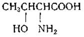
Threonine was obtained from acid hydrolysates of fibrin in 1935 by Rose et al. [83]. The work of these researchers was aimed at isolating a factor present in protein hydrolysates that is essential for the growth of rats. The discovery of threonine made it possible to demonstrate for the first time that rats can grow on a diet containing purified amino acids. Upon chemical reduction of threonine, Rose and his coworkers obtained L-a-aminobutyric acid, and by oxidation converted threonine into D-lactic acid. Threonine was synthesized by Carter [84], and subsequently West and Carter [85] obtained the four stereoisomers of this amino acid. Like serine, threonine occurs in the form of its phosphoric ester [86]. Serine and threonine react with periodic acid to yield glyoxylic acid, ammonia, and formic or acetic aldehyde, respectively [87]:
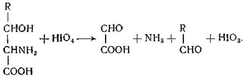
This reaction is used for the Quantitative determination of the aforementioned amino acids.
L-Tryptophan (a-amino-β-3-indolepropionic acid)
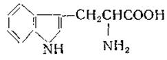
Tryptophan was isolated in 1901 by Hopkins and Cole from the products of casein Digestion with pancreatic juice [88]. Prior to this, Adamkiewicz [89] had observed that the action of sulfuric acid on a mixture of glacial acetic acid and albumin produces a violet color. Hopkins and Cole found that the development of this color is due to the presence of glyoxylic acid in the glacial acetic acid preparations. The authors attempted to isolate from protein hydrolysates the substance responsible for this color and ultimately obtained tryptophan. The structure of tryptophan was established in 1907 by Ellinger and Flamand [90]. This amino acid is present in many proteins, but usually in small amounts.
The dietary tryptophan requirement of animals compared to their requirement for other amino acids is relatively small (see p. 124).
L-Phenylalanine (a-amino-β-phenylpropionic acid)
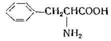
Phenylalanine was isolated by Schulze and Barbieri [73] in 1879 from etiolated lupine sprouts. Subsequently, the same authors obtained this amino acid from hydrolysates of plant proteins. As a result of the chemical synthesis of phenylalanine carried out in 1882 by Erlenmeyer and Lipp [74], Schulze and Barbieri were able to establish the identity of The amino acid they had isolated with the synthetic product [73].
L-Cysteine (a-amino-β-mercaptopropionic acid) and L-cystine [β, β',-dithiodi(a-aminopropionic acid)]

Cystine was isolated by Wollaston [27] in 1810 from urinary calculi.
In 1899, Mörner [28] obtained cystine from horn hydrolysates. Cystine is a major component of Keratins and is also present in many other proteins. There is compelling Evidence for the presence of cysteine in proteins, but only cystine—the oxidation product of cysteine—is usually found in acid hydrolysates of proteins. When a protein contains a large amount of tryptophan, the formation of cysteine during acid hydrolysis is possible [29]; at the same time, both cystine and cysteine are destroyed upon treatment with alkali.
The presence of cysteine in certain proteins is indicated by the fact that these proteins give a positive color (red) reaction with sodium nitroprusside. In neutral or alkaline solution, especially in the presence of Metal Ions, cysteine is rapidly oxidized to cystine. Baumann [30] in 1884 described the reduction of cystine to cysteine by treatment with tin and hydrochloric acid. Erlenmeyer [31, 32] established the structure of cystine and cysteine through the synthesis of these amino acids.
Cysteine reacts with formaldehyde to form thiazolidinecarboxylic acid:

This reaction, which is atypical for cystine, was used for the separate determination of cystine and cysteine [33].
A highly specific color reaction for cysteine is the Sullivan reaction: cysteine produces a characteristic reddish-brown color with sodium 1,2-naphthoquinone-4-sulfonate in a strongly reducing medium [34].
Last update: 06/08/2026
Editorial and Educational Adaptation: This material has been compiled based on the primary/original source text. The project team performed an editorial review, corrected technical inaccuracies, structured sections, and adapted the content for an educational format.
What was processed:
- elimination of formatting defects (OCR errors, structural breaks, corrupted characters);
- editorial organization of content;
- standardization of terminology in accordance with academic sources;
- verification of factual statements against the original source text.
All mentions of the author, publication year, and origin of the primary text have been preserved in accordance with the source.