Chemistry and Biology of Proteins - F. Haurowitz 1953
Sources of Amino Acids for Protein Biosynthesis
Enzymatic Protein Hydrolysis
Proteolytic Enzymes were discussed in Chapter XII. As seen from the data presented therein, many Enzymes can be isolated in a highly purified, crystalline form. The function of proteolytic enzymes located in the gastrointestinal tract is to break down dietary Proteins and convert them into Amino Acids, which are subsequently rapidly absorbed. Proteolytic enzymes are also found in all body Cells, where their presence is manifested by autolysis, i.e., the Breakdown of Proteins following Cell death. Such degradation occurs even under strictly sterile conditions and is consequently not caused by Bacteria; it differs fundamentally from putrefaction, which is frequently accompanied by proteolysis. Proteolysis during putrefaction is brought about by the proteolytic enzymes of proliferating bacteria, whereas autolysis is mediated by tissue enzymes identical or closely related to cathepsin.
Proteolysis consists in the hydrolytic conversion of compounds of the RCONHR' type into RCOOH + H2NR' via the Cleavage of peptide bonds. The extent of proteolysis is assessed by the increase in carboxyl and amino groups (see Chapter III). The hydrolytic cleavage of peptide bonds undoubtedly constitutes the initial phase of proteolysis; however, it remains unknown whether the action of a proteolytic enzyme is restricted solely to this cleavage or whether other processes also occur during this phase. This uncertainty stems from the anomalous volume contraction of the solution frequently observed in the initial phase of proteolysis [2]. The Hydrolysis of peptide bonds is accompanied by the binding of free Water molecules and should lead to a volume reduction of 15–25 mL/mol (see Chapter III). In reality, however, the volume contraction found during lactoglobulin hydrolysis proved to be 50 mL/mol [2].
The Physical Properties of protein solutions also undergo drastic Changes in the initial phase of proteolysis: viscosity decreases, and proteins lose their ability to coagulate upon heating. This is usually attributed to The conversion of protein into lower-molecular-weight Peptides. For instance, during the Pepsin Digestion of egg albumin, The amount of protein losing its heat-coagulability is proportional to the number of liberated amino groups [3]. On the other hand, in the initial phase of gelatin or casein Hydrolysis by Trypsin, no increase in free amino groups is observed [4]; consequently, the sharp drop in viscosity is not caused by peptide bond cleavage. It is possible that at this stage of enzymatic action, very weak bonds between individual groupings of the protein molecule are broken. Therefore, one might say that the enzyme acts as a depolymerase.
It was noted previously that many Globular proteins, which are resistant in their Native State to the action of proteolytic enzymes, readily undergo cleavage by these enzymes following Denaturation. Thus, for example, denatured Hemoglobin is cleaved by Papain 100 times faster than native hemoglobin [5]. Native Collagen is attacked by trypsin with difficulty, whereas heat-treated collagen is easily hydrolyzed [6]. The same applies to egg albumin and serum globulins [7]. Serum albumin, which is not cleaved by trypsin In aqueous solutions, is hydrolyzed by this enzyme when dissolved in 30% ethyl alcohol [8]. The resistance of native proteins to proteolytic enzymes is probably related to the absence in these proteins of sites accessible to enzyme molecules. Upon denaturation of the protein molecule, however, the peptide chains unfold, rendering the structures required for enzyme action accessible, thereby enabling proteolysis to take place. Such groups for trypsin are the alkaline side chains of Arginine or Lysine (see p. 293). In native proteins, these groups are concealed within the globule and are therefore inaccessible to the enzyme; small hydrogen ions, however, can penetrate to these groups, causing them to be titrated by acids. The appearance of these groups On the surface of unfolded denatured molecules explains not only the hydrolysis of these molecules by trypsin, but also The formation of intermolecular salt-like bonds and insoluble coagulates (see p. 154).
If we designate the native protein as N and the denatured protein as D, The addition of a proteolytic enzyme will shift the equilibrium N ⇄ D to the right, since enzymes preferentially cleave the denatured protein and thereby stimulate the further conversion of N to D [9, 10]. It is possible that The first phase of enzyme action consists in accelerating the N ⇄ D interconversion, and that the enzyme thus acts as a denaturase.
The concept that The breakdown of denatured proteins is facilitated by the appearance of unfolded peptide chains upon denaturation is supported by the fact that wool keratin becomes susceptible to digestion after purely mechanical Treatment; for instance, powdered wool, unlike native wool, is cleaved by trypsin [11]. This is also evidenced by experiments with monomolecular protein films formed at the water-air interface; such expanded protein films are readily cleaved by proteolytic enzymes, whereas solutions of the same proteins remain resistant to these enzymes [12].
The ability of denatured proteins to undergo enzymatic cleavage more readily than native proteins may seem somewhat surprising to a biologist, given that all animals, except humans, ingest native proteins in their diet. It should not be assumed, however, that Protein Denaturation during cooking promotes better digestion and assimilation. The point is that native proteins are also rapidly denatured under the strongly acidic conditions of gastric juice (pH 1–2) and are subjected to pepsin action already in their denatured state. Nevertheless, protein denaturation during food preparation is significant in pathological cases where gastric juice has a neutral rather than an acidic reaction. In such instances, the denatured protein will undoubtedly be cleaved by digestive tract enzymes faster than native proteins [7].
The work of Bergmann and his coworkers demonstrated that the Enzymatic hydrolysis of peptides requires the presence of side chains of a specific Structure. For example, the proteolytic action of trypsin requires the presence of alkaline side chains in the peptides, whereas pepsin exerts its effect in the presence of side chains containing aromatic or acidic amino acids. Below, using the tetrapeptide tyrosyllysylglutamyltyrosine as an example, the individual groups required for the action of various enzymes are illustrated [13].
Class="center">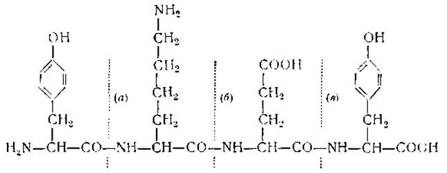
Bond (a) is cleaved by Chymotrypsin because its CO group belongs to the aromatic amino acid Tyrosine. Bond (b) is split by trypsin, which acts on peptide bonds formed by the carboxyl group of basic amino acids. Pepsin and carboxypeptidase cannot cleave bonds (a) and (b), but they are able to break bond (c). The action of pepsin is due to the fact that the NH group in (c) belongs to an aromatic amino acid. The Cleavage of the bond by carboxypeptidase depends on the terminal carboxyl group adjacent to the bond [14].
As already mentioned in Chapter XII, not only peptide bonds are susceptible to enzymatic action. Hydrolytic cleavage can occur, for example, even when a peptide bond is replaced by an ester bond. It remains unknown how extensively The structure of peptides destined for cleavage can be altered without disrupting the action of proteolytic enzymes. Minor variations that affect only the Rate of Enzymatic hydrolysis are, of course, possible; thus, at pH 4, pepsin is capable of cleaving not only carbobenzoxyglutamyltyrosine, but also, to a certain extent, cysteinyltyrosine or tyrosylcysteine [15]. Extensive experimental work must still be carried out to finally clarify the limits of Specificity of various proteolytic enzymes [16].
Another unresolved problem of great importance is the structure of intermediates formed during the enzymatic degradation of proteins. Two pathways for this cleavage can be envisaged: (1) the sequential detachment of amino acids from the site directly attacked by the enzyme, resulting in a stepwise shortening of the peptide chain comprising the protein molecule until its complete breakdown into amino acids; (2) the cleavage of protein molecules first into large fragments, then into smaller pieces, and finally into amino acids. It is also conceivable that Protein Cleavage by proteolytic enzymes proceeds simultaneously via both pathways or through some intermediate route. Elucidating The Mechanism of proteolysis is important not only from a theoretical standpoint, but also from biological and physiological Perspectives. If proteolysis proceeds via the first pathway, the protein is directly converted into amino acids, and only transiently are small amounts of intermediate Polypeptides formed. If, however, proteins are broken down into large fragments, the formation of significant amounts thereof should cause marked changes in the osmotic pressure and other PHYSICOCHEMICAL PROPERTIES OF the cellular environment.
As early as the studies of Kühne (1885), as well as Hofmeister and his coworkers, it was shown that the action of enzyme mixtures of gastric or pancreatic juice on proteins yields a series of intermediate products termed albumoses and peptones. The former are salted out upon saturation with ammonium sulfate but are not heat-coagulable, whereas the latter are neither salted out nor heat-coagulable. These substances are not homogeneous, but represent mixtures of higher and lower polypeptides, as well as Certain amino acids such as cystine and tyrosine, which also coprecipitate during the salting-out of albumoses. Although studies on albumoses and peptones are now of purely historical interest, further investigation of Protein Hydrolysis intermediates is of great significance, as it represents one of the most convenient experimental approaches for elucidating Protein Structure itself.
The few attempts undertaken to date to elucidate the Mechanism of Enzymatic protein breakdown have not yielded unambiguous results. Upon the action of pepsin on egg albumin, about 35% of the protein is converted into Peptides with a molecular weight below 1,000, 25% into peptides with a molecular weight between 1,000 and 10,000, 10% into fragments with a molecular weight between 10,000 and 30,000, and 30% into even larger fragments [3]. On the other hand, the action of pepsin on egg albumin dissolved in acetic acid yields only small peptides with a Molecular Weight of about 1,000, with no formation whatsoever of intermediates having molecular weights ranging from 1,000 to 44,000 [17]. In this case, proteolysis can be compared to an explosion, in which a large molecule breaks down instantly into a multitude of small compounds without the accumulation of intermediates. In other words, this process involves the rapid cleavage of a limited number of peptide bonds rather than the slow cleavage of many.
An "all-or-none" type of cleavage has also been observed during the action of crystalline trypsin on crystalline serum albumin or globulin, or during the Pepsin Hydrolysis of serum albumin or fibrin [18].
The action of a crude pancreatin preparation on casein or other proteins produces a large amount of free amino acids, whereas the action of crystalline trypsin on casein yields no free amino acids [19]. The pepsin hydrolysis of fibrin also liberates free amino acids [18]. It follows from these contradictory reports that the course of the proteolytic process depends on both the enzyme used and The Nature of the substrate, and we still know very little about the regularities governing this process. Nor is there certainty that proteolytic enzymes are capable of cleaving any peptide bonds other than those linking L-amino acids together; the existence of a D-peptidase has been postulated [20, 21], but has not been convincingly proven [22].
It was noted above that the Introduction/43.html">Action of Certain peptidases requires the presence of divalent Metal Ions. Cathepsin and papain are activated by the addition of cyanides and sulfides. Reduced Glutathione or similar reducing agents may be essential for cellular autolysis [23].
Proteolytic breakdown occasionally depends on the presence of substances functioning as co-substrates. For example, glycylanilide, which is not cleaved by papain, undergoes degradation upon the addition of acetylphenylalanylglycine as a co-substrate. The explanation for this remarkable phenomenon is that the added co-substrate forms an intermediate compound—acetylphenylalanylglycylglycylanilide—which is subsequently cleaved into the unchanged co-substrate, aniline, and Glycine [24].
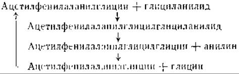
This reaction sequence demonstrates that proteolysis and Protein Synthesis are frequently closely interrelated and that, at least in some cases, hydrolytic degradation involves the intermediate synthesis of larger molecules. It must be remembered, however, that peptide cleavage does not necessarily imply peptide hydrolysis, and peptides can be oxidized directly without the formation of free amino acids. The possibility of such a degradation pathway is supported by the presence of dehydropeptidases in almost all Organs [25]. These dehydropeptidases catalyze the hydrolysis of dehydrated peptides:

The reaction products are ammonia, an α-keto acid, and an amino acid or peptide. It should be noted that dehydropeptidases are hydrolyzed by organ extracts faster than peptides [25], and they are also cleaved by pancreatic juice enzymes [26, 27]. These facts suggest that The oxidation of PROTEINS AND PEPTIDES may sometimes occur without the hydrolytic cleavage of the peptide bond or may precede such cleavage.
Last update: 06/08/2026
Editorial and Educational Adaptation: This material has been compiled based on the primary/original source text. The project team performed an editorial review, corrected technical inaccuracies, structured sections, and adapted the content for an educational format.
What was processed:
- elimination of formatting defects (OCR errors, structural breaks, corrupted characters);
- editorial organization of content;
- standardization of terminology in accordance with academic sources;
- verification of factual statements against the original source text.
All mentions of the author, publication year, and origin of the primary text have been preserved in accordance with the source.