BIOCHEMISTRY AND MOLECULAR BIOLOGY - W. ELLIOTT - 2002
CHAPTER 15. AMINO ACID METABOLISM
On a normal diet, the Digestion of Proteins in the Small Intestine results in the absorption of a fairly large amount of Amino Acids. These amino acids enter the portal vein and are carried directly by the bloodstream to the Liver. All Cells, with the exception of a few highly specialized ones (such as erythrocytes, for example), use amino acids for the synthesis of proteins and a multitude of other substances: membrane components, Neurotransmitters, heme, and so on. Consequently, all cells possess specialized Amino Acid Transport systems, since amino acids in their ionized state cannot cross Cell/29.html">The Lipid Bilayer of Plasma Membranes.
There is no specific storage form for amino acids, unlike high-molecular-weight Polysaccharides, which are broken down into monomers when needed. Instead, all functional proteins serve as a reserve for amino acids, with Muscle Proteins being the primary source simply because they are the most abundant. However, these proteins are involved in Muscle contraction, and when they are intensively broken down to meet amino acid demands—for instance, during hepatic Gluconeogenesis—muscle atrophy ensues. Such a situation poses significant problems for humans, since in the course of evolution we have lost The ability to synthesize half (10 out of 20) of the amino acids (referred to as Essential Amino Acids). Curiously, these include amino acids whose synthesis involves a particularly high number of steps and requires numerous specialized Enzymes encoded by many genes. In other words, these are amino acids that are "expensive" to produce.
One can only guess why this came to be. It is possible that this is a purely economic measure, as it is more advantageous to obtain the most complex amino acids from food, synthesizing only the simplest ones ourselves. At the same time, if humans obtained all necessary provisions from the wild, their diet would likely contain all the required amino acids. In such a scenario, the inability to synthesize Certain amino acids is not nearly as critical compared to the complete absence of food.
The Development of agriculture provided a rapidly multiplying human race with a source of chemical energy in the form of plant CARBOHYDRATES.
Plant-based foods allow people to survive, but they do not always satisfy their requirements for essential amino acids, since the proteins of certain cultivated plants, such as maize, are deficient in Lysine and Tryptophan. However, even with a prolonged intake of a diet rich in high-quality proteins, humans cannot store essential amino acids for future use. Only a minor fraction of them is utilized for its intended purpose, while the unrequested surplus is simply oxidized or converted into fats or Glycogen. In some situations, this is equivalent to burning diamonds for warmth.
The result of an inadequate diet, deficient in even a single essential amino acid, is a pathological condition known as kwashiorkor. Since virtually all proteins contain a full Complement of amino acids, a shortage of any one of them leads to the synthesis of defective protein molecules. This causes wasting, apathy, stunted growth, and a decrease in serum protein levels in the Blood. The latter leads to a reduction in the oncotic pressure of the blood and appears to be the direct cause of edema.
A vicious cycle ensues. The cells lining the small intestine must constantly renew themselves and produce digestive enzymes. In kwashiorkor, their ability to do so is severely impaired. As a result, even the food that is available is poorly digested and absorbed. Children suffer particularly from kwashiorkor because a growing Organism needs to synthesize large amounts of protein.
It turns out that the evolutionary loss of the ability to synthesize certain Amino Acids and the failure to store their surpluses brings major troubles for humans. Admittedly, it cannot be ruled out that evolution had good reasons for this. For instance, intermediates in the Biosynthesis of Essential amino acids may be toxic to higher organisms. In particular, they can disrupt Brain development or function.
Nitrogen Balance in the Body
Amino acids are involved in maintaining the nitrogen balance of the body. If the body consumes as much nitrogen (primarily in the form of amino acids)
as it excretes, this steady state is referred to as nitrogen balance. During periods of growth or wound healing, more nitrogen is consumed than is excreted—the balance is considered positive. Nitrogen balance becomes negative when less nitrogen is consumed than is excreted; this is observed, for example, during starvation, when muscle proteins are converted into glucose and nitrogen is excreted in the urine. In animals, proteins are in a state of continuous turnover: they are constantly being degraded and resynthesized. Although the majority of amino acids released during protein breakdown are reutilized in subsequent Protein Synthesis, we lose about 0.3% of our body's nitrogen daily in the form of urea.
Amino acids are considered essential if their removal from the diet leads to a negative nitrogen balance and, consequently, to growth arrest in experimental animals. Some of these amino acids are strictly essential: for humans, these are lysine, phenylalanine, and tryptophan. Below is a list of dispensable and essential amino acids for humans.
Class="center">Dispensable amino acids |
Essential amino acids |
Asparagine |
|
Aspartic acid |
Isoleucine |
Cysteine** |
Leucine |
Glutamic acid |
Lysine |
Glutamine |
|
Phenylalanine |
|
Tryptophan |
|
Tyrosine*** |
Valine |
* Required only during growth.
** Can be synthesized from phenylalanine.
*** Can be synthesized from methionine.
Tyrosine is not a strictly essential amino acid because it can be derived from phenylalanine, provided the latter is supplied in sufficient quantities. Similarly, mammals can synthesize cysteine from another essential amino acid, methionine. Overall, it is impossible to formulate rigid rules governing the body's requirements for amino acids. "First-class" proteins are rich in essential amino acids, whereas plant proteins are low in certain essential amino acids. Since the Amino Acid Composition of different proteins varies greatly, a vegetarian diet must inherently be more diverse.
General Amino acid METABOLISM
The General scheme of amino acid metabolism is shown in Fig. 15.1.
Fig. 15.1. Amino Acid Catabolism. Some amino acids are partly ketogenic and partly glucogenic

Aspects of Amino Acid Metabolism
Excess amino acids (those not used in protein synthesis or for other specific needs) are degraded to produce energy or to create energy reserves (fats or glycogen). This raises A number of important questions that need to be addressed separately: How does the body synthesize dispensable amino acids? How are certain amino acids converted into other physiologically important low-molecular-weight compounds? In this chapter, we will Touch upon the following issues.
1. How Amino groups are removed from amino acids (in other words, how deamination occurs). Although Amino acids differ in Structure, most of them undergo deamination in a similar way, which significantly simplifies The Study of this process.
2. What happens to the keto acids (carbon skeletons) produced during the Deamination of Amino acids. Each of them follows its own metabolic pathway, and here we will consider only selected Examples that are especially interesting or important for understanding Metabolism as a whole.
3. How the nitrogen removed from amino acids is converted into urea.
4. How amino acids are synthesized. In animals, this naturally applies only to non-essential amino acids. If the corresponding keto acid is available (i.e., the carbon Skeleton of the amino acid), synthesis often boils down to the reversal of deamination. We will examine only a few such examples. In Bacteria and plants, all amino acids are synthesized via individual pathways, with starting Materials supplied from general metabolic reactions. The synthetic capacity of animals is limited by the existence of essential amino acids. Studying the biosynthetic pathways of all amino acids involves a massive Amount of Information. Here we will limit ourselves to the most interesting examples.
5. Some amino acid transformations are quite fascinating and crucial for building a comprehensive picture of amino acid metabolism. Some of these will also be discussed in this chapter.
Deamination of amino acids
First, a bit of chemistry. Schiff bases are formed as a result of a reversible reaction between the carbonyl group of an aldehyde or ketone and a free amino group:

This reaction is reversible; consequently, the Schiff base is readily hydrolyzed. If R = H, Hydrolysis will release ammonia (NH3). Thus, the removal of two hydrogen atoms from an amino acid leads to deamination, whereas The addition of a pair of hydrogen atoms results in the synthesis of an amino acid from a keto acid and ammonia (non-ionized forms of the compounds are shown for simplicity):

Now let us return to biological deamination. One of the most important amino acids in metabolism is glutamic acid, the deamination of
which is catalyzed by Glutamate dehydrogenase. It uses either NAD+ or NADP+ as an oxidizing agent (at physiological pH values, NH3 is protonated and exists in its ionized form, NH4+):
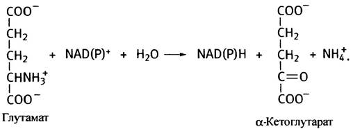
Glutamate dehydrogenase is a key enzyme in deamination and participates in The oxidation of many amino acids. It is allosterically inhibited by ATP and GTP (which serve as indicators of high energy levels: energy reserves are abundant—fuel is not needed) and activated by ADP and GDP. An increase in the levels of the latter indicates The Need for Oxidative Phosphorylation (energy reserves are low—fuel is required).
α-Ketoglutarate participates in The Citric Acid Cycle, making it possible for glutamic acid to be oxidized (following deamination) to H2O and CO2. Since α-ketoglutarate can be converted into oxaloacetate (see Fig. 8.12), glutamic acid can, under certain conditions, participate in glucose synthesis (see Chapter 11, Fig. 11.3). Thus, glutamic acid belongs to the category of glucogenic amino acids.
Other Amino Acids lack corresponding dehydrogenases. How then are they deaminated? Several specialized mechanisms exist for this purpose, but most of them rely on The transfer of an amino group from an amino acid to α-ketoglutarate, yielding the corresponding keto acid and glutamate. The latter is then deaminated by glutamate dehydrogenase. In other words, the deamination of other amino acids proceeds in two stages. The first is called Transamination, and the second is deamination.
The transamination stage can be represented as follows:
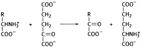
Suppose that R = CH3, meaning The amino acid is alanine. The deamination of alanine proceeds According to the following scheme:
Alanine + α-Ketoglutarate —> Pyruvate + Glutamate (1)
Glutamate + NAD+ + Н2O —> α-Ketoglutarate + NADH + NH+4 (2)
Overall:
Alanine + NAD+ + Н2O —> Pyruvate + NADH + NH+4.
Together, both stages of this process—transamination and glutamate deamination—are understandably referred to as transdeamination. Enzymes that catalyze transamination are called transaminases or aminotransferases. Many transaminases specific to various amino acids are known, and Most amino acids can be deaminated with their participation.
The reversibility of the transaminase reaction means that an amino acid can be synthesized from its corresponding keto acid (however, the keto acids of corresponding essential amino acids are not synthesized in the body). An example of transamination is the malate-aspartate shuttle (see p. 116), which involves this reversible reaction:

Mechanism of transamination reactions
The active centers of all transaminases contain a tightly bound coenzyme, pyridoxal-5'-phosphate (PLP). It participates in numerous enzymatic Transformations of Amino acids, acting as an electrophilic intermediate. Its primary Functions are first to accept an amino group from an amino acid as an acceptor, and then to transfer it to a keto acid as a donor. All of this takes place with the participation of a single enzyme. As is often the case, the coenzyme turns out to be
I a vitamin derivative (in this case, vitamin B6). In reality, the vitamin B6 group comprises three related substances: pyridoxal, pyridoxine, and pyridoxamine. All of them are converted into Pyridoxal phosphate within the organism (Fig. 15.2).
Fig. 15.2. Structure of compounds of the Vitamin B6 group and the transaminase cofactor, pyridoxal phosphate
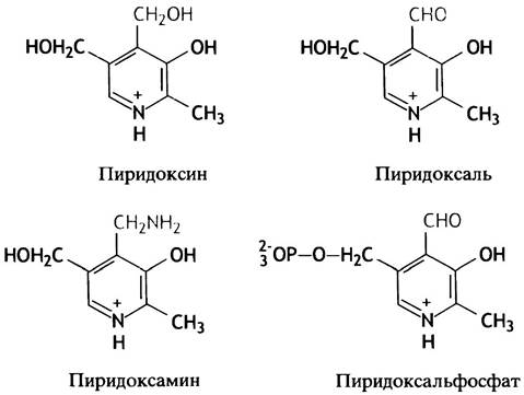
The "working group" of pyridoxal phosphate is the aldehyde group -CHO. The transamination reaction in general terms can be represented as follows:
E-PLP + amino acid 1 <-> E-PLP-NH2 + keto acid 1
E-PLP-NH2 + keto acid 2 <-> E-PLP + amino acid 2
Here PLP is pyridoxal phosphate, and PLP-NH2 is pyridoxamine phosphate.
The Mechanism of this reaction is illustrated in Fig. 15.3. Both reactions occur within the active center of the transaminase, and pyridoxal phosphate and pyridoxamine phosphate are tightly bound to this enzyme.
Fig. 15.3. Mechanism of transamination (simplified representation). R-CH = NH- is the complex of pyridoxal phosphate with the amino group of a protein lysine residue. R-CH2-NH3+ is pyridoxal phosphate. The forward reaction (red arrows) leads to The conversion of the amino acid into a keto acid, and pyridoxal phosphate into pyridoxamine phosphate. The reverse reaction (black arrows) represents transamination between a keto acid and an amino acid

The pathway of transdeamination considered above is the most general for amino acids; however, some of them lose their amino groups in a different manner.
Special deamination mechanisms
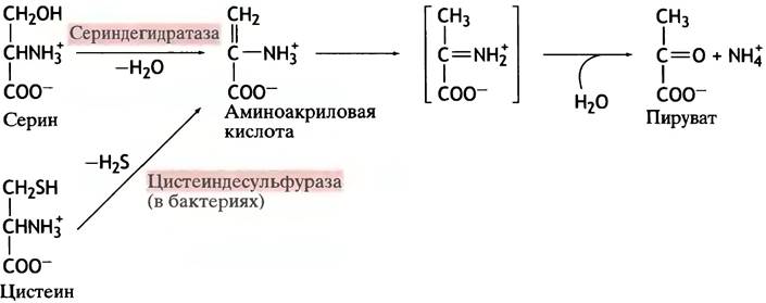
Serine (a hydroxyl-containing amino acid) undergoes deamination via a dehydration reaction catalyzed by a specific dehydratase. Cysteine, which differs from serine by containing a thiol group instead of a hydroxyl group, is deaminated following the Cleavage of H2S. In both cases, the reaction product is pyruvate (Fig. 15.4).
Fig. 15.4. Conversion of serine and cysteine into pyruvate
The Fate of the keto acid, or the carbon skeletons of deaminated amino acids
Considering The Role of amino acids in overall metabolism, some of them are termed glucogenic, while others are ketogenic. The term ketogenic amino acid implies that acetyl-CoA is formed during its metabolism, which is subsequently converted into Ketone Bodies. This definition outwardly contradicts the assertion that ketone bodies arise only under conditions of excessive fat metabolism (see p. 85), whereas under normal conditions, acetyl-CoA is not expended on their synthesis. In reality, the term ketogenic originated long ago when the fate of dietary amino acids was studied using starved animals as a test system. It was precisely under these conditions that any increase in acetyl-CoA levels was accompanied by a rise in easily detectable ketone bodies in the blood. Under normal conditions, ketone bodies are not formed from ketogenic amino acids, and the true meaning of the term is that these amino acids are converted not into pyruvate, but into acetyl-CoA. Glucogenic amino acids stimulate an increase in blood glucose levels (in diabetics, in urine).
Aspartic acid is deaminated to oxaloacetate—one of the metabolites of The Citric Acid cycle, which can be converted into glucose. Consequently, aspartic acid is a glucogenic amino acid. This category also includes glutamic acid and alanine, which are converted into α-ketoglutarate and pyruvate, as well as serine, cysteine, and a number of other amino acids.
Some amino acids are both Ketogenic and Glucogenic. For instance, the Metabolism of Phenylalanine yields both acetyl-CoA and fumarate, which participates in the citric acid cycle. Of the 20 amino acids, only two — leucine and lysine — are exclusively ketogenic. Regarding the degradation of isoleucine, methionine, threonine, and valine, see page 136.
Phenylalanine metabolism
Phenylalanine is an aromatic amino acid, an excess of which is normally converted into tyrosine by the enzyme phenylalanine hydroxylase (Fig. 15.5). This enzyme is notable for the fact that the two hydrogen atoms in the reaction are supplied by an electron donor—the coenzyme tetrahydrobiopterin.
Fig. 15.5. Normal and abnormal metabolism of phenylalanine. RH4 stands for tetrahydrobiopterin; RH2 stands for dihydrobiopterin

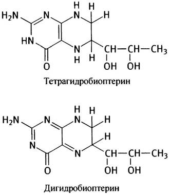
It may seem strange that this reaction requires both oxygen and a reducing agent, but such phenomena are quite common and we will encounter them again. The point is that one oxygen atom is used here to form a hydroxyl group on the aromatic ring. The second oxygen atom is reduced to an H2O molecule, with two hydrogen atoms supplied by tetrahydrobiopterin. A specialized enzyme system using NADH as a reductant converts dihydrobiopterin back into the starting tetrahydrobiopterin, so that the latter acts catalytically.
Phenylalanine hydroxylase belongs to the class of Monooxygenases (enzymes that incorporate a single oxygen atom into a substrate) or mixed-function oxygenases, since two compounds—the Amino Acid and a pair of hydrogen atoms—are oxygenated (see p. 210 for a Discussion of the difference between oxidation, the removal of electrons, and oxygenation, the Introduction of an oxygen atom into a molecule).
Phenylalanine itself normally does not undergo deamination; it must first be converted into tyrosine. A relatively common genetic defect is known in which the conversion of phenylalanine to tyrosine is impaired or completely blocked due to a deficiency of the enzyme or, less frequently, tetrahydrobiopterin. The accumulating phenylalanine is consequently forced to undergo deamination into an abnormal metabolite, phenylpyruvate, which is immediately excreted in the urine (see Fig. 15.5). This disorder is called phenylketonuria (PKU). It is especially dangerous for infants because phenylpyruvate causes irreversible brain damage and early death. If the condition is detected right after birth (by urinalysis or, even better, by a blood test for phenylpyruvate), the child is placed on a low-phenylalanine diet that still contains adequate amounts of tyrosine. This prevents the disease and normalizes development. Why phenylpyruvate has such a destructive effect on the brain remains unknown.
Another quite fascinating genetic disorder is known as maple syrup urine disease. In this condition, ketoacids corresponding to three aliphatic amino acids—leucine, isoleucine, and valine—accumulate in the body. Named for the characteristic odor of the urine, this disease is also associated with biochemical abnormalities in the brain and is much rarer than phenylketonuria.
Another genetic disorder, alkaptonuria, manifests externally through the rapid darkening of urine left exposed to air. The cause is a defect in tyrosine degradation, resulting in the accumulation of an intermediate product, homogentisic acid. This compound is a diphenol that, upon oxidation by atmospheric oxygen, turns into a dark pigment.
Methionine and Methyl group transfer
Methionine is one of the essential amino acids.

The most crucial part of methionine is its methyl group. These groups play a tremendous role in cellular life. Many compounds undergo methylation, with methionine itself serving as the donor of the methyl group transferred to other substances. Methionine as such is a stable compound and cannot act as a methylating agent. However, its methyl group is activated when methionine reacts with ATP to form the sulfonium cation S-adenosylmethionine, or SAM (Fig. 15.6).
The presence of a highly reactive group makes its transfer from this substance to Other Compounds thermodynamically favorable. The three phosphate groups of ATP, which provide the energy for the synthesis of S-adenosylmethionine, are converted into pyrophosphate PPi and inorganic Pi, after which PPi is hydrolyzed into two molecules of Pi (see Fig. 15.6). The transfer of the methyl group from S-adenosylmethionine to other compounds is catalyzed by transmethylases. In the process, S-adenosylmethionine is converted into S-adenosylhomocysteine. The latter is hydrolyzed to homocysteine—a methionine variant in which the S-CH3 group is replaced by an -SH group. Next, the thiol group is transferred from homocysteine to serine, yielding cysteine (see Fig. 15.6). An intermediate in this pathway (cystathionine) accumulates in the body under certain genetic defects and is excreted in the urine. This condition is called cystathioninuria.
Fig. 15.6. Synthesis of S-adenosylmethionine from methionine (a), and of cysteine from homocysteine (b)

Where Are Methyl Groups Transferred?
The products of methylation by S-adenosylmethionine include creatine (see p. 380), phosphatidylcholine (see p. 55), epinephrine (see p. 200), and the nitrogenous bases of Nucleic Acids (except thymine).
Only non-essential amino acids are synthesized within the body. We will not delve into all the details of these processes here, as their chemistry is as diverse as it is specific. Our goal is to highlight the most interesting and important aspects of this problem and demonstrate how amino acids are synthesized from intermediates of Glycolysis and the citric acid cycle.
In fact, just five such compounds—3-phosphoglycerate, phosphoenolpyruvate, pyruvate, oxaloacetate, and α-ketoglutarate—along with two Monosaccharides of the Pentose Phosphate Pathway, serve as precursors for all 20 amino acids synthesized in bacteria and plants.
Synthesis of Glutamic Acid
The deamination of glutamic acid is carried out by glutamate dehydrogenase, which utilizes NAD+ or NADP+ as Cofactors. The reaction is reversible, but in animals, The formation of glutamate from the amino acid is arguably less significant than the transamination of α-ketoglutarate. The amino group Donors in this process are other amino acids, such as alanine or aspartic acid. Prokaryotes possess an alternative pathway for the amination of α-ketoglutarate, which operates at low NH4+ concentrations and requires an energy input.
Synthesis of Aspartic Acid and Alanine
These amino acids are formed in cells via the transamination of oxaloacetate (R = -CH2COO-) and pyruvate (R = -CH3):

Synthesis of Serine
Serine is synthesized in three steps from the glycolytic intermediate 3-phosphoglycerate. First, 3-phosphoglycerate is oxidized to the keto acid 3-phosphohydroxypyruvate:
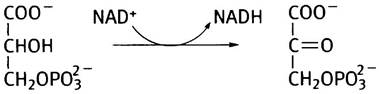
This keto acid then undergoes transamination with glutamic acid to yield 3-phosphoserine, which is subsequently hydrolyzed to serine and Pi.

Synthesis of Glycine
Glycine (NН3+-СН2-СОО-) is the simplest of the α-amino acids. Its synthesis is achieved by the removal of the terminal hydroxymethyl group of serine. The reaction proceeds with the participation of a coenzyme, tetrahydrofolic acid, which acts as a carrier of single-carbon units. Such transfers play a crucial role in nucleotide synthesis (see Chapter 18).
Synthesis of Other Compounds from Amino Acids
A wide range of low-molecular-weight, physiologically active compounds are synthesized in the body from amino acids. Amines are formed as a result of the Decarboxylation of amino Acids:
RСН2NН3+СОO- —> RСН2NН3+ + СO2.
Catecholamines, which include the Hormones dopamine, adrenaline (epinephrine), and noradrenaline (norepinephrine), get their name from their structural similarity to catechol (1,2-dihydroxybenzene).
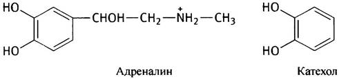
Among the compounds derived from amino acids are neurotransmitters: γ-aminobutyrate (GABA), 5-hydroxytryptamine, and the aforementioned catecholamines. The hormone thyroxine is synthesized from tyrosine. This far-from-exhaustive list demonstrates that amino acids serve as precursors for many biologically vital substances.
What Happens to Amino Groups After Their Removal from Amino Acids? The Urea Cycle
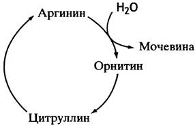
The amino nitrogen of catabolized amino acids is excreted from the mammalian body in the urine as urea—an inert, Water-soluble, non-toxic substance. Urea is formed in the liver by the Cleavage of the guanidino group from arginine via the hydrolytic enzyme arginase. Concurrently, Ornithine is produced—a non-protein amino acid.

To convert ornithine back into arginine, a carbon atom from CO2 and the amino nitrogen released during the degradation of all 20 catabolized amino acids are utilized. The arginine-ornithine cycle was the first metabolic cycle to be described. Krebs (who later discovered the citric acid cycle) and Henseleit discovered that if arginine was added to liver cells, The amount of urea produced significantly exceeded the amount of the added amino acid. The same phenomenon occurred upon the addition of ornithine. The scientists concluded that the interconversion of both amino acids is a cyclic process involving both arginine and ornithine.
The formation of arginine from ornithine proceeds in several stages, with another non-protein amino acid, citrulline, serving as an intermediate.
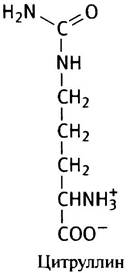
Like arginine and ornithine, citrulline stimulates urea synthesis by liver cells. Thus, the famous urea cycle was finally deciphered (Fig. 15.7).
Fig. 15.7. The cycle of urea formation from arginine. The incorporation of nitrogen and CO2 into the cycle is discussed in the text
Mechanism of Arginine Synthesis
The first step in arginine synthesis—the formation of citrulline—takes place in the mitochondrial matrix. It begins with the Condensation of ammonium ions and CO2 to form carbamoyl phosphate, which is a mixed anhydride of carbamic acid (NН2COOH) and phosphoric acid. Carbamoyl phosphate is a high-energy compound and a potent acylating agent.
Its synthesis is catalyzed by carbamoyl phosphate synthetase in the following reaction:
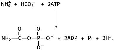
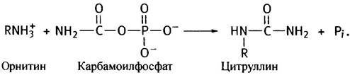
The synthesis of each of its molecules consumes 2 ATP molecules. One of them, cleaved to ADP and Pi, drives the formation of carbamate from ammonium and CO2, while the second participates in its phosphorylation. Next, the carbamoyl group of carbamoyl phosphate is transferred to ornithine by the enzyme ornithine transcarbamylase, converting it into citrulline (ornithine is represented as R-NH3+):
Conversion of citrulline to arginine
The Second Stage of arginine synthesis takes place in the Cytosol and involves the Conversion of the carbonyl group (C = O) in citrulline into the imido (C = NH) group of arginine. The carbonyl of citrulline is significantly less reactive than the ketone carbonyl in keto acids; therefore, this conversion cannot be achieved via transamination. In reality, it proceeds in two stages. In The First stage, citrulline condenses with aspartate in an ATP-dependent reaction, yielding argininosuccinate:
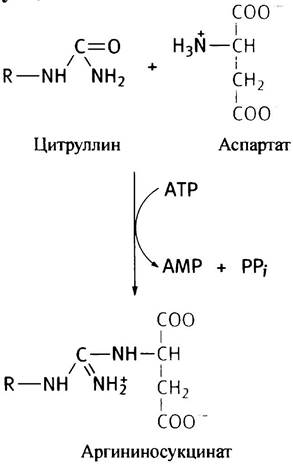
The name of this reaction product stems from the fact that its molecular structure resembles an arginine derivative of succinate, although its cleavage by argininosuccinate lyase yields arginine and fumarate rather than succinate:
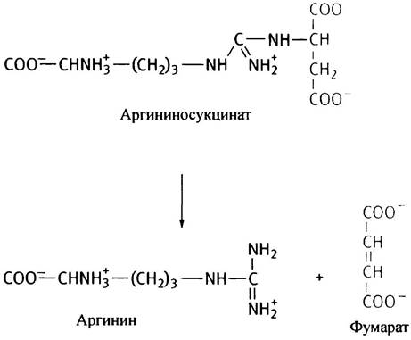
The complete urea cycle is illustrated in Fig. 15.8. It is primarily regulated through changes in enzyme levels. Their amounts increase both in response to a high dietary amino acid intake and during starvation, when muscle proteins are degraded.
Fig. 15.8. Enzymes of the urea cycle. Enzyme levels correlate with the amount of dietary protein consumed. The cycle is allosterically regulated at the carbamoyl phosphate synthetase step (presumably by N-acetylglutamate). Ornithine is converted to citrulline within the Mitochondria, whereas the remaining steps of the cycle occur in the Cytoplasm

In addition, carbamoyl phosphate synthetase is allosterically regulated by N-acetylglutamate, the level of which correlates with amino acid concentration.
How Amine Nitrogen is Transported to the Liver for Processing into Urea
Glutamine as the Transport Form of Ammonia in the Blood
Ammonia generated during AMINO ACID DEAMINATION is toxic. Consequently, its blood concentration must be kept extremely low; otherwise, brain dysfunction and coma may ensue. For this reason, it is not transported to the liver in free form. Ammonia reacts with glutamic acid, and through the action of the enzyme Glutamine Synthetase, glutamine (the amide of glutamic acid) is formed. This pathway proceeds via the intermediate γ-glutamyl phosphate, a mixed anhydride of glutamic and phosphoric acids. This is a high-energy compound capable of reacting with ammonium ions (both reactions occur within the Active Site of the synthetase).

The source of glutamate for this process is α-ketoglutarate from the citric acid cycle, which undergoes transamination with other amino acids. Glutamine is transported via the blood to the liver, where it is hydrolyzed by glutaminase, and the released ammonia is utilized for urea synthesis.
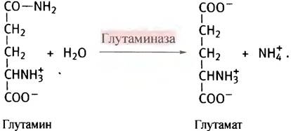
Glutaminase is also present in the Kidneys, where its function is to neutralize acids eliminated from the blood using ammonia. Aside from serving as an ammonia carrier and one of the 20 standard amino acids building proteins, glutamine participates in the synthesis of several other metabolites. In these cases, it is utilized in the form of glutaminamide as a nitrogen source.
Alanine as a Transport Form of Amine Nitrogen in the Blood
Approximately 30% of the amine nitrogen delivered to the liver following muscle protein degradation is carried by alanine. Naturally, the proportion of alanine in proteins is much lower; alanine is primarily formed via the transamination of pyruvate by other amino acids. Upon reaching the liver, alanine undergoes deamination. The ammonia is used for urea production, and the pyruvate is converted to glucose. The latter is returned via the bloodstream to the Muscles, thereby completing the glucose-alanine cycle of Ammonia Transport (Fig. 15.9).
Fig. 15.9. The glucose-alanine cycle delivers nitrogen (in the form of alanine) from muscles to The Liver and returns glucose from the liver to the muscles
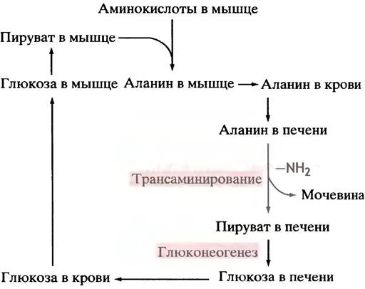
During starvation, this cycle assumes particular importance. Once glycogen stores are depleted, the liver must supply glucose to the brain and other Tissues in need. The primary source of metabolites utilized by the liver for gluconeogenesis is amino acids derived from muscle protein breakdown. The regulation of this degradation will be discussed in Chapter 22. Many amino acids are converted into pyruvate while still in the muscles, which is then transported to the liver in the form of alanine. To reiterate, the glucose-alanine cycle does not increase the net amount of glucose; rather, it serves as a mechanism to divide amino acid metabolism between the muscles and the liver, as well as a safe transport vehicle for amine nitrogen to the liver.
Chapter 15 Questions
1. Explain how the oxidation of Amino acids can lead to their deamination.
2. Which amino acids undergo Oxidative Deamination?
3. Describe in detail the deamination of alanine.
4. Which coenzyme is involved in transamination? What is its structure and how does the transamination process occur?
5. How are serine and cysteine deaminated?
6. What do the terms " ketogenic" and "glucogenic" mean with respect to amino acids? Name strictly ketogenic amino acids.
7. What is phenylketonuria as a medical condition?
8. What is the role of tetrahydrobiopterin in the hydroxylation of phenylalanine?
9. Methionine serves as a source of methyl groups in various biochemical processes. Explain how methionine is activated in biological methylation reactions.
10. Describe the reactions involved in the urea cycle.
11. Why do the levels of urea cycle enzymes increase during both high-protein diets and starvation?
12. How are ammonia and amino nitrogen transported from peripheral tissues to the liver for urea synthesis?
Last update: 06/08/2026
Editorial and Educational Adaptation: This material has been compiled based on the primary/original source text. The project team performed an editorial review, corrected technical inaccuracies, structured sections, and adapted the content for an educational format.
What was processed:
- elimination of formatting defects (OCR errors, structural breaks, corrupted characters);
- editorial organization of content;
- standardization of terminology in accordance with academic sources;
- verification of factual statements against the original source text.
All mentions of the author, publication year, and origin of the primary text have been preserved in accordance with the source.