Human Biochemistry, Volume 1 - Murray R. 1993
Protein and Amino Acid Metabolism
Catabolism of Amino Acid Nitrogen
Ammonia Transport
Although ammonia can be excreted from the body as ammonium salts—especially during metabolic acidosis—most of it is eliminated as urea, the principal nitrogenous component of urine. Ammonia is constantly produced in Tissues, yet it is present in the peripheral Blood only in trace amounts (10–20 µg/100 mL); it is rapidly cleared from the Circulatory system by the Liver, where it is incorporated into glutamate, glutamine, or urea.
Class="center">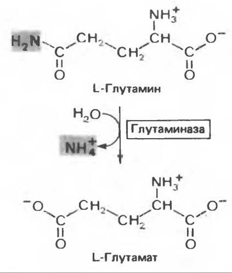
Fig. 30.7. The reaction catalyzed by glutaminase is virtually irreversible and proceeds in the direction of glutamate and NH+4 formation. It should be emphasized that the amide nitrogen of glutamine is removed, rather than the nitrogen of the a-amino group.
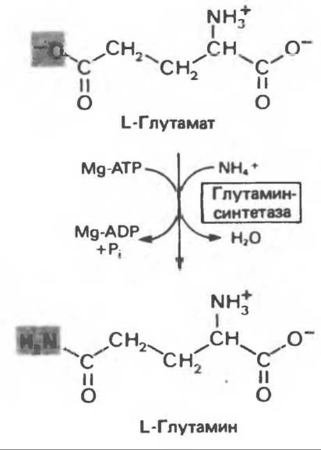
Fig. 30.8. The reaction catalyzed by Glutamine Synthetase. The reaction equilibrium strongly favors glutamine synthesis.
The removal of ammonia via Glutamate dehydrogenase has already been mentioned. The formation of glutamine is catalyzed by glutamine synthetase (Fig. 30.8), a mitochondrial enzyme present in high concentrations in renal tissue. The Synthesis of the amide bond of glutamine is driven by the Hydrolysis of one equivalent of ATP to yield ADP and Pi. The equilibrium of this reaction lies far in the direction of glutamine synthesis (see also Chap. 29).
The release of the amide nitrogen of glutamine as ammonia occurs through the hydrolytic Cleavage of ammonia catalyzed by glutaminase (Fig. 30.7). Unlike the reaction catalyzed by glutamine synthetase, the glutaminase reaction proceeds without the involvement of Adenine NUCLEOTIDES and is heavily shifted toward the formation of glutamate; it does not operate in the direction of glutamine synthesis. Thus, glutamine synthetase and glutaminase catalyze the interconversion of the free ammonium ion and glutamine (Fig. 30.9); this is analogous to the interconversion of glucose and glucose-6-phosphate by glucokinase and glucose-6-phosphatase (see Chap. 17).
A reaction analogous to the one catalyzed by glutaminase is mediated by L-asparaginase, which is found in animal tissues, plants, and microorganisms. The potential use of asparaginase and glutaminase as antitumor agents has been explored because certain tumors exhibit an abnormally high demand for glutamine and asparagine.

Fig. 30.9. The interconversion of ammonia and glutamine catalyzed by glutamine synthetase and glutaminase. Both reactions proceed predominantly in the directions indicated by the arrows. Thus, glutaminase serves exclusively for the deamidation of glutamine, whereas glutamine synthetase serves exclusively for the Synthesis of Glutamine from glutamate. Glu - glutamate.
While the primary pathway for ammonia removal in Brain tissue is the formation of glutamine, urea synthesis is the most important pathway in the liver. Urea formation can also occur in brain tissue, but it plays no significant role in ammonia detoxification. The formation of glutamine in the brain must be preceded by the synthesis of glutamate, since the blood-borne supply of glutamate proves insufficient when blood ammonia concentrations are high. The direct precursor of glutamate is a-ketoglutarate. The conversion of ammonia to glutamine can lead to a rapid depletion of Citric Acid Cycle intermediates unless they are replenished by the conversion of Pyruvate to oxaloacetate, accompanied by CO2 fixation (see Chap. 17). Brain tissue indeed exhibits substantial incorporation of CO2 into Amino Acids, presumably after the latter enters The Citric Acid Cycle; following ammonia administration, additional oxaloacetate is channeled into glutamine synthesis (via the a-ketoglutarate stage).
Last update: 06/08/2026
Editorial and Educational Adaptation: This material has been compiled based on the primary/original source text. The project team performed an editorial review, corrected technical inaccuracies, structured sections, and adapted the content for an educational format.
What was processed:
- elimination of formatting defects (OCR errors, structural breaks, corrupted characters);
- editorial organization of content;
- standardization of terminology in accordance with academic sources;
- verification of factual statements against the original source text.
All mentions of the author, publication year, and origin of the primary text have been preserved in accordance with the source.