The Evolution of Viruses - Zhdanov, V. M. 1990
Special Section
Hepadnaviruses
The hepadnavirus family (hepar + DNA — etymologically referring to the Liver, which is targeted by these Viruses, and DNA, the viral genetic material) comprises several well-characterized viruses: human hepatitis B virus (HBV), woodchuck hepatitis virus (WHV), ground squirrel hepatitis virus (GSHV), and duck hepatitis virus (DHV). Later, other viruses of this group were discovered: snake hepatitis virus from Taiwan, tree squirrel hepatitis virus [Feitelson M. et al., 1986], marmot hepatitis viruses from northern Ukraine and Kazakhstan, the Norway rat hepatitis virus, voles Apodemus sylvaticus, harvest mice Micromys minutus, shrews Crocidura suaveolens, and moles Talpa europaea [Krivanec K. et al., 1984]. Data on insectivore hepatitis viruses are currently derived solely from serological assays. Given that previously studied viruses typically have a narrow host range—often restricted to a single species—this family already encompasses pathogens affecting mammals, birds, and reptiles. Breaking once again with the general style of the book, we shall begin with The history of the Discovery of the human hepadnavirus, the CAUSATIVE AGENT OF hepatitis B.
The METABOLISM/13.html">History of the discovery and investigation of the human hepatitis B virus dates back to the last century, when an outbreak of hepatitis was described in Germany among factory workers vaccinated against smallpox using humanized vaccine (i.e., vaccine that had passed through The Human Body). Although this outbreak did not attract much attention, it already became clear at that time that such Vaccines should be avoided, as cases of transmission of other diseases, particularly Syphilis, were known.
The possibility of parenteral transmission of hepatitis gained prominence in the 1930s and 1940s due to The Use of certain vaccines (against yellow fever and sandfly fever), as well as the mass Treatment of syphilis patients, predominantly military personnel. These observations have been described in detail elsewhere [Zhdanov V. M., 1948], so we shall limit ourselves to A brief Overview. It is worth noting that by this time, the scientific consensus was beginning to take shape that infectious hepatitis—identified as an independent nosological entity in the previous century by S. P. Botkin—was a viral infection.
Extensive outbreaks of hepatitis B, which had not yet been classified as an independent nosological entity, were observed in connection with mass vaccinations against yellow fever. A thorough analysis by G. Findlay and F. McCallum (1937) led to the entirely correct Conclusion that the virus had been introduced into the vaccine preparation via human serum contained within it. A similar hepatitis outbreak was identified following immunizations against sandfly fever [Sergiev P. G. et al., 1940], as this vaccine also contained human serum. By this time, accumulating data pointed to hepatitis transmission among individuals receiving Blood transfusions or parenteral administration of blood products. Thus, the risk of hepatitis transmission via parenteral administration of human blood, plasma, serum, and other blood derivatives was definitively established, with latent human virus carriers serving as the likely sources of infection. A striking feature in these observations was the prolonged incubation period. Whereas ordinary infectious hepatitis—which occurred in peacetime and frequently accompanied wars—had an average incubation period of 3 to 4 weeks, in the described cases of transmission via blood or its derivatives, the incubation period most commonly spanned 3 months, ranging from 1 1/2 to 6 months. Also notable were the more severe clinical course and certain symptomatic differences between parenterally transmitted hepatitis and infectious hepatitis.
Crucial for recognizing the nosological independence of the disease were observations of jaundice among predominantly military personnel during World War II (primarily in Britain and the United States) who received multiple injections of salvarsan and other drugs. The illnesses developed several months after THE START OF repeated injections. Examination of these patients ruled out syphilitic liver damage, and specially designed experiments demonstrated that transmission occurred via minute quantities of blood or Lymph remaining not only on needles but also within syringes. Therefore, merely changing needles did not prevent infection in patients receiving parenteral anti-luetic treatments if even microscopic traces of blood entered the syringe. It was roughly estimated that introducing as little as 0.01 ml of blood from a putative virus carrier was sufficient to cause infection. Based on these observations, strict protocols for the thorough sterilization of syringes and needles were developed, which rapidly halted the iatrogenic spread of hepatitis.
Since observations of hepatitis transmitted via blood products and blood-contaminated instruments pointed to an independent disease caused by a pathogen distinct from the putative infectious hepatitis virus, experiments on volunteers were undertaken to determine the presence or absence of cross-Immunity. While some of these experiments yielded ambiguous results, subsequent rigorous and carefully controlled trials delivered conclusive findings: the two diseases did not induce cross-immunity, confirming that their causative agents are distinct.
Attempts to reproduce parenterally transmitted hepatitis in laboratory animals during the 1970s revealed that only a single animal species—the chimpanzee—is susceptible to the disease. Until very recently, efforts to cultivate the virus in either tissue or organ cultures had also been unsuccessful.
Studies by B. Blumberg et al. (1964, 1967) were of pivotal importance for understanding the Etiology of the disease. Investigating blood-circulating Antigens in Australian aborigines, they discovered a previously unknown antigen, which for several years was referred to as the Australian antigen. However, it was soon demonstrated that this antigen is widespread across various countries and ethnic groups, with carrier rates among examined populations ranging from a fraction of a percent to tens of percent depending on the region. The "Australian antigen" has a distinct Morphology and can be detected not only serologically but also via Electron Cell/15.html">Microscopy. It consists of spheres with a diameter of 22 nm or filamentous structures of varying length sharing the same diameter. Additionally, blood preparations from patients and antigen carriers contain larger particulate formations [Dane D., 1970] with a diameter of 42 nm. This is the actual virus of this hepatitis form. Morphologically, it consists of an icosahedral core (nucleocapsid) enclosing packaged DNA. The outer envelope is composed of Lipids and protein molecules of the surface antigen, abbreviated as HBsAg. The Blumberg antigen ("Australian antigen") represents HBsAg molecules densely packed within lipids. The core of this antigen is the "a" determinant, which possesses protective properties; additional determinants include "d", "y", "r", and "w". Combinations of these—adr, adw, ayr, ayw—form serological subtypes distributed across different Regions of the globe. Subsequently, additional antigenic variants of this glycoprotein were identified.
In accordance with WHO recommendations (1973), a nomenclature was introduced whereby the fecal-orally transmitted hepatitis caused by a picornavirus is designated as hepatitis A (with its virus termed HAV), while the form discussed here is designated as hepatitis B (with its virus termed HBV). To be frank, these designations are far from optimal, as Latin letters are conventionally used to denote serological Variants of the same virus, whereas here we are dealing with evolutionarily distant entero- and hepadnaviruses. Furthermore, it soon became evident that hepatitis A and B did not encompass all forms of Viral Hepatitis, necessitating the Introduction of the awkward term "non-A, non-B hepatitis," which was later subdivided further into groups with fecal-oral and parenteral transmission mechanisms.
Before outlining theories on the evolution of hepadnaviruses, it is useful to briefly recall that hepatitis B is a global public health challenge. This is reflected in the sustained attention given to the issue by both national health authorities and the WHO.
Currently, hepatitis B, including HBsAg carriage, is widespread globally. According to WHO estimates, over 200 million people are HBsAg carriers [WHO,
1984], and the carrier state is typically chronic. Hepatitis B frequently (in 65–68% of cases) takes a chronic course—specifically chronic active hepatitis—culminating in liver cirrhosis. Up to 80% of primary liver Cancer cases are caused by the hepatitis B virus. Virtually all mortality and almost all instances of chronic liver disease following acute hepatitis are associated with hepatitis B.
Hepatitis B has an uneven geographical distribution. In Western Europe, North America, parts of South America, and Australia, the HBsAg carrier rate ranges from 0.05% to 0.2%, while the prevalence of anti-HBs Antibodies (indicating population exposure levels) ranges from 4% to 6%. In Eastern Europe, Japan, the Middle East, and several South American countries, HBsAg carriage ranges between 2% and 7%, with anti-HBs antibodies at 20–55%. In China, Southeast Asia, tropical Africa, and certain South American nations, HBsAg carriage reaches 8–20%, and anti-HBs antibodies reach 70–25%. It should also be emphasized that alongside blood-borne and iatrogenic transmission, natural transmission routes exist, including close household and sexual contact. The frequency of this natural transmission pathway is reflected in pediatric morbidity and neonatal infection rates. In countries of the first group, children are rarely affected; In the second group, pediatric morbidity is high, but neonatal infection is uncommon; in the third group, children are infected very frequently, and The rate of neonatal infection is high [WHO, 1985].
Let us also briefly review the Pathogenesis of hepatitis B. Following infection, the disease develops on average after 3 months and runs a severe course (compared to hepatitis A). The virus and HBsAg are detectable during the final weeks of the incubation period and for the first 3 weeks of clinical illness. With more sensitive diagnostic Methods, the period of HBsAg carriage extends to 7–8 weeks. Viremia ceases and is replaced by The production of antibodies against viral Proteins: the core HBcAg and its derivative HBeAg. During the acute phase of the disease, viral polymerase activity is detectable (all these virus-specific proteins are discussed below). In acute cases, HBsAg carriage also ceases, and corresponding antibodies appear. However, the disease frequently assumes a chronic active course leading to liver cirrhosis and occasionally primary liver cancer. Even in cases of acute illness resulting in recovery, chronic and often lifelong HBsAg carriage may develop [see Ananyev V. A., 1983; Zhdanov V. M. et al., 1986].
Aside from potential coinfections of hepatitis B with other bacterial and viral infections, including hepatitis A and non-A, non-B hepatitis, a very common companion to hepatitis B is delta infection, or delta hepatitis. The delta agent, or delta virus as it is now properly called, is one of the smallest known viruses. Its genome consists of a single-stranded RNA with a Molecular Weight of approximately 0.5×10⁶. The RNA encodes a single protein. Virions measure 32 nm in diameter and contain RNA enclosed within an icosahedral capsid composed of delta virus protein molecules. The capsid is surrounded by an outer envelope consisting of lipids and HBsAg. The delta virus is defective and replicates only in the presence or with the assistance of the hepatitis B virus. It is incapable of causing disease on its own, but when coinfected with the hepatitis B virus or acting as a superinfection, it severely exacerbates the clinical course and promotes a higher frequency of severe forms of chronic infection. In Western Europe and North America, it accounts for 30–40% of chronic hepatitis cases among HBsAg carriers, while in South America (Venezuela, Colombia), mortality from delta infection reaches 20%.
In the United States and several other countries, high-risk groups for hepatitis infection include homosexuals, individuals with multiple sexual partners, and intravenous drug users. Furthermore, guidelines from the U.S. Centers for Disease Control and Prevention classify healthcare workers, staff and residents of institutions for the intellectually disabled, hemodialysis patients, recipients of specific blood product batches, certain ethnic groups, correctional facility inmates, and individuals residing in endemic regions as high-risk groups.
In the USSR, hepatitis B distribution was uneven. Based on rough estimates, the entire country harbored up to 2 million HBsAg carriers. In the European part of the USSR, Siberia, and the Far East, HBsAg carriage did not exceed 0.5%, whereas in the Central Asian republics and Moldova it reached 4–6% and even 9%. The frequency of delta infection in chronic active hepatitis B reached 40–60%. Primary liver cancer was relatively rare. Although official statistics indicated that hepatitis B accounted for no more than 10% of all registered cases, studies suggested its true share among viral hepatitis cases could be up to 30%. For comparison, analogous estimates for the U.S. stood at approximately 60%. In the Central Asian republics, pediatric morbidity was quite high, and "natural" transmissions—occurring independently of iatrogenic Procedures or blood products—were very frequent. Overall, THE CONTRIBUTION OF the latter to hepatitis B transmission was relatively low nationwide.
In economically developed countries, the primary preventive measures against hepatitis B include screening blood Donors for HBsAg carriage (or screening collected blood samples accordingly), preventing iatrogenic transmission—primarily through the use of disposable syringes and needles—and others. In regions of high endemicity as well as among high-risk groups, prophylactic immunization is advisable. Corresponding vaccines, prepared either from the Blood Plasma of HBsAg carriers or using Recombinant DNA technology, are produced in several countries, including the USSR.
As previously mentioned, hepadnavirus virions are spherical with a diameter of 42 nm. Viral DNA is associated with virion polymerase and proteins. The DNA and proteins are enclosed within a capsid, forming the virion core. The core is enveloped by a double-layered lipid membrane embedded with HBsAg and pre-S region proteins (see below). The virion envelope is fragile and easily disrupts upon interaction with The Cell membrane. Unlike virions ("Dane particles"), supramolecular structures ("Australian antigen") are dense formations resistant to external factors: they can be disrupted only by treatment with strong detergents followed by pronase Digestion. These differences between HBsAg in virions and 22-nm particles stem from the stability of intra- and intermolecular Disulfide Bonds [Zaydes V. M. et al., 1985] and the absence of pre-S region proteins—which are required for virus adsorption onto susceptible Cells—in the 22-nm particles. Consequently, the "half-life" of 22-nm particles is long, and HBsAg accumulating in the blood remains virtually undegraded. The number of 22-nm particles can exceed that of Viral Particles by 105–106-fold, reaching massive concentrations (109–1011 per 1 ml of blood).
The hepadnavirus genome is a double-stranded circular DNA with a molecular weight of approximately 2×10⁶ (Fig. 48). The DNA size ranges from 3,021 Base Pairs (duck hepatitis virus) to 3,320 base pairs (woodchuck hepatitis virus), 3,311 base pairs (ground squirrel hepatitis virus), and 3,188 base pairs (human hepatitis virus) [Kodama K. et al., 1985]. The genomic Structure of all these viruses is roughly similar (Fig. 49). The double-stranded genomic DNA is incomplete: the light strand is missing across 15–45% of The Genome. In the double-stranded region, a protein is covalently attached to the 5' end of the full-length strand, similar to Picornaviruses, SV 40, or Adenoviruses (Gerlich W., Robinson W., 1980; Yamashita T. et al., 1984]. Upon phenol extraction, hepadnavirus DNA is non-infectious, but when cloned into Plasmids as DNA with two complete strands, it is capable of inducing hepatitis in the sole experimental animal model—the chimpanzee [Will H. et al., 1982, 1985]. As seen in Fig. 49, the c, pre-S-S, and X genes are separated from one another, while the polymerase Gene occupies the major portion of the genome, partially or completely overlapping all three aforementioned genes [Sinisky J. et al., 1979]. All genes, particularly the X gene, are functionally active and encode the synthesis of proteins against which antibodies are produced [Meyers M. et al., 1986]. The hepadnavirus genome contains four open reading frames corresponding to the C, pre-S and S, X, and polymerase proteins. Despite differences in the Primary Structure of hepadnaviruses, their Secondary structure is conserved [Schaeffer E., Sinisky J., 1984].
Class="center">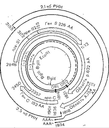
Fig. 48. Genome STRUCTURE OF THE hepatitis B virus.
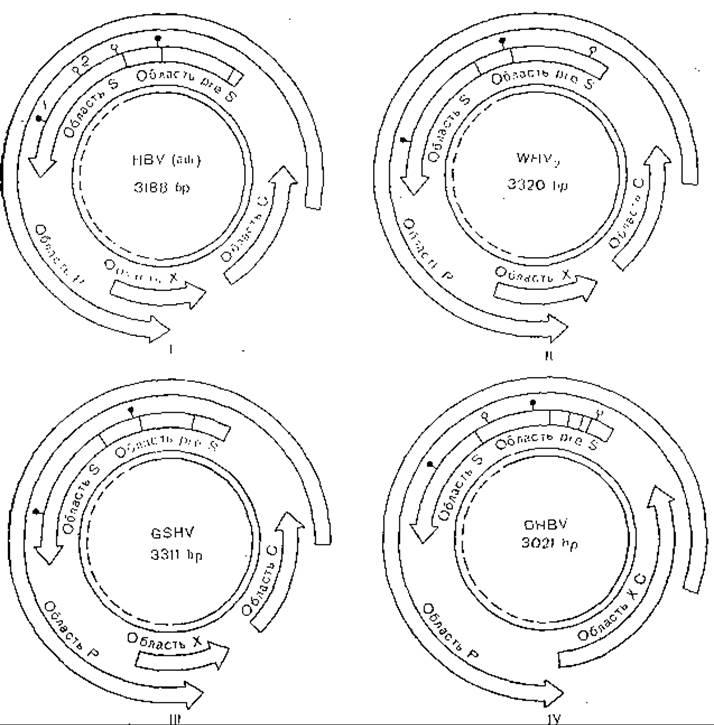
Fig. 49. Genome structure of hepadnaviruses,
I — human hepatitis B virus; II–IV — animal viruses. The inner double circle represents viral DNA, with the single-stranded region indicated by a dashed line; 1 — conserved glycosylation regions; 2 — non-conserved glycosylation regions; HBV — hepatitis B virus; WHV — woodchuck hepatitis virus; GSHV — ground squirrel hepatitis virus; DHBV — duck hepatitis virus.
The HBsAg of human and woodchuck hepatitis viruses show the closest Homology, reaching up to 74% [Galibert F. et al., 1981]. Both proteins are immunologically related [Mill-man I. et al., 1982]. Similarly, the HBcAg of all known mammalian hepadnaviruses exhibits immunological cross-reactivity, which is not the case for HBsAg. The duck hepatitis virus shares no immunological relationship with mammalian hepadnaviruses.
The Replication of hepadnaviruses is quite complex. It involves virion adsorption, endocytosis and membrane fusion, completion of the incomplete DNA strand, gene Transcription and Translation, genome replication via an RNA intermediate, as well as the assembly and maturation of progeny virions. The attachment protein (pre-S) interacts with hepatocyte receptors, which also serve as receptors for polymerized serum albumin. However, the hepatotropism of hepatitis B viruses is not absolute. In experiments with chimpanzees infected with hepatitis B, the virus was detected in peripheral blood lymphocytes during The Development of chronic disease, while in woodchucks infected with woodchuck hepatitis virus, it was regularly found in T AND B lymphocytes and in the Bone Marrow [Korba B. et al., 1986].
The entry of virions into the cell via endocytosis and the fusion of the viral envelope with the cell membrane apparently differ little from similar processes in other enveloped viruses. Reaching the Cytosol first, the viral cores subsequently penetrate the cell nuclei, where viral gene transcription occurs—presumably catalyzed by cellular polymerase—followed by their transport into the Cytoplasm, the synthesis of virus-specific proteins, and the retrograde transport of some of these proteins back into the nuclei. Unfortunately, these processes remain poorly understood, and the proposed mechanisms are rather speculative than experimentally grounded. However, certain facts evidencing the Processing of virus-specific proteins are undeniable. HbsAg undergoes extensive glycosylation, and HBcAg undergoes proteolytic Cleavage to yield HBeAg [Petit M., Pillot J., 1985]. The pre-S region can be read from several initiation codons, resulting in the production of the p24, p27, and p35 proteins [Rutter W. et al., 1984; Persing D. et al., 1985] as well as pre-S itself [Schaeffer E. et al., 1986]. Significant divergence of the pre-S gene among different hepadnaviruses has been noted [Neurath A. et al., 1984], which presumably reflects differences in hepatocyte receptors for these viruses and accounts for the narrow host range of each hepadnavirus. Additionally, the pre-S1 and pre-S2 regions within this domain possess their own initiation codons.
Gene transcription is preceded by the completion of the incomplete strand, which is mediated by virion polymerase immediately after the removal of the outer envelope, making the core accessible to nucleotide penetration. The subsequent fate of such circular double-stranded DNA can follow one of two paths: either productive infection proceeds further, or an integrative process is established [Zhdanov V. M., 1974]. It is hypothesized that in the latter case, the region of the X gene serves as an integration site, and upon translation following integration with the cellular genome, fusion proteins of viral and cellular genes are formed, akin to the gag-onc genes in Retroviruses [Meyers M. et al., 1986]. The hepadnavirus genome or its fragments integrate into multiple sites of cellular DNA. Concurrently, genes—especially the gene HBcAg, with the exception of the HBsAg gene—become methylated, which prevents their transcription. This is presumably associated with the predominant or exclusive expression of the HBsAg gene in transplanted hepatoma lines as well as in HBsAg carrier organisms [Miller R., Robinson W., 1983]. The genome integration region corresponds to a single-stranded segment of the genome; therefore, the completion of this strand must necessarily precede integration [Koshy R. et al., 1983]. This process is accompanied by fragmentation and rearrangement of the integrated viral DNA segments. Among all genes, only the complete transcriptional unit of the HBsAg gene remains "intact," which is also likely the reason for the expression of solely this gene from the integrated provirus [Shaw Y. et al., 1984]. Virus-specific repeat sequences are absent from the flanks of the integrated genes. At the same type, cellular repeats have been detected, along with the integration of only a portion of the pre-S region alongside the Integration of the full HBsAg gene [Yaginuma K. et al., 1985]. The integration of the hepadnavirus genome underlies the development of primary liver cancer, although its mechanisms remain unclear. It is worth noting that known hepadnaviruses exhibit varying oncogenic potentials in this regard. These potentials are most pronounced in woodchuck hepadnaviruses: 50% of infected animals develop primary liver cancer.
Several transplantable mouse primary liver cancer cell lines containing integrated hepatitis B virus genomes have already been established. Cells from one such line, PLC/PRF/5, were inoculated into nude mice, which subsequently developed tumors. Along with HBsAg, these tumors began to produce HBcAg. Thus, the genes for HBcAg were retained in the cancer cells and their expression was triggered upon transplantation into nude mice [Marquardt A. et al., 1984].
Hepadnavirus replication is unusual. A replication model involving The formation of an RNA intermediate has been proposed [Summers J., Mason W., 1982; Summers J., 1984] (Fig. 50). Following the entry of virions into cells and the disintegration of the outer envelope, virion polymerase completes the light DNA strand (genes encoding virus-specific Protein Synthesis reside on the heavy DNA strand), rendering the DNA accessible for transcription. This process generates a full-length plus-strand and an RNA replicative intermediate, or pregenome. Concurrently, genes from 4 reading frames are transcribed and translated to yield virus-specific proteins, including the virus-specific polymerase. The pre-nucleocapsid (immature nucleocapsid), formed presumably via self-assembly, incorporates the pregenomic RNA and the polymerase. The latter drives reverse transcription and the Synthesis of the full-length minus-strand, while the pregenomic RNA is simultaneously degraded. Subsequently, the plus-strand is synthesized and a signal for nucleocapsid maturation is triggered. At some preceding stage, a terminal protein covalently linked to the 5' end of the full-length DNA strand penetrates the immature nucleocapsid, forming the circular conformation of the viral genome. The plus-strand, as is well known, is synthesized incompletely, and upon completion of synthesis, an outer lipid envelope embedded with HBsAg supramolecular structures is formed.
Such are the MAIN STAGES OF the complex replication cycle of hepadnaviruses. The core tenets of this hypothesis can be considered proven. In particular, an RNA intermediate has been detected in infected hepatocytes, the replicative complex has been isolated, its products obtained in in vitro assays, the actinomycin D resistance of minus-strand synthesis on an RNA intermediate template demonstrated, and nucleocapsid precursors even visualized via electron microscopy. Certain details of hepadnavirus replication are conveniently studied using the duck hepatitis virus (Peking duck hepatitis virus) because it is transmitted vertically (transovarially), with the entire synthesis process completing during embryo maturation within the fertilized egg [O’Connel A. et al., 1983; Urban M. et al., 1985]. Replication of this virus has also been reproduced in primary duck hepatocyte cultures [Tuttleman J. et al., 1986]. Other models for studying hepadnaviruses have also been developed. Experiments on chimpanzees infected with human hepatitis B virus and on woodchucks infected with woodchuck hepatitis virus revealed the accumulation of viral DNA and RNA in peripheral blood lymphocytes during the chronic course of the disease. DNA was present in the form of episomes in both lymphocytes (B AND T cells) and bone marrow, but not in macrophages. These findings reveal new aspects of hepatitis B pathogenesis [Korba B. et al., 1986]. Reports by E. Liang et al. (1986) on the cultivation of human hepatitis B virus in chicken embryos treated with non-toxigenic streptococci require further verification.
Thus, multiple nucleic acid synthesis steps occur during hepadnavirus replication: completion of the incomplete DNA strand (DNA-dependent DNA Synthesis), transcription from 4 open reading frames, synthesis of a full-length RNA strand (pregenome, RNA intermediate), reverse transcription (RNA-dependent DNA synthesis), synthesis of the incomplete light chain (DNA-dependent DNA synthesis), and, finally, degradation of the RNA intermediate. To this must be added two further processes occurring in cells infected with human or woodchuck hepatitis virus: the synthesis of the replicative intermediate (RNA?) of the defective delta virus [Rizzetto M. et al., 1984], which frequently accompanies hepatitis B, and the synthesis of progeny DNA strands of this virus.

Fig. 50. Replication of the hepatitis B virus genome (hypothetical diagram).
a — DNA polymerase; b — covalently linked protein; 1 — hepatitis B virion; 2 — cell entry and DNA maturation; 3 — transcription, formation of full-length RNA plus-strand (pregenome); 4 — pregenome packaging, formation of immature core; 5 — reverse Transcription of DNA minus-strand, degradation of pregenome, formation of intermediate cores; 6 — plus-strand synthesis and packaging signal; 7 — virion maturation and egress from the cell.
The putative polymerase is encoded within the relatively small hepadnavirus genome. This region occupies the major part of the genome, completely overlapping the pre-S-S region and partially overlapping the C and X regions. There is good reason to believe that the Open Reading Frame of this gene is shifted relative to the reading frame of the pre-S-S region and those of the other two partially overlapping genes. The protein encoded by this gene consists of 832 amino acid residues [Ohno S., 1984] and has been identified in hepatocarcinoma cells [Will H. et al., 1986]. The polyfunctional nature of this protein is unquestionable, if only because it mediates at least 5 processes: completion of the light DNA strand in the virion, or more precisely, in the nucleocapsid (DNA-dependent DNA synthesis), synthesis of the RNA intermediate, or pregenome (DNA-dependent RNA Synthesis), synthesis of the full-length heavy DNA strand (reverse transcription), synthesis of the incomplete light strand (DNA-dependent DNA synthesis), and degradation of the DNA intermediate (RNase H function). It is hardly surprising that these Functions bring hepadnavirus polymerase close to retroviral Reverse Transcriptase, perhaps even indicating a phylogenetic relationship between these polyfunctional proteins.
Gene transcription from the 4 open reading frames is likely mediated by cellular polymerases [Rutter W. et al., 1984]. The Role of hepadnaviruses in the RNA Replication of their defective satellite—the delta virus—remains entirely unclear.
Later, researchers successfully cultivated the delta virus in the presence of duck hepatitis virus. Fertilized duck eggs were infected with duck hepatitis virus, and 2- to 6-month-old birds were subsequently inoculated with serum containing the delta virus. Delta antigen and delta RNA were detected in the majority of the birds [Ponzetto A. et al., 1986]. It is also pertinent to note here that The Mechanism of the damaging effect of hepatitis B virus on hepatocytes undergoing replication remains unclear. It is possible that these lesions are not caused directly by the virus, but rather result from cellular immune responses against viral antigens expressed on cell membranes. We previously suggested that acute liver dystrophy represents the rejection of an entire organ As a result of these immunopathological processes. However, viral integration accompanied by HBsAg production and its secretion into the bloodstream does not lead to hepatocyte damage. It appears, firstly, that such cells proliferate intensively and thereby enhance the regenerative capacity of the liver parenchyma, while liver cancer is a peculiar penalty—neither obligatory nor frequent. Secondly, the presence of an integrated virus with disrupted genes, aside from the "intact" and even amplified HBsAg gene, serves as a sort of defense against progressive infection caused by viral replication. Thus, we arrive at a paradoxical conclusion: prolonged antigen carriage guarantees that the blood is non-infectious. These considerations are well illustrated by the hepatitis B virus transgenesis model in mice [Chisari F. et al., 1985].
HBsAg carriage is unique in character; assuming that an HBsAg particle consists of several hundred protein molecules and the particle titer can reach 1012–1013 per 1 ml of blood, these concentrations are comparable to those of the most abundant blood proteins. Besides the aforementioned blocking, fragmentation, and rearrangement of other viral genes, this is likely explained by the fact that HBsAg gene integration occurs in a chromosomal region subject to intensive Gene Expression. However, this explanation was discarded after HBsAg particles were shown to be capable of reacting and binding with polymerized albumin.
Up to this point, the Discussion has focused on vertebrate hepadnaviruses. Cauliflower mosaic virus has been shown to share similarities with hepadnaviruses [Marsh L. et al., 1985]. Although the genome of this virus is 3 to 4 times larger than that of hepadnaviruses, it is organized on the same principle: it possesses double-stranded DNA with an incomplete second strand. Its putative replication model involves the formation of an RNA intermediate and reverse transcription [Pfeiffer P., Hohn T., 1983]. Further experimental verification revealed a replicative RNA intermediate with a sedimentation coefficient of 35S, encapsidated into DNA-containing virion-like structures, RNA-DNA hybrids, as well as heterogeneous DNA (minus strands) 6,000–8,000 NUCLEOTIDES in length (representing the full genome), actinomycin D-resistant RNA-DNA syntheses, and antibiotic-sensitive DNA-DNA syntheses—that is, the core stages of virus-induced syntheses characteristic of hepadnaviruses.
Cauliflower mosaic virus, which infects members of the family Cruciferae, possesses two reading frames: one directs the synthesis of a 1.9 kb transcript (region VI) encoding an inclusion body protein (presumably analogous to the hepadnavirus HBsAg), while the other directs the synthesis of an 8.2 kb transcript of unclear function. These data were obtained from Experiments on the integration of the complete cauliflower mosaic virus genome into plant Chromosomes using the Ti plasmid [Shewmaker C. et al., 1985]. Furthermore, the replication of this virus is strikingly similar to that of retroviruses, which in turn is explained by the resemblance between the DNA and RNA genomes of these viruses [Dixon L. et al., 1986]. This similarity was demonstrated in recombination experiments between weakly divergent (5%) cauliflower mosaic virus strains. It is worth noting that the pol gene of the AIDS virus, murine oncoviruses, and avian oncoviruses shares homology with the putative polymerase gene of hepatitis B virus and cauliflower mosaic virus [Seiki M. et al., 1983]. At the same time, cauliflower mosaic virus and other viruses belonging to this group (9 viruses are currently known) exhibit significant differences from hepadnaviruses. Virions are isometric (like hepadnavirus capsids), and the capsid is composed of a single protein with a molecular weight of 42,000. Virion diameter is approximately 50 nm. Transcription yields either a full-length RNA strand (replicative intermediate) or 7 fragments likely corresponding to 7 genes, including a protein with a molecular weight of about 60,000 that accumulates in the cytoplasm as inclusions of unknown function. It was subsequently shown that the 5' end of the minus-strand serves as the initiation site for reverse transcription, primed by a protein covalently bound to the 5' end of the DNA [Molnar-Kimber K. et al., 1984].
Hepatitis B virus DNA synthesis initiates in the region of the first direct repeat (DR1), with the terminal protein presumably acting as the primer. In contrast, second-strand synthesis initiates in the region of the terminal repeat (DR2), but its 5' end is attached to an oligoribonucleotide containing DR1. Fig. 51 illustrates the mechanisms of hepadnavirus genome replication and transcription based on these considerations, while Fig. 52 demonstrates the Similarities and differences between hepadnavirus and retrovirus replication.
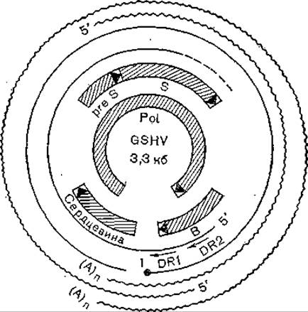
Fig. 51. Transcriptional and translational maps of the mammalian hepadnavirus (GSHV) genome. DNA strands are represented by thin lines; 1 — protein at the 5' end of the minus-strand; two classes of intracellular RNAs are represented by wavy lines.
While the 3 mammalian hepatitis B viruses display substantial similarity, duck hepatitis virus is less closely related to them. For instance, it lacks the gap between genes 5 and 8, which form a single reading frame [Mandaray E. et al., 1984]. Moreover, experiments specifically with this virus demonstrated structural homology between protein P products and avian reverse transcriptase (we will return to this issue later). In subsequent studies [Miller R., Robinson W., 1986], it was revealed that the Amino Acid Sequence of the putative hepatitis virus reverse transcriptase shares homology with retroviral Amino acid sequences. In particular, this was uncovered when comparing the genomes of human, woodchuck, ground squirrel, and duck hepadnaviruses with corresponding genomic regions of type C retroviruses, endogenous DNA sequences of type C retroviruses, and endogenous human DNA sequences. Especially pronounced is the homology of 67 nucleotides in region 45 of human hepatitis B virus with the long terminal repeats of retroviruses, whereas the homology of short heptanucleotide segments reaches 90–97%. A high degree of homology (41%) was also found between 98 amino acid residues of the hepatitis B virus core protein and the C-terminal portion of the gag gene P30 protein of murine type C retroviruses. All these data lead to the conclusion that hepatitis B virus and retroviruses share a common origin, with hepatitis B virus representing a branch of the retroviral evolutionary tree.
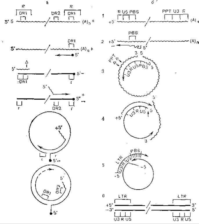
Fig. 52. Model of viral DNA synthesis on an RNA template in hepatitis B viruses (a) and retroviruses (b).
Wavy line — RNA; straight line — DNA, R — terminal sequences, terminal repeats, PBS — primer binding site (+RNA), PPT — polypurine tract, r — complementary DNA sequence forming short duplexes.
In conclusion, it is appropriate to mention the scheme by [Varmus H., 1983], which demonstrates the high degree of similarity in the life cycles of hepadna-, caulimo-, and retroviruses. A genealogical tree, based on the monophyletic origin of vertebrate hepadnaviruses, was constructed by K. Kodama et al. (1985). The further evolution of hepatitis B virus (of various serological subtypes) was determined by comparing plasmid pHB320 carrying the polymerase gene of hepatitis B virus subtype ayw [Pumpen P. et al., 1982] with previously obtained nucleotide sequences for various serological subtypes. Frequency analysis of synonymous nucleotide substitutions in the non-overlapping frequent genes C and P (regions 840–1373, 1903–2307, 2462–2854) was chosen as the research method. Based on the obtained results, V. Bychko (1986) constructed a Phylogenetic Tree (Fig. 53), which differs somewhat from the scheme of K. Kodama et al. According to V. Bychko's scheme, all hepatitis B virus variants diverge into 3 phylogenetic lineages, with subtypes ayw and adw being as phylogenetically close as the WHV and GSHV viruses, whereas subtype adr is even more distant phylogenetically from the first two (Fig. 54).
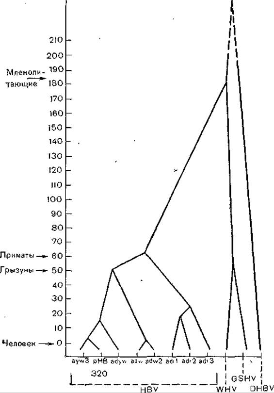
Fig. 53. Phylogenetic tree of hepadnaviruses constructed based on the frequency analysis of synonymous nucleotide substitutions in non-overlapping regions of genes C and P. The ordinate represents time (million years).
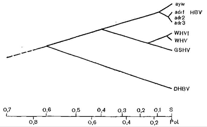
Fig. 54. Phylogenetic tree of hepadnaviruses. The relative distances between hepadnaviruses reflect the substitution rate of Pol and S ORFs. The abscissa represents map units.
It is now evident, of course, that this scheme does not encompass the entire group of viruses, and new members will undoubtedly be discovered in the coming years. What is the genealogical relationship between animal- and plant-infecting viruses? Are the multifunctional enzyme of these viruses and the reverse transcriptase of retroviruses products of divergent evolution or the result of convergence? What ecological links (perhaps mediated by Arthropods) have enabled these viruses to occupy such disparate ecological niches? Why has narrow specialization (a restricted host range) not hindered the spread of these viruses among evolutionarily distant host species? All these and other questions demand comprehensive and multifaceted research—ranging from the ANALYSIS OF GENE structures and their encoded proteins to sophisticated ecological analysis unconstrained by established traditions.
Last update: 13/08/2026
Editorial and Educational Adaptation: This material has been compiled based on the primary/original source text. The project team performed an editorial review, corrected technical inaccuracies, structured sections, and adapted the content for an educational format.
What was processed:
- elimination of formatting defects (OCR errors, structural breaks, corrupted characters);
- editorial organization of content;
- standardization of terminology in accordance with academic sources;
- verification of factual statements against the original source text.
All mentions of the author, publication year, and origin of the primary text have been preserved in accordance with the source.